Anatomy Lecture #7 - Thorax, Heart, and Circulation
 Call Kai
 Learn
Learn Practice Test
 Spaced Repetition
 Match
Match Flashcards
 Knowt Play
1/71

There's no tags or description
Looks like no tags are added yet.
Name | Mastery | Learn | Test | Matching | Spaced | Call with Kai | Chat |
|---|
No analytics yet
Send a link to your students to track their progress
72 Terms
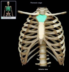
Manubrium
What is this bone?

Sternum
What is this bone?

Sternal Angle
What is this bony landmark?

Costal Cartilage
What is this structure?

Xiphoid
What is this bony landmark?

1-7
How many true ribs?

8-12
How many false ribs?

Costovertebral Joint
What is this bony articulation?

Costotransverse Joint
What is this bony articulation?

Costovertebral: Head of rib
Costotransverse: Tubercle of a rib
What do these joints articulate with?

Supraspinous Fossa
What is this?

Superior Border
What is this?

Coracoid Process
What is this?

Acromial Process
What is this?

Glenoid Cavity
What is this?

Spine
What is this?

Infraspinous Fossa
What is this?

Lateral Border
What is this?

Medial Border
What is this?

Neck of Scapula
What is this?

Pulmonary
What cavity is this?

Mediastinum
What cavity is this?

Abdominal Cavity
What cavity is this?
Phrenic Nerve - C3, C4, C5
What nerve innervates the diaphragm and at what level?
External Intercostal, Inspiration
What is this muscle and it's function?

Internal Intercostal, Deep forced expiration
What is this muscle and it's function?

Pectoralis major
What is this muscle?

Pectoralis Minor
What is this muscle?

Serratus Anterior; Holds scapula to ribcage
What is this muscle? What does it do?

Scalenes
What is this muscle?

Diaphragm; Musculotendinous sheet dividing the thoracic cavity from the abdominal cavity
What is this/what does this do?

Caval, Esophageal, Aortic
What are the 3 apertures of the diaphragm?
Left Lung
Which lung is smaller?

Lingula; Moves into costomediastinal recess during inspiration
What is this structure? What does it do?

Cardiac Notch; Accommodates heart
What is this structure? What does it do?

Hilum of Lungs; Doorway in and out of lung tissue, connects lung to mediastinum, and contains pulmonary vessels, nerves and bronchi
What is this structure? What is it?

Bronchopulmonary Segments
What are these segments?
Pleura
Fibroserous membrane surrounding each lung and lining pulmonary cavities

Parietal Pleura
More superficial

Visceral Pleura
Deeper, closer to lungs

Pleural Cavity
Space within the pleural sac between visceral and parietal

Pleuritis
inflammation of the pleura
Pathway of air through mouth
- Oral cavity
- Oropharynx
- Laryngopharynx
- Larynx
- Trachea
- Primary Bronchi
- Secondary Bronchi
- Tertiary Bronchi
- Conducting Bronchioles
- Terminal Bronchioles
- Respiratory Bronchioles
- Alveolar sac
- Alveoli
Pathway of air through nose
- External nares
- Nasal cavity
- Internal nares
- Nasopharynx
- Oropharynx
- Laryngopharynx
- Larynx
- Trachea
- Primary Bronchi
- Secondary Bronchi
- Tertiary Bronchi
- Conducting Bronchioles
- Terminal Bronchioles
- Respiratory Bronchioles
- Alveolar sac
- Alveoli
Hyaline Cartilage
What cartilage does secondary bronchi have most of?
Bronchial Artery
What artery supplies the lung with oxygenated blood?

Epiglottis
What is this structure?
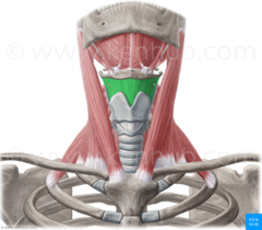
Thyrohyoid muscle
What is this structure?

Cricothyroid muscle
What is this structure?

Hyoid bone
What is this structure?

Cricoid Cartilage
What is this structure?

Tracheal membrane
What is this structure?

Cricothyroid membrane
What is this structure?
Carina
What is the internal split of the bronchi?
Tracheal Bifurcation
What is the external split of the bronchi?
Type II pneumocyte
Which pneumocyte secretes surfactant?
Type I pneumocyte
Which pneumocyte absorbs surfactant?
Emphysema
Destroyed or damaged alveoli
Heart, great vessels, pericardium, esophagus, trachea, thymus
What does the mediastinum contain?
Epicardium
outermost layer of the heart
Myocardium
Thickest and middle layer of heart
Endocardium
Most deep layer of heart
Phrenic nerve
Innervates pericardium

Trabeculae Carnae
What muscle is this?
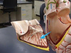
Pectinate
What muscle is this?

Papillary
What muscle is this?

Myocardium
What muscle is this?

Auricle
What structure is this?
Cardiac skeleton
Dense fibrous connective tissue that separates chambers and provides anchoring points (framework for valves)
Auscultation
Heart sounds
Blood flow through heart
1-Superior & Inferior Vena Cava, 2-R Atrium, 3-Tricuspid Valve, 4- R Ventricle, 5-Pulmonary Valve, Pulmonary Trunk 6-Pulmonary Artery, Arterioles, Capillaries 7- Lungs-pick up oxygen, Venules 8-Pulmonary Veins, 9- L Atrium, 10- Mitral Valve (Bicuspid), 11-L Ventricle, 12- Aortic Valve, 13-Aorta, 14- Body
Conduction pathway
SA node, internodal bundle, AV node, bundle of his, R and L bundle branches, Purkinje fibers