14. Nervous System: Cerebral Vasculature, Ventricles & CSF
 Call Kai
 Learn
 Practice Test
 Spaced Repetition
 Match
 Flashcards
 Knowt Play
1/67

There's no tags or description
Looks like no tags are added yet.
68 Terms
arch of aorta branches
branchiocephalic trunk → short, bifurcates into
right common carotid artery
right subclavian artery
left common carotid → neck + head
left subclavian artery → neck + head

vertebral arteries
branches of left + right subclavian arteries - ascend within neck
travel within transverse foramina of cervical vertebra
foramina = holes that they run through
enter skull via magnum foramina
makes right angle turn after exiting last hole
right + left converge to form basilar artery

vertebro-basilar system
provides blood to brain
left + right vertebral arteries join to form single basilar artery
basilar artery → on anterior surface of pons

basilar artery
supplies arterial branches to brainstem structures
on anterior surface of pons
formed by convergence of vertebral arteries
already inside skull
divides to form posterior cerebral arteries → contribute to Circle of Willis
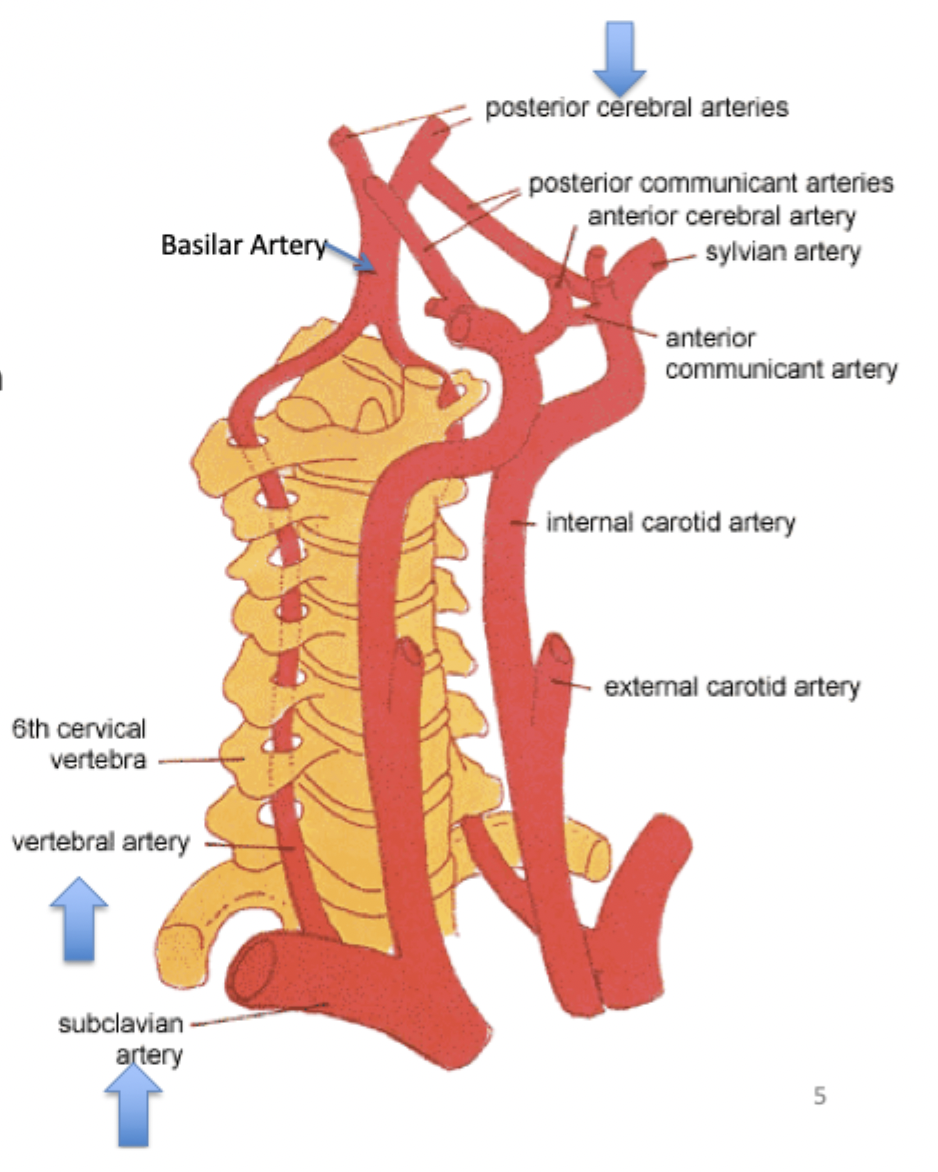
common carotid artery branches
bifurcates into 2 arteries
external
supplies blood to neck, lower jaw, face
internal
enters skull via carotid canal → supplies cerebrum
3 major branches
ophthalmic artery
anterior cerebral artery
middle cerebral artery

basilar artery
(1)
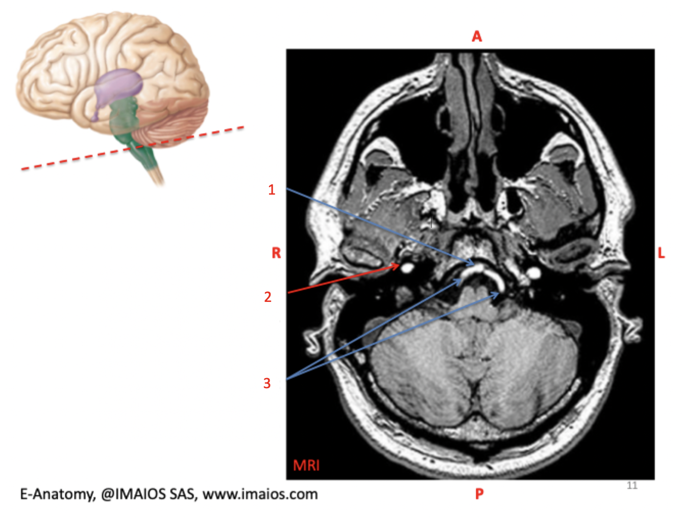
internal carotid artery
(2)

vertebral arteries (left and right vertebral arteries)
(3)

internal carotid arteries (left and right internal carotid arteries)
(1) bookended by temporal lobes

basilar artery
(2)

basilar groove
(3) contains basilar artery

fourth ventricle
(4)

Circle of Willis
main blood supply to brain via vertebral + internal carotid arteries
anastomoses b/w 2 major cerebral arterial systems
anterior cerebral arteries connected by anterior communicating artery
blood flow joins + mixes
surrounds optic chasm + infundibular stalk (to pituitary gland)
posterior to optic chiasm
anterior to midbrain

anterior cerebral artery
(1)

optic chiasma (optic chiasma)
(2)

infundibulum
(3)

right posterior cerebral artery
(4)

basilar artery
(5)
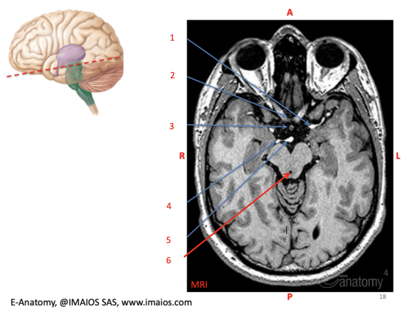
cerebral aqueduct
(6)

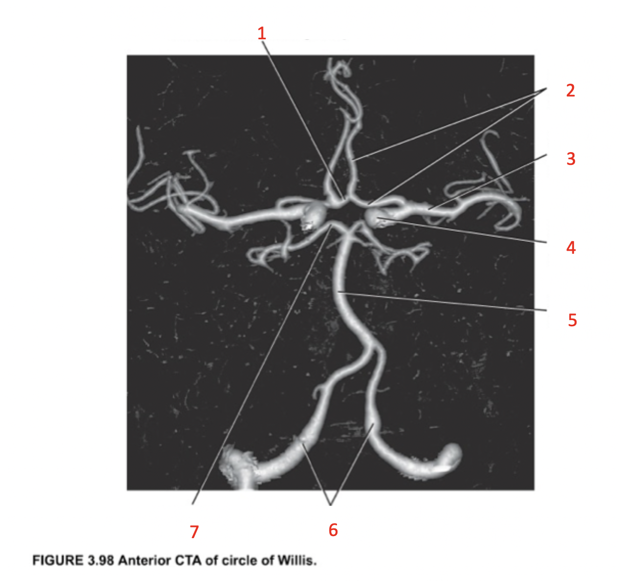
anterior communicating artery
(1)
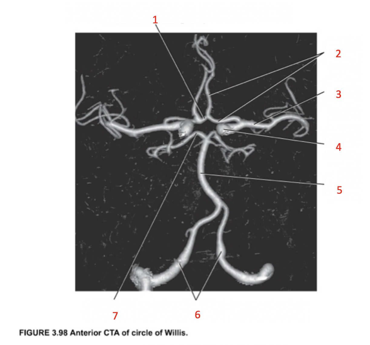
anterior cerebral artery
(2)
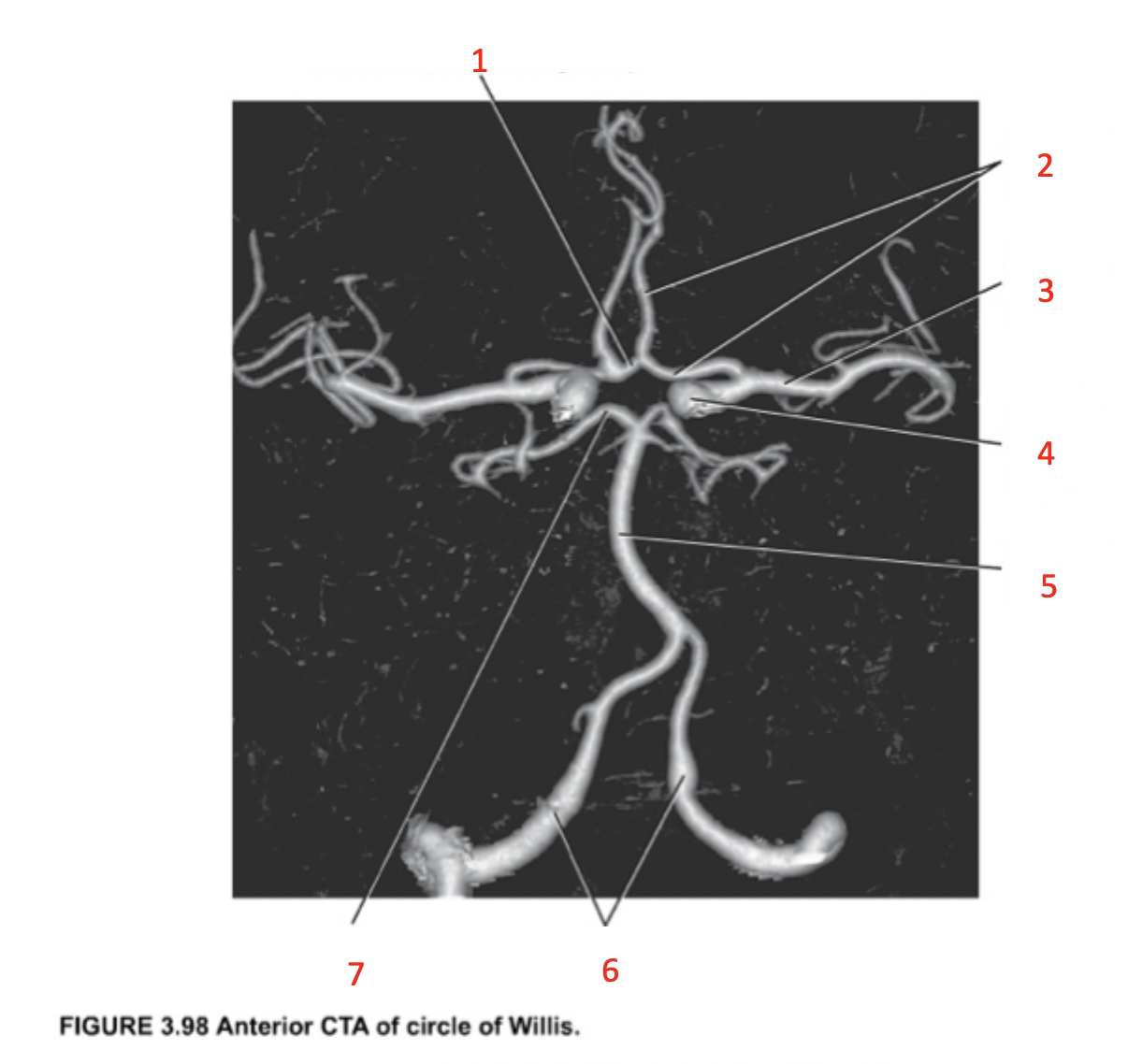
middle cerebral artery
(3)
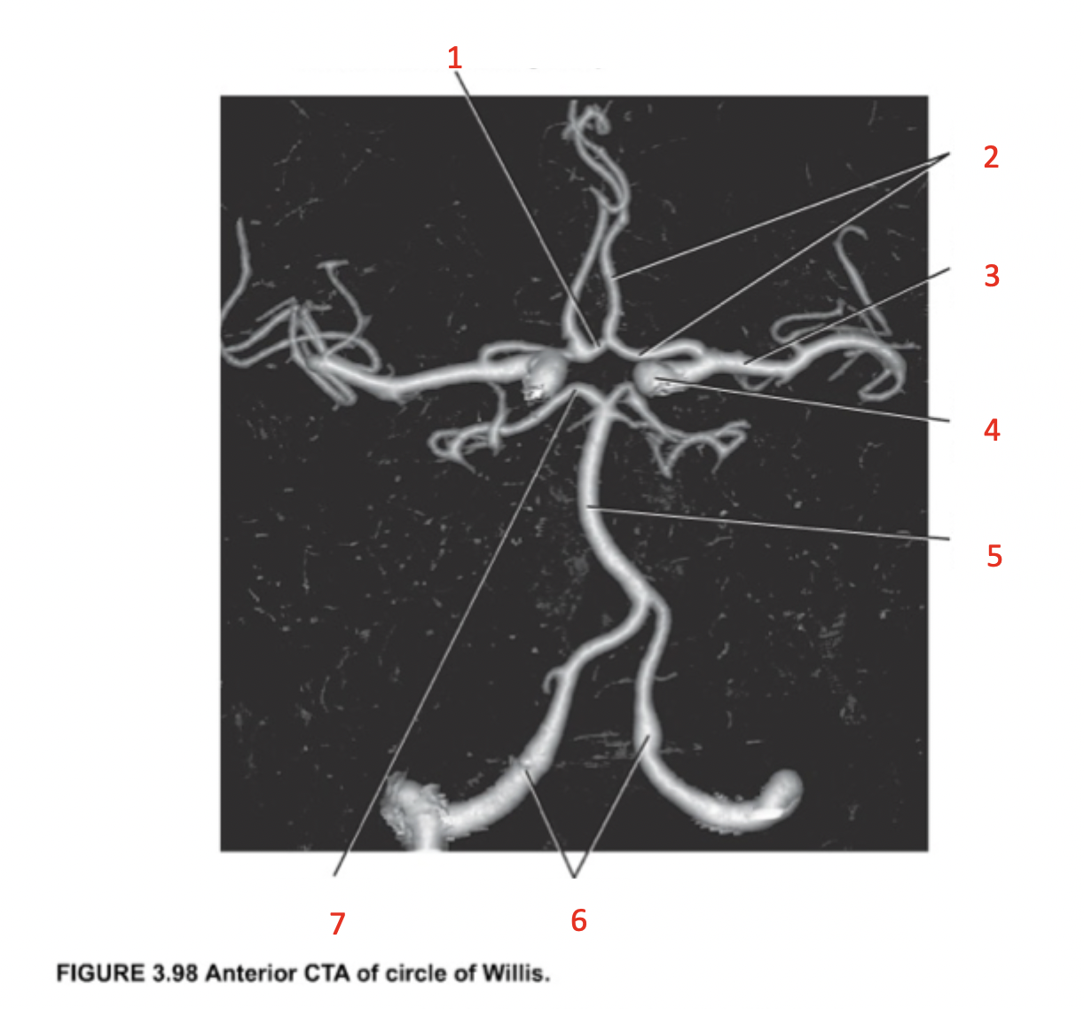
internal carotid artery
(4)

basilar artery
(5)

vertebral arteries
(6)

posterior cerebral artery
(7)
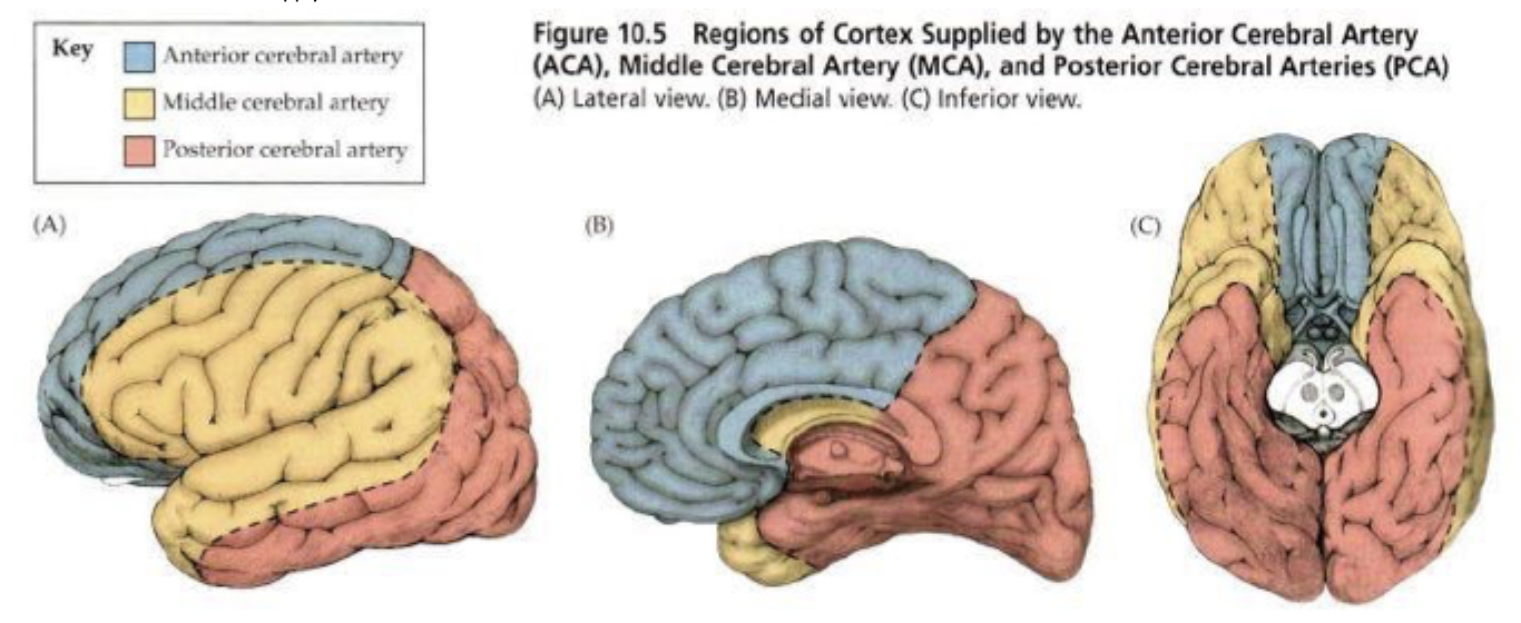
cerebral artery distribution
distribution of where cerebral arteries will supply blood to
middle cerebral artery
runs through crevice b/w temporal + frontal lobes
numerous branches burrow out from Circle of Willis from crevice
burrows in to supply blood to brain
middle cerebral artery
(1)
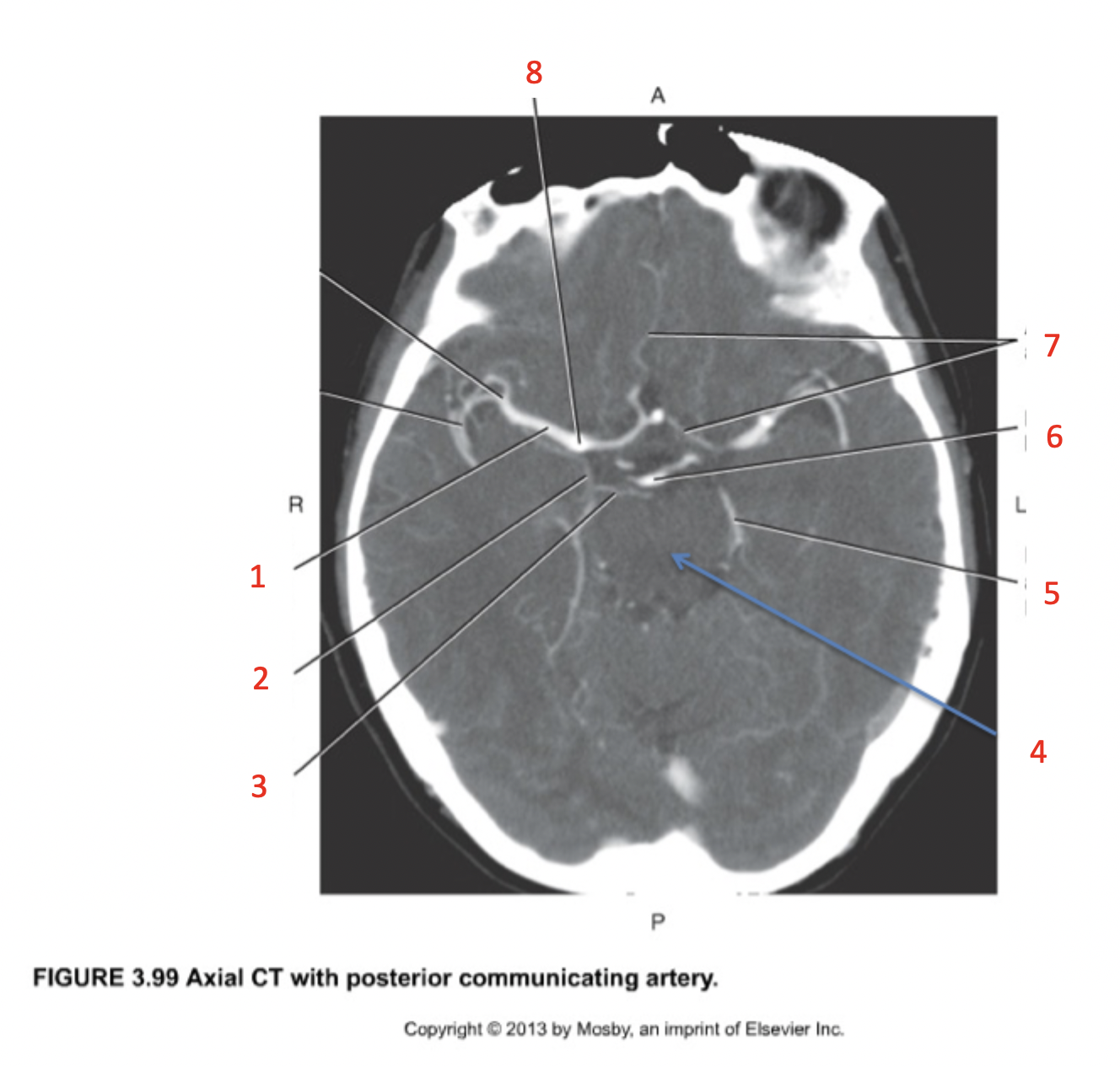
posterior communicating artery
(2)
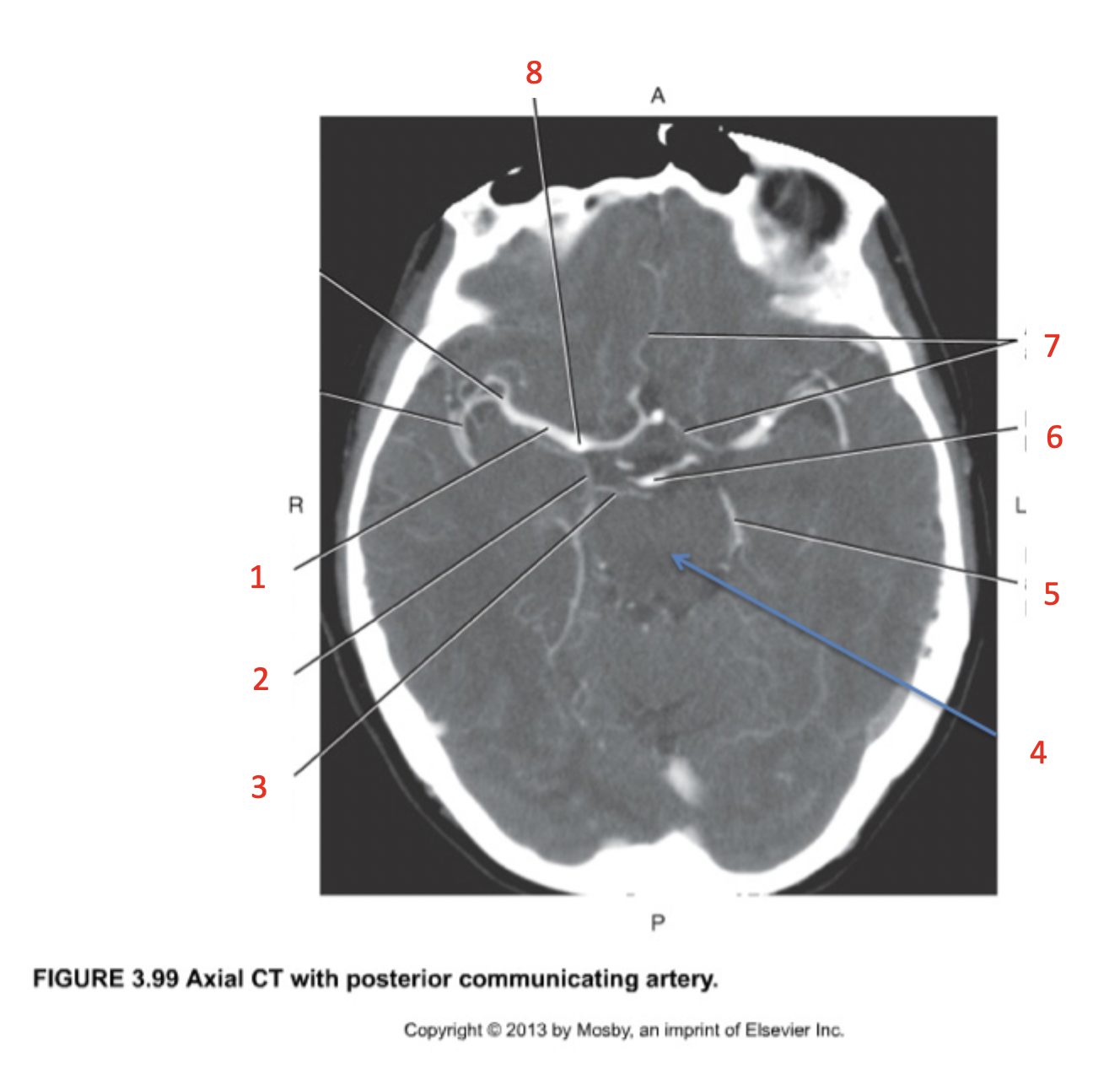
posterior cerebral artery
(3)

midbrain
(4)
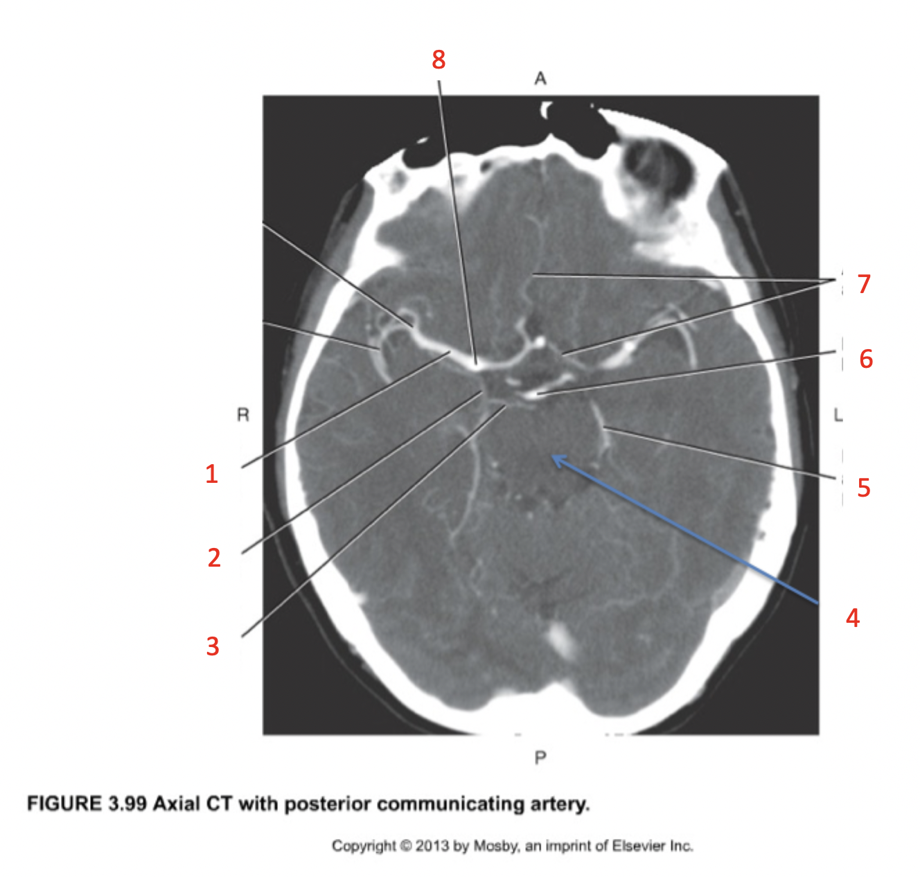
posterior cerebral artery
(5)

basilar artery bifurcation
(6)

anterior cerebral artery
(7)

internal carotid artery
(8)

brain blood supply
brain = 2% body weight → consumes 20% of circulating O2 + glucose
highly vascularized
doesn’t have capacity to store glucose → needs constant supply to produce ATP
gas/nutrient/waste exchange occurs at capillary level
only specific substances able to pass capillaries into cerebral tissue → BBB
EXCEPTION = choroid plexus
blood-brain barrier (BBB)
protects brain tissue from contact w/ harmful substances + pathogens
allows only specific substances to pass from capillaries into cerebral tissue
continuous capillaries
have no pores + endothelial cells connected by tight junctions
astrocyte layer
surrounds capillaries to influence permeability to selected substances
substances allowed
rapidly → diffusion
O2
CO2
H2O
hydrophobic substances
slowly → membrane transport
hydrophilic substances
glucose
amino acids
ions
specific proteins + peptides
too large = generally cannot cross → requires transcellular pathway
choroid plexus
capillary network within ventricle walls of brain → creates bulk of CSF via filtration event
continuous capillaries lined w/ ependymal cells → NOT astrocytes
decides what substances can enter/exit
bidirectional movement/transfer of substances
blood plasma filtered via ependymal cell layer → becomes CSF within ventricles
total volume at given time = 150mL
volume produced each day = 500mL
CSF circulates + returns to systemic circulation
metabolites from nervous tissue returned to blood

CSF (cerebrospinal fluid)
filtered blood plasma
constantly produced by ependymal cell layer within ventricles
constantly circulating through ventricular system + into subarachnoid space
functions
facilitates nutrient + metabolite exchange b/w blood + nervous tissue
circulates through connected spaces
cavities in brain = ventricles
spinal cord = central canal
subarachnoid space
maintenance of optimal chemical environment
maintains correct [glucose, ion, etc.] for efficient neuronal signalling
mechanical protection
bathes entire cerebrum + forms layer surrounding brain = shock absorption for brain
CSF within subarachnoid space (b/w skull + brain) = protective fluid cushion for brain
ventricles
cavities in brain within cerebrum filled w/ CSF
1st + 2nd = lateral
within cerebral hemispheres
separated by septum pellucidum, anteriorly
interventricular foramina of Monro → drain into 3rd
3rd
b/w thalami - right in center
CSF drains into 4th via cerebral aqueduct of Sylvius (within midbrain)
4th
posterior to pons
anterior to cerebellum
drains into subarachnoid space via lateral + medial apertures
continuous w/ spinal cord central canal

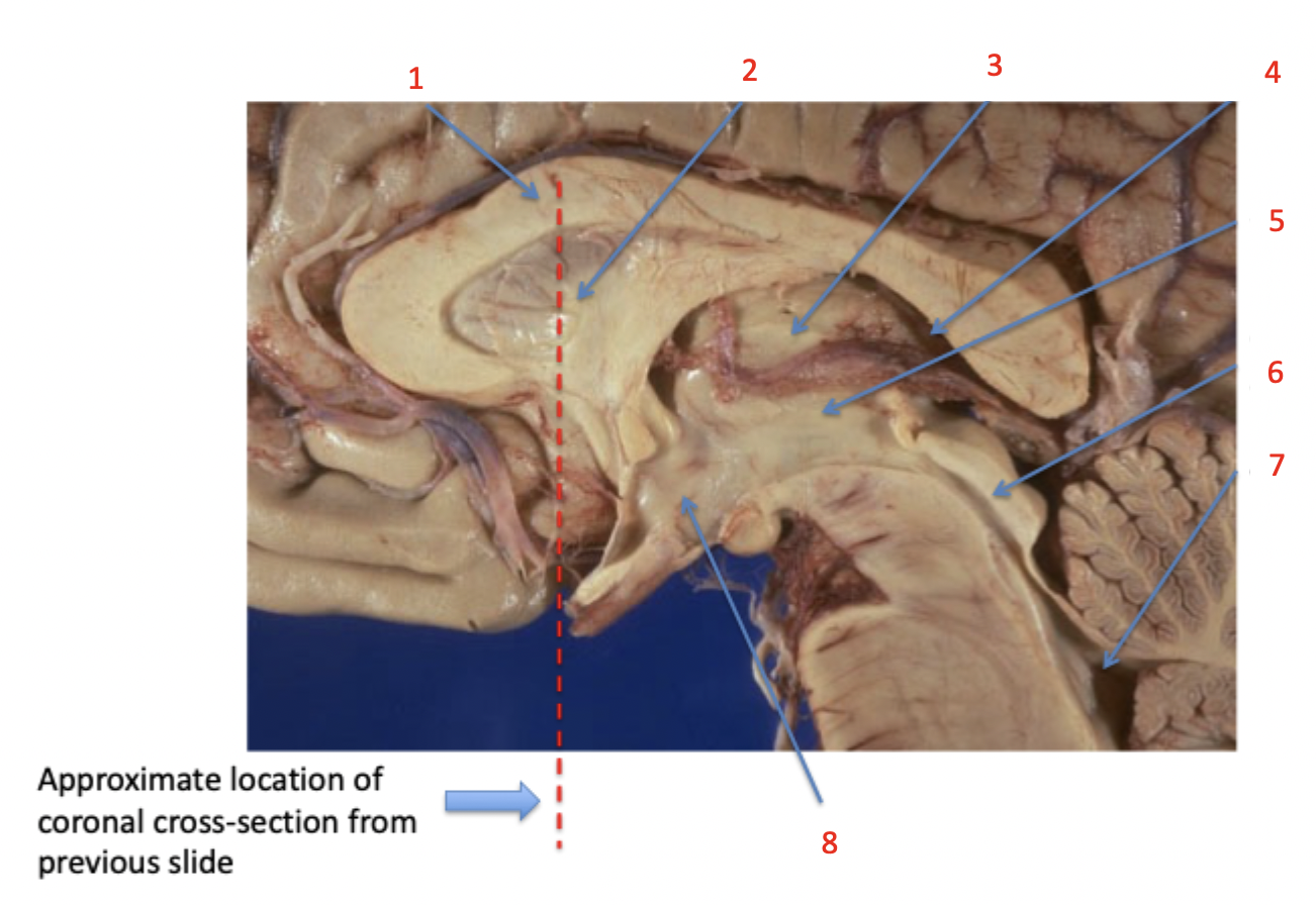
corpus callosum
(1)

septum pellucidum
(2) partition b/w 2 frontal horns of lateral ventricles

thalamus
(3)

lateral ventricle
(4)

third ventricle
(5)

cerebral aqueduct
(6)

fourth ventricle
(7)
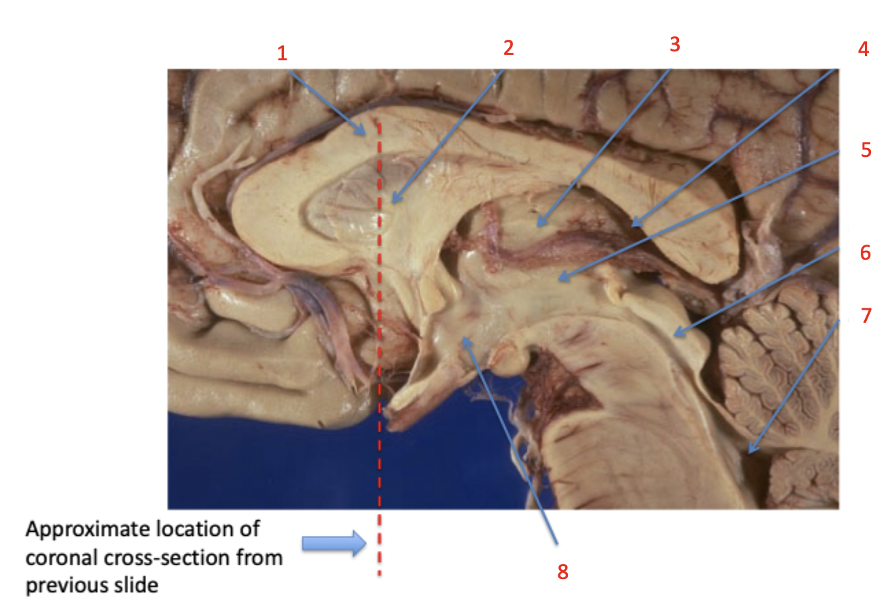
hypothalamus
(8)
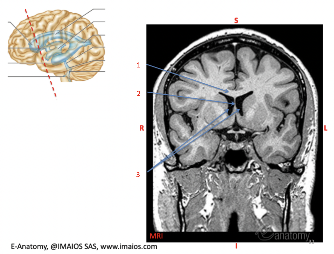
corpus callosum
(1)

septum pellucidum
(2)

lateral ventricle
(3)
caudate
(1)

thalamus
(2)

lateral ventricle posterior horn (occipital horn)
(3)
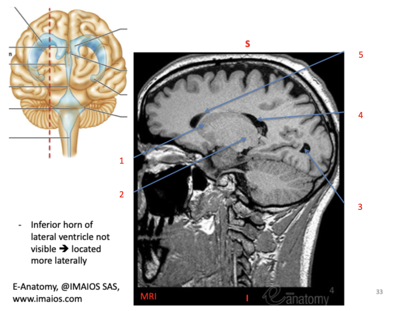
lateral ventricle body
(4)

lateral ventricle anterior horn (frontal horn)
(5)

lateral ventricle anterior horn (frontal horn)
(1)
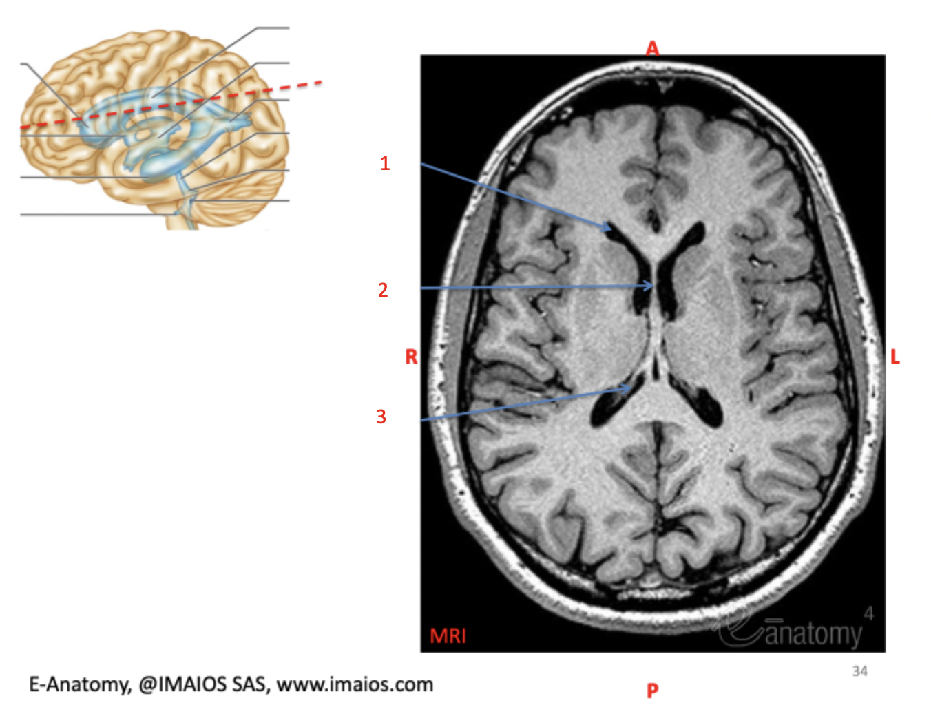
septum pellucidum
(2)

lateral ventricle body
(3)
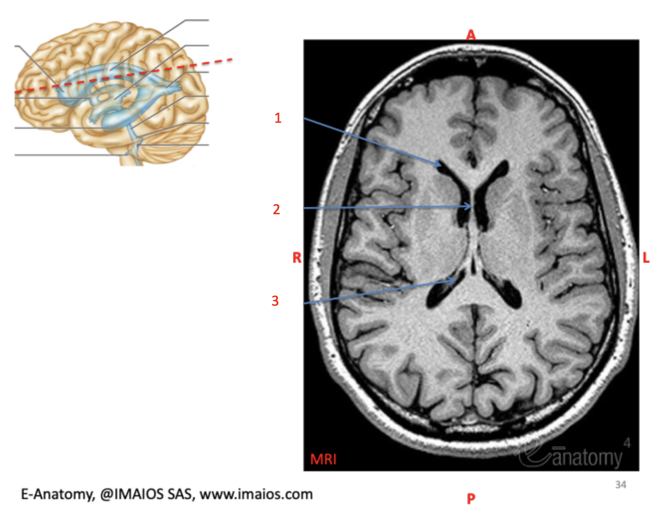
septum pellucidum
(1)
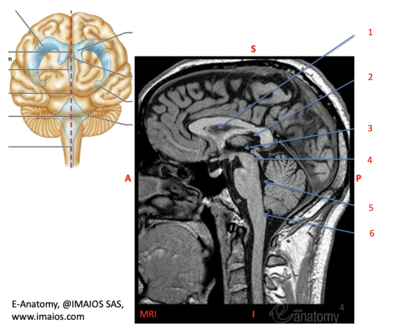
lateral ventricle body (foramina of Luschka)
(2)

third ventricle
(3)

cerebral aqueduct
(4)

fourth ventricle
(5)

median aperture (foramen of magendie)
(6)

third ventricle
(1)
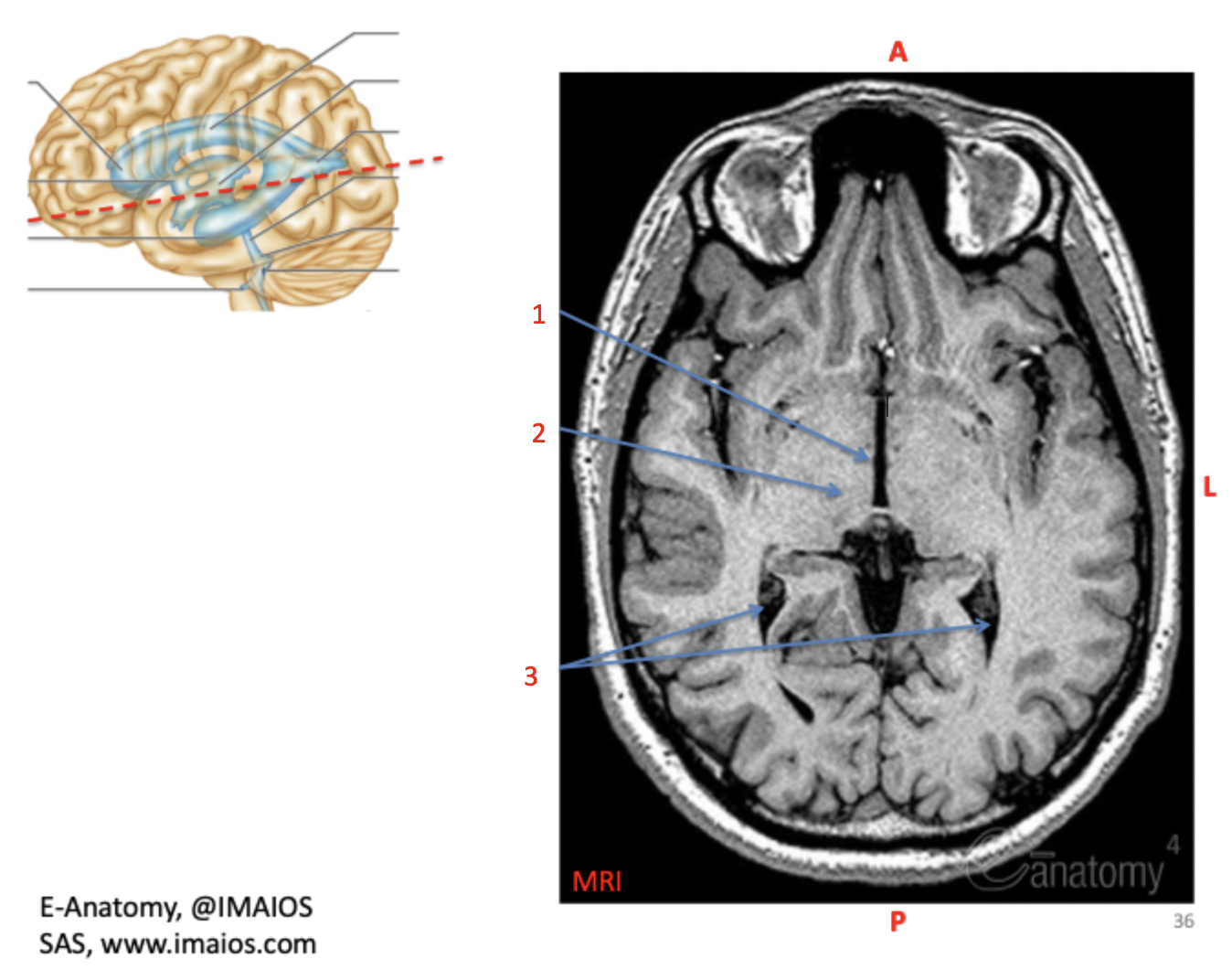
thalamus
(2)

lateral ventricle posterior horn (occipital horn)
(3)
