MOLBIOL 4RR3 TEST 2
 Call Kai
 Learn
 Practice Test
 Spaced Repetition
 Match
 Flashcards
 Knowt Play
1/87

There's no tags or description
Looks like no tags are added yet.
88 Terms
DNA amplification

PCR
selective DNA amplification
prior seq infor = required to design 2 primers specific for the target
primers + target seq + heat stable DNAP + dNTP = amplification of a dna target fragment
why is heat stable polymerase required for PCR?
rxn made of 3 cycles
denaturation - 93-95°C
reannealing - 50-65°C
dna synthesis - 70-75°C
specificity of amplification and primer design
nested primers
initial amplification useed as a target source for a second rxn
hot-start pcr
leave the enzyme or primers until the first denaturation step
lowers background resulting from nonspecific binding of primers
touchdown pcr
annealing temp = lowered gradually
to increase specificity early in amplification
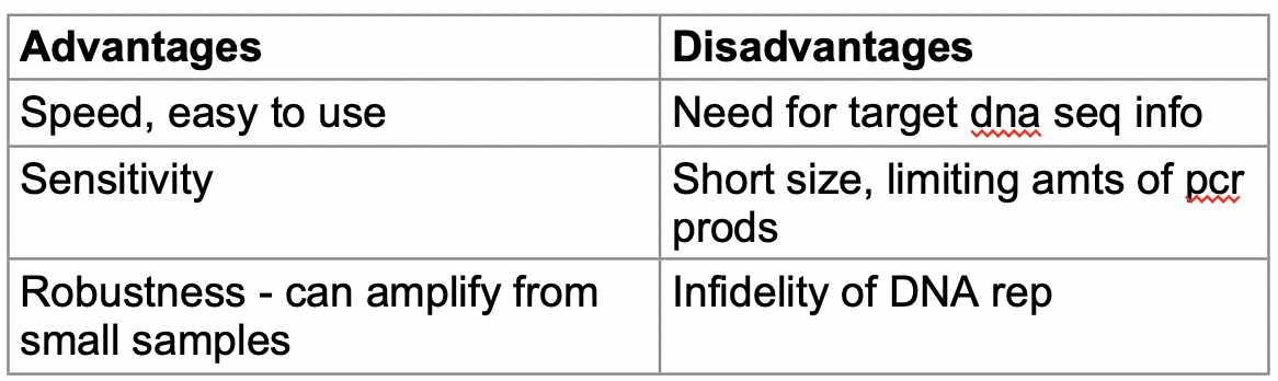
PCR pros and cons
Applications of PCR
for screening uncharacterized mutations
permits rapid genotyping for polymorphic markers
assaying for known mutations
allelic discrimination by size or susceptibility to RE
allelic discrimination by susceptivity to an artificially introduced RE site
allele-specific PCR = primers are designed so as to discriminate bw target DNA seqs that differ by a single nt in the region of interest
degenerate oligonucleotide-primed PCR
linker-primed PCR - ligation adapter pcr
anchored pcr - target-specific primer + universal primer
Cell-based DNA cloning - VECTOR SYSTEMS
Plasmids
Cosmid vectors
Vectors based on bacteriophage P1 and F factor plasmids
bacterial artificial chromosomes (BAC) vectors
bacteriophage P1 vectors and PACs
yeast artificial chromosomes (YACs)
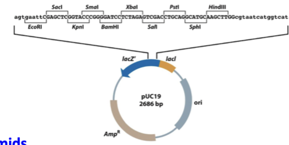
Plasmids
Origin: natural bacterial plasmids
Important features;
multiple cloning site (MCS)
antibiotic resistance gene
screening system for recombinant plasmids
DNA inserts: small fragments
Cosmid vectors
Structure: plasmid containing bateriophage λ cos sequence
Important features:
combine plasmid and phage features
used to clone larger DNA fragments
DNA inserts: 30-44 kb
Bacterial Artificiakl Chromosomes (BAC) vectors
origin: E. coli F factor - fertility plasmid
important features:
low-copy-number replicon
provides greater stability for large inserts
can maintain large DNA fragments
DNA inserts: up to 300 kb
bacteriophage P1 vectors
origin: bacteriophage P1
important features:
P1 components incorporated into a circular plasmid
P1 packages its genome inside a protein coat
DNA inserts: large fragments
P1-derived artificial chromosomes PACs
structure: combination of P1 and F-factor systems
important features:
Combines features of both systems
Used for cloning large DNA fragments
DNA inserts: large fragments
yeast artificial chromosomes (YACs)
origin: artificial yeast chromosome
important features:
contains essential chr components
centromeres → required for disjunction of sister chromatids in mitosis
telomeres → required for complete replication and protection of the chromosome ends
autonomous replicating seq elements → required for autonomous replication of chromosomal DNA, thought to act as specific rep origin
allow replication and stable maintenance of large DNA fragments
DNA inserts: large fragments
“4 SHORT SEQS REQUIRED → 2 TELOMERERS, 1 CENTROMERE, 1 ARS”
Nuclear acid hybridization
ssDNA binds to its complementary dna seq → forms dsDNA
purpose: uses a labeled nucleic acid probe to find matching DNA and RNA molecules in a mixture of unlabeled nucleic acid mols
Preparation of nucleic acid probes
conventional dna probes
labeled by adding labeled dNTPs
isolated by cell-based dna cloning or by PCR
rna probes
generated from DNA within specialized plasmid containing a phage promoter sequence
oligonucleotide probes
short ssDNAs
NA hybrization - Factors affecting dna strand separation
strand length
longer strands = more H+ bonds
require more energy to separate them
base composition - GC content
GC pairs = 3 H bonds
AT pairs = 2 H bonds
higher GC content = stronger bonding = harder to separate
chemical envr
Na+ ions - stabilize DNA strands
formamide and urea - destabilize dna strands = easier to separate
DNA and RNA labeling
during in vitro synthesis
labeling of DNA
nick-translation
random primed labeling/PCR mediated labeling
labeling of RNA
in vitro transcription system
end-labeling
involves the addition of labeled group to the terminal nts
southern blot hybridization (SBH)
target DNA = digested with one or more restriction endonucleases
size-fractionated by agarose gel electrophoresis
denatured and transferred to NC for hybridization
immobilized ssDNA seqs = allowed to associate with labeled ss probe DNA

Southern Blotting Hybridization - Application
Restriction enzyme (RE) mapping
detects pathogenic point mutations
identifies changes in RE recognition sites
Restriction fragment length polymorphism (RFLP)
detects differences in dna fragment lengths
mutations can create or remove RE sites, changing fragment sizes
VNTR-basex RLFPs (DNA fingerprinting)
detects differences in the number of repeated DNA seqs
VNTR = variable number tandem repeats
also called hypervariable minisatellite DNA (5-64 bp)
DNA probes detect these differences
Gene deletion detection
restriction mapping can identify missing DNA sections
Tissue in situ hybridization
a labeled probe is hybridized against RNA in tissue sections
Colony blot and plaque lift hybridization
grow colonies → bacteria containing plasmids are grown on an agar palte
transfer → colonies are transferred onto nitrocellulose filter by direct contact
dna denaturation → colonies treated with alkali to separate dsDNA into ss
hybridization → a labeled nucleic acid probe binds to complementary DNA seqs
detection → filter is washed, dried, and exposed to x-ray film (autoradiography) to identify positive colonies
Gridded high density arrays of DNA clones
uses robotic gridding devices
automatically transfer DNA clones from microtiter plates onto a membrane
each dna clone = placed at a specific, predetermined linear position (coordinate)
purpose: allows rapid and efficient screening of large DNA libraries to identify specific dna clones
DNA microarray technology
Purpose: allows large-scale, rapid and automated DNA hybridization
Main advantage: miniaturization and automation allow thousands of DNA samples to be analyzed at once
Three main types:
DNA clone microarrays → dna clones are placed onto a surface using microspotting
DNA probe microarrays → dna probes are placed onto a surface using microspotting
Oligonucleotide microarrays → short DNA seqs are synthesized directly on the surface (in situ)
Application: identify rare target seqs, analyze gene exp
Detection: laser scanning detects fluorescent signals from hybridized probes
CRISPR-Cas9 Technology
CRISPR → clustered regularly interspaced short palindromic repeats
Discovery
identified by francis mojica, 1996
Function
acts as a primitive immune system in bacteria by storing DNA seqs from previous viral invaders
Cas9 protein
recognizes matching DNA seqs and cuts the target DNA
Potential applications for CRISPR-Cas9 technology
human therapeutics → treat genetic diseases by correcting or modifying faulty genes
sickle cell disease
beta-thalassemia
duchenne muscular dystrophy
cancer
nonhuman apps
agriculture → improve crops and livestock
livestock → modify animals to improve desirable traits
industrial biotechnology → produce useful biological products
gene editing processes
guide rna directs cas9 toa specific dna seq
cas9 cuts target dna
cell repairs dna → allow the gene to be disrupted or corrected
First CRISPR-edited babies
Researcher: he jiankui
November 2018: announced the birth of the world’s first genetically edited babies → lullu and nana
target gene: CCR5, codes for a protein that HIV uses to enter certain immun cells
goal: disable the CCR5 gene to make the babies more resistant to HIV infection
outcome: gene was deited in human embryos = successful pregnancy and birth of twins
RAISED MAJOR ETHICAL CONCERNS
disabling CCR5 does not guarantee complete immunity to HIV
long term effect unknown
Single cell RNA sequencing (scRNA-seq)
Purpose: measures gene exp in individual cells to identify differences bw cell types
Workflow
collect tissue
dissociation → break down tissue into individual cells
rna extraction
reverse trancription → convert RNA into cDNA
amplification
PCR to amplify cDNA
In vitro transcription → to produced amplified RNA
sequencing library preparation
sequencing → determine rna-derived seqs
gene expression profiling → analyze which genes are expressed in each cell
clustering → group cells with similar gene expression patterns to identify different cell types

Constructing trancript maps
Purpose: identify genes and expressed regions within cloned gDNA
Methods:
Hybridization to RNA/cDNA
DNA clone is hybridized against a northern blot containing a panel of mRNA isolated from different tissues
positive signal = cloned DNA contains a gene expressed in that tissue
Zoo blot hybridization
compares DNA seqs across different species
coding regions are highly conserved between species
noncoding regions are less conserved
CpG island identification
short (~1 kb), GC-rich, hypomethylated DNA regions
found near the 5’ end of genes
approximately 56% of genes are associated with CpG islands
Exon trapping
exons can be identified in cloned gDNA
dna is inserted into an expression vector (eg pSPL3)
vector is introduced into eukaryotic cells
RT-PCR used to detect and retrieve trapped exons
transcript maps
RNA-seq data or ESTs (expressed seq tags) are mapped to specific genomic regions
mapping can be done using computer analysis or PCR
computer analysis
homology searches identify similar DNA seqs
exon prediction programs identify potential exons
Expression Studies at Different Levels

Data integration

General Nucleotide and Protein Sequence Repositories

Eukaryotic genome database

Program for sequence homology search

Organization of the human genome
Nuclear genome
24 linear ds DNA molecules, ~3200 mB, ~30000 genes
~45% transposon-based repeats
~44% other non-conserved
~6.6% heterochromatin
~3% highly conserved (other)
1.5% highly conserved (coding)
Mitochrondrial genome
1 circular double-stranded DNA molecule, 16.6 kb, 37 genes
~93% highly conserved (coding)
~5% highly conserved (other)
<2% other non-conserved
Human Nuclear DNA categorization
single sequence DNA/low copy DNA
~45% of total genome
cds = ~1.5% of total genome
DNA from introns = 53.5%
intermediate repeated DNA
present at between 10² and 10^5 copies per genome (~45% in total)
Highly repetitive DNA
present at up to 10^6 copies per genome (~10% of the total)
Human genes
about 30-35000 in total
some genes (ie histone genes) do not contain introns
alpha globin gene
size: 0.8 kb
introns count: 3, account for 30% of total genomic locus
dystrophin
gene defective in DMD
size: 2.4 Mb
introns count: 79, account for 99.4% of total genomic locus
gene mean size: 27 kb
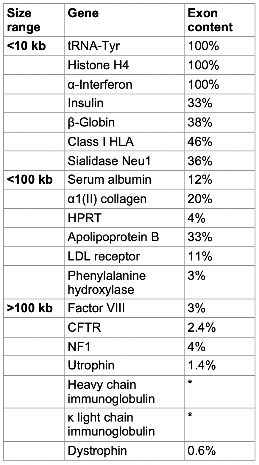
Gene length and exon content
Overlapping genes within the class III region of HLA complex
Genes within genes: Neurofibromatosis type 1 gene (NF1)
intron 26 of NF1 gene contains 3 interal two-exon genes


Gene families
arise by gene duplication and divergence from an ancestral gene
because a family has multiple copies, a loss of function in one may be tolerated
led to evolution of pseudogenes - non-functional copies
Clustered and interspersed multigene families

Pseudogenes
defective gene copies found in gene families
unprocessed pseudogenes in a gene cluster
tandem gene duplication, inappropriate termination codons
truncated genes and internal gene fragments
eg. class I HLA
nonprocessed pseudogenes in a dispersed gene family
eg. NF1 gene fragments over 7 chrs
processed pseudogenes in a dispersed polypeptide-encoding gene family
cDNA + oligo dA/dT. copied by retroptransposition
processed pseudogenes in an RNA-encoding gene family
retrotransposition from RNAP III transcripts
Processed pseudogenes
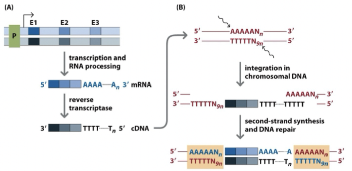
gene transcribed into mRNA
introns are spliced out = mRNA has poly-A tail in 3’ end
reverse transcriptase = RNA to cDNA
cDNA gets pasted into the genome at random spot
cell repairs and fills in the dna, leaving short repeated sequences on each side
Non-protein coding genes
bulk of cellular RNA consists of rNA and tRNA required for mRNA translation
additional functions of noncoding RNAs
snRNA invovled in spliceosome function
snoRNA - control site specific base modifs in rRNA and U6 snRNA
7SL rna molecule - forms part of the signal recognition particle required for the translocation of proteins across the ER
miRNAs - 22 nts long can function as antisense regulators of genes to inhibit translation
lncRNA - rna molecules >200 nts, can modulate gene expression
Genes
dna sequence that contributes to the phenotype of an organism in a way that depends on its sequence
RNA polymerases
Prokaryotes (E. coli)
one RNAP transcribes all genes
control region has two promoter elemts
at -35 and -10, upstream of the transcription start site (+1)
Eukaryotes
3 RNAPs
Pol I - transcribes rRNA
Pol II - transcribes polypeptide-encoding genes (mRNA) and some RNAs
Pol III - transcribes tRNAs, 5S rRNAs, and small RNAs
Promoter
typical eukaryotic gene has a promoter region, extends about 200 bp upstream of the TSS (nt + 1)
promoter contains one or more of these elements:
enhancers
response elements
silencers
most human promoters have TATA box
centered around position -30 relative to the TSS
binds general transcription factors, designated TFIIX (X = placeholder for specific factor)
Pol II transcription initiation
TFIID (containing TBP) binds the TATA box upstream of the TSS
TFIIB joins the complex
RNAP II arrives together with TFIIF and other TFs
TFIIE and TFIIH are then added = complete initiation complex
the nts (ATP, UTP, CTP, GTP) are added
RNA synthesis begins
phosphate groups added to the tail of RNAP II
RNAP II moves along the DNA as RNA is made, leaving some general factors behind at the promoter
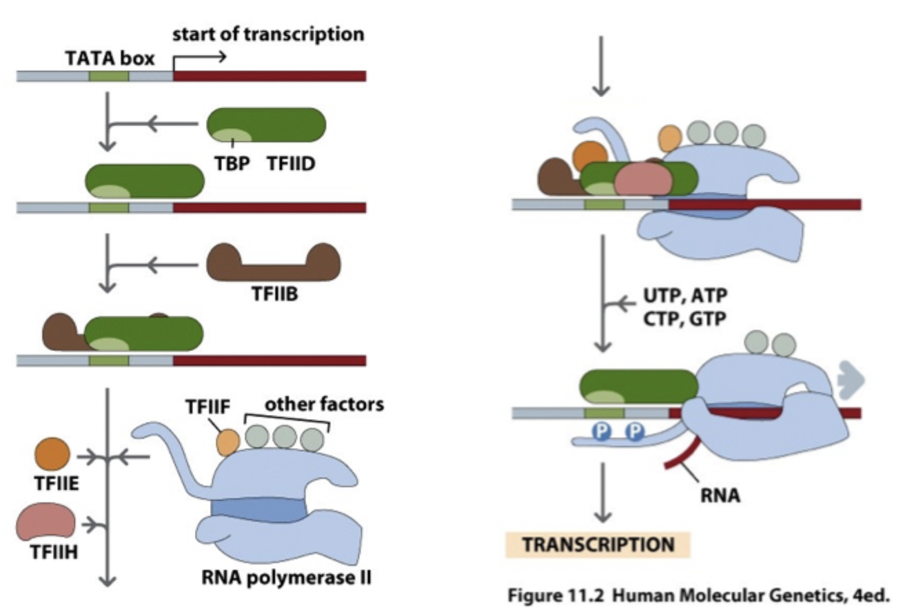
Transcription factors for transcription by RNAP II

Regulatory elements within the promoter
GC box (GGGCGG → binds the TF SP1
CAAT box (GGCCAATCT) → binds the TFs CTF and NF1
Oct box (ATTGCAT) → binds the TFs Oct-1 and Oct-2
Enhancers → operationally distinguished from promoters by 3 criteria
located a considerable distance from the TSS
located upstream or downstream
action not dependent on orientation
Eukaryotic structural gene

DNA looping
a type of gene regulation
brings proteins bound to enhancers or silencers into direct contact with proteins bound to the promoter

Response Elements
What? induce genes in response to signals
Where? within promoter region, closely upstream of the promoter or in more distant locations
Examples
cAMP response element (CRE)
serum response element (SRE)
heat-shock response element (HRE)
Silencers
elements that bind certain proteins, resulting in negative control of gene expression
Housekeeping genes
expressed in most tissues most of the time
responsible for functions likely to be necessary in any cell
cAMP response
a hormone binds its receptor on cell mem
receptor activates a G protein (⍺ subunit separates from the β and γ subunits)
G protein activates adenylate cyclase → converts ATP into cAMP (+PPi)
cAMP binds the regulatory subunits of PKA → releases the active catalytic subunits (before PKA is inactive complex of 2R and 2C subunits)
free C subunits move into the nucleus
C subunit phosphorylates CREB
phosphorylated CREB activates target genes that contain a CRE

Transcrption
RNAP transcribes the DNA template
mRNA already has a 5’ cap and polyadenylation (AAUAAA) signal appears in the transcript
a spscific endonuclease cleaves the mRNA downstream of the AAUAAA signal
poly A polymerase adds adenine residues to the new 3’ end, using ATP (releasing PPi)
result: mRNA with a 5’ cap, the AAUAAA signal, and a poly-A tail (~100-250 adenine res) at 3’ end
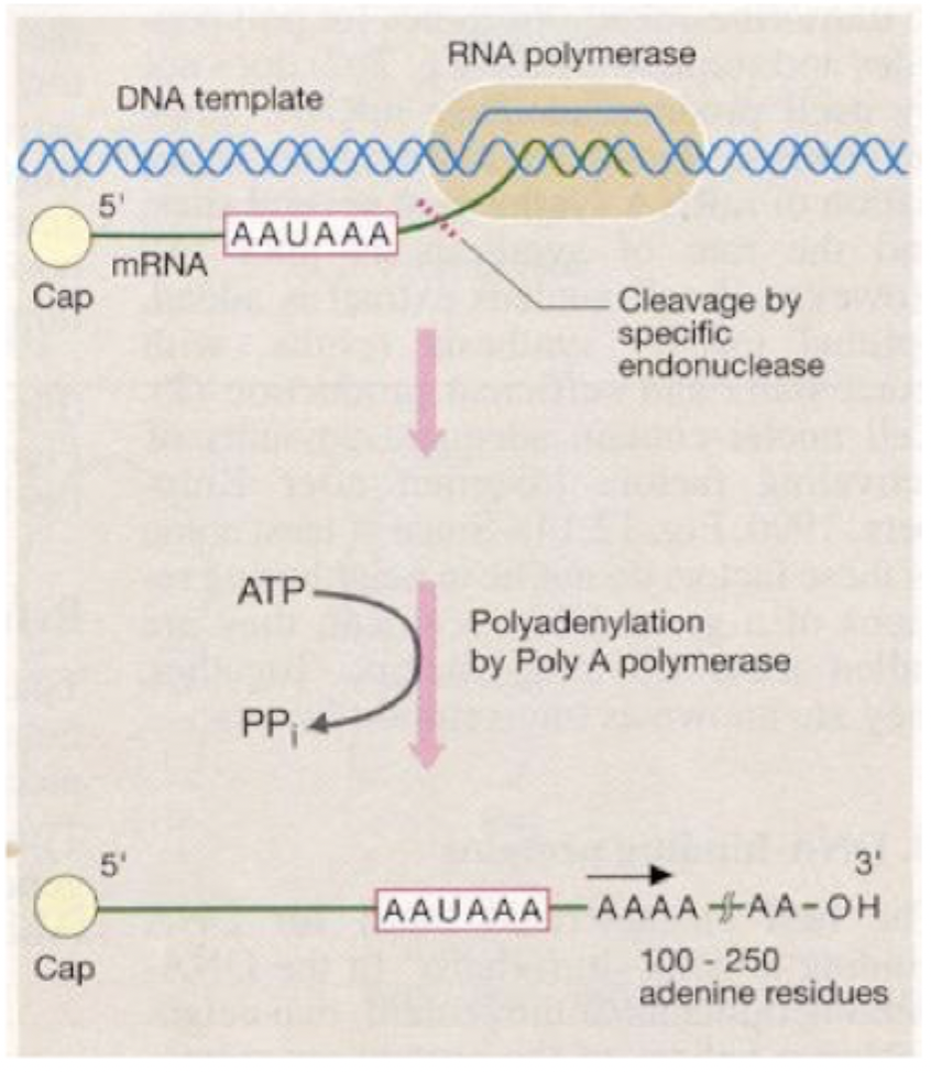
Cap formation
as soon as transcription starts
5’ end of nascent mRNA molecule is covalently modified to form a cap
by addition of 7-methylguanosine to 5’ nt of nascent mRNA in a 5’-5’ triphosphate linkage
cap is essential for initiation of translation
Transcription termination
sequence AAUAAA in the nascent RNA triggers termination a few hundred bps downstream
an endonuclease cuts the RNA downstream of this signal
5’ → 3’ exonuclease degrades the RNA still attached to the polymerase
transcription stops when exonuclease meets the polymerase

Polyadenylation

RNA is cleaved about 20 bp downstream of the AAUAAA signal
3’ end is then covalently modified by adding 100-200 adenine residues = poly-A tail
One gene, more than one protein
alternative transcription and processing of genes can generate multiple protein products
alternate promoters → generate tissue-specific isoforms
alternative splicing and polyadenylation → human genes are prone to both
RNA editing → specific cytosine or adenine residues are substituted by deamination (C→U editing in APOB gene)s
Dystrophin gene - multiple promoters
has at least 7 promoters that drive tissue- and cell type-specific expression
each promoter has its own first exon, which is spliced onto the shared downstream exons (numbered 2 to 79)

Types of alternative splicing
A. exon skipping (cassette exon)
B. alternative 5’ splice site - exon is included in a longer or shorter form, depending on which 5’ splice site is used
C. alternative 3’ splice site - same idea, but with 3’
D. intron retention 0 blue region bw the two pink exons = either spliced out as an intron or kept in the matuer mRNA
E. mutually exclusive exons - blue exon or brown exon included, never both

Neuroxin-3 transcript in CNS - alternative splicing

combines alternate promoters with alternative splicing, so one gene can produce a very large number of different transcripts and protein isoforms in the CNS
Tissue-specific RNA editing of the APOB gene
Gene
APOB has a large exon 26 → contains a CAA codon at nt 6666
Liver
no editing occurs, CAA codon is read normally
translation continues to downstream stop codon = full length ApoB100 protein
Intestine
cytidine deaminase APOBEC1 edits the mRNA
change C → U at p6666
turns CAA into UAA = premature stop codon
translation stops early = shorter ApoB48

Cholesterol cycle
intestine → liver (dietary route)
dietary lipids absorbed in the intestine → packaged into chylomicrons, travel to the lievr
in the enterocyte, MTP helps load lipid onto ApoB48 to form chylomicron
liver → periphery (VLDL → LDL)
in hepatocyte, MTP loads lipid droplets onto ApoB100 to form VLDL
VLDL → LDL, delivers cholesterol to peripheral tissues
periphery → liver (reverse cholesterol transport, HDL)
pre-beta HDL picks up cholesterol from peipheral cells
matures into HDL3, then HDL2
HDL2 returns to the liver, where cholesterol can be removed in bile
CETP transfers lipids bw HDL and VLDL/LDL

CpG Islands
DNA can be modified by methylation of cytosine to form 5-methylcytosine
occurs at the dinucleotide CpG
CpG
occurs less frequently in the genome, because
accidental deamination of cytosine produces uracil
whereas deamination of 5'-methylcytosine produces thymine
uracil not found in DNA, replaced with cytosine by glycolase
thymine is mispaired with G res
DNA mehtylation
generally associated with repression of transcription
CpG islands are about 1kb in length
tend to extend over promoters of expressed genes
About 56% of human genes are estimated to be associated with such seqs
Changes in DNA methylation during mammalian development
Germ line (before fertilization)
primordial germ cells (PGCs) start with a high degree of methylation
around gonadal differentiation → erasure of parental epigenetic settings (methylation drops at low level)
as germ cells develop → de novo methylation and establishment of imprinting marks occur
producing methylated sperm and egg
After fertilization
early embryo → demethylation
blastocyst stage → methylation level is lowest
After blastocyst
de novo methylation begins again in somatic cells, then maintenance methylation keeps the pattern in adults.
Placenta and yolk sac (trophoblast lineages) are methylated too, but less than the embryo proper.
Some cells become PGCs (primordial germ cells), and the cycle starts over.

Chromatin conformation - ChIP
Crosslink proteins to DNA, lyse the cells, and isolate the chromatin.
Fragment the chromatin and mix it with an antibody specific for just one protein.
Precipitate the antibody-bound protein-DNA complexes.
Reverse the crosslinking, degrade the protein, and purify the DNA fragments.
Analyze the purified DNA in one of these ways:
Standard ChIP: test specific target regions.
Genomewide ChIP:
ChIP-chip: hybridize the fragments to a microarray.
ChIP-PET or STAGE: tag the sequence fragments.
ChIP-seq: sequence the fragments.