6. Diagnostic Methods
 Call Kai
 Learn
 Practice Test
 Spaced Repetition
 Match
 Flashcards
 Knowt Play
1/45

There's no tags or description
Looks like no tags are added yet.
46 Terms
sign
objective findings by examiner, eg.
rash
lump in breast
changes in skin (colour, asymmetrical borders, size)
symptom
subjective indication of disease or change in condition as perceived by patient - impacts on daily life, eg.
pain
loss of appetite
cramping
dysphagia
numbness
syndrome
set of signs or symptoms that arise from common cause
many diseases share same signs + symptoms
need tests + procedures to narrow down
diagnosis
identification of disease or condition - can be objective or subjective
subjective = based on medical opinion from patient complaints + history
objective = based on test/exam results
eg. biopsy, lab results
B symptoms
common signs/symptoms that can be confused w/ other diseases/conditions
often associate w/ lymphomas
eg. cough, aches, fever, unexplained weight loss, etc.
medical record
legal public document that includes all past medical experiences, eg. Cerner
available to medical staff, insurance companies + can be requested by court of law
includes:
medical history
lab tests
medical procedure results
progress notes
consent forms
may be valid for certain periods of time → would need patient reconsent
medical images
Cerner
health record system used across BC + includes
documentation
pathology reports
operative reports
referrals
medical orders + history
consent forms
appointment schedules
patient info
set-up + positioning information, including modifications
other treatments in addition to radiation therapy, eg. chemo, pre-meds, clinical trial
female medical history
free estrogen in body decreases w/ increased pregnancies + breastfeeding
gravida = # of pregnancies
para = # of births
menses + birth control info → lifetime exposure to estrogen
eg. menstruation at 9 more at risk than at 16
oncology consult
part of medical history - patient interview w/ oncologist
establish medical history
form relationship + trust w/ patient
determine patient’s treatment goals
review treatment options, outcomes, potential side effects
used to refer back to if patient’s have questions or confusions arise
confirm patient info + treatment details
medical history
can be risk factor for developing certain diseases
premalignant condition
high risk of becoming cancer
eg. colon polyps, moles
paraneoplastic syndrome
condition of symptoms resulting due to production of substances/hormones from tumour itself
symptoms can occur remotely from tumour
family history
important for hereditary cancers
demographic data
insight into prevalent diseases for certain populations
includes
age
gender
race
nationality
physical examination
organized + detailed exploration of patient’s anatomy
inspection = observation
colour of skin
ulcers
abrasions
moles
edema
palpation = touch
lymph nodes
superficial + deep tumours
percussion = tapping
determine sites of pain
auscultation = listening
lung, heart, arteries, stomach, bowels
vital signs
temperature
pulse
respiratory rate
blood pressure
pain assessment
diagnostic methods
ways to identify disease or condition
medical record + medical history
physical examination
screening
laboratory studies
medical imaging
biopsy
screening
helps detect cancers early before they are symptomatic
laboratory studies
baseline taken first + deviations from baseline monitored
baseline is different for everyone → look for deviations specific to patient
patient self-assessment can help determine if anything is off
includes
blood studies
blood chemistry
microbiologic studies
urine studies
fecal studies
immunologic studies
serology studies
blood studies
laboratory study looking at blood counts
ex. high WBC could mean infection
blood chemistry
laboratory study looking at chemical in blood
electrolytes
proteins
glucose
microbiologic studies
laboratory study looking at organisms in blood → infections
urine studies
laboratory study looking at urine composition + concentration
urinary system
kidney disease
endocrine system dysfunction
fecal studies
laboratory study looking at digestion + metabolism
GI diseases, eg. bleeding, obstruction, parasite
immunologic studies
laboratory study looking at antigen-antibody reactions
serology studies
laboratory study looking for
neoplastic disease
infectious disease
allergic reaction
electronic pulses
determines functionality of heart, brain, muscles
2 main tests
electrocardiogram (ECG)
can determine cardiotoxicity effects of chemotherapy
see if heart is healthy enough to undergo other/further treatments
electroencephalogram (EEG)

positron emission tomography scan (PET scan)
type of nuclear medicine imaging
radionucleotide + sugar injected → emits gamma rays at tumour site
areas w/ high metabolism always light up
ie. brain, heart bladder
used to identify primary tumour sites + metastasis
especially helpful for bone → determine bone metastasis

radiographic studies
used to visualize many structures, eg. x-ray
especially effective for lung imaging
lungs full of air → anything not supposed to be there easily visualized
other structures include
mammogram
abdomen
kidneys
bladder
skull
spine
bones

computer tomography (CT)
visualization of internal anatomy
rotating x-ray tube allows images to be reconstructed on several planes
w/ or w/o contrast depending on structure of interest
can indicate angiogenesis
blood vessels w/ high contrast enhancement
can also be used to
make sure positioning is correct
see if bladder is full → BUT higher radiation dose so use bladder scan instead

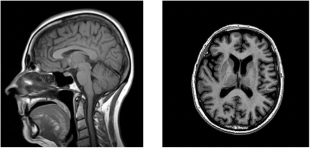
magnetic resonance imaging (MRI)
radiofrequency waves + strong magnetic field creates images that allow for delineation of soft tissue
contrast is magnetic
bright tissues = lots of blood vessels
tumours will appear bright due to angiogenesis
eg. PI-RADS used for prostate cancer screening
diagnostic ultrasound (US)
uses high frequency sound waves to acquire images
helps guide biopsies
eg. bladder scan → RTs can use
used to look for fluid → see if bladder is full or not
sometimes required before radiation therapy treatment
biopsy
examines histologic grade of tumour → determines cancer diagnosis
different methods include
exfoliative cells
incisional biopsy
excisional biopsy
fine needle aspiration (FNA)
core needle
endoscopic
image guided
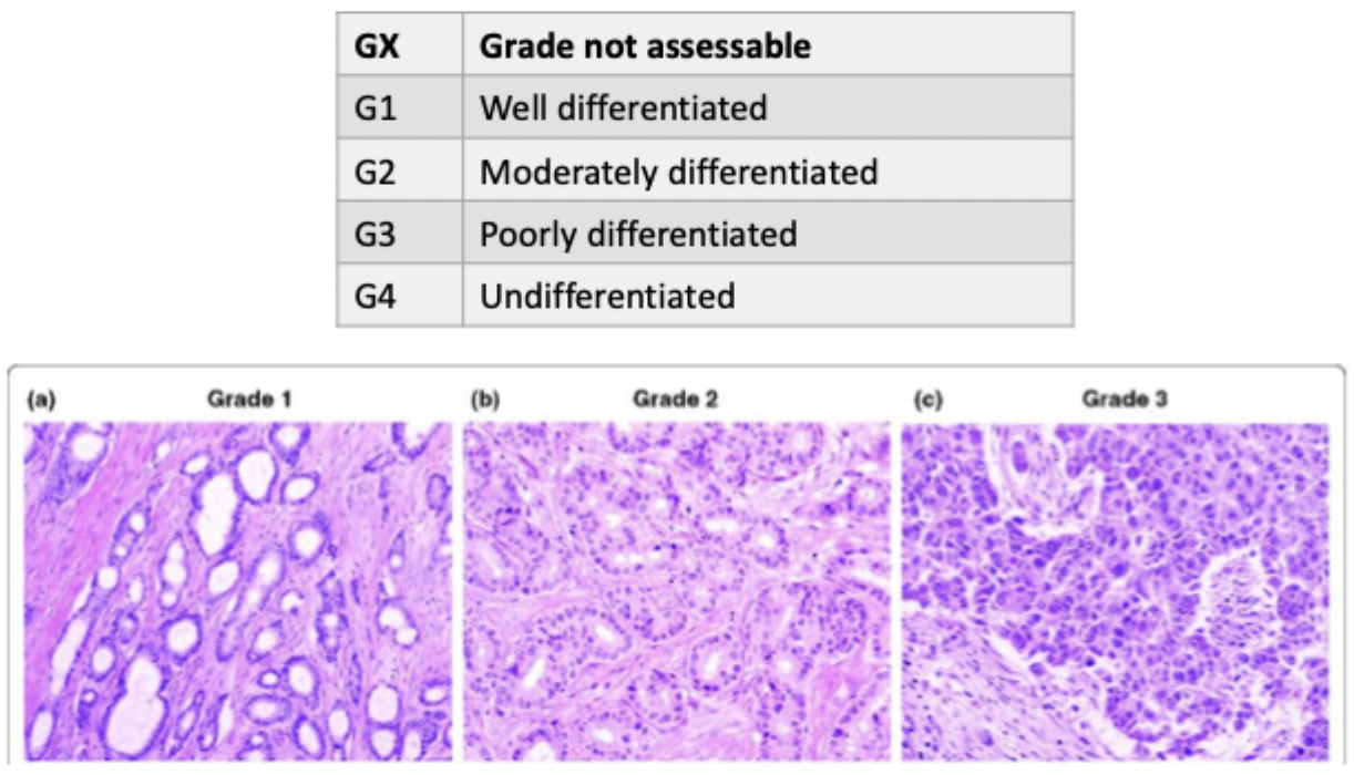
grade
level of differentiation of cells in tumour
determined by biopsy
ranges from G1 (better prognosis) - G4 (worse prognosis)
exfoliative cells
naturally or deliberately sloughed off - common for cavities/linings
found in urine, mucous, feces, sputum
helps identify cervic, colon, lung cancers
incisional biopsy
needle removes small amount of tissue
often for solid tumours
excision biopsy
removal of whole tumour for diagnosis
edges sampled to determine clear margins
often for solid (skin) tumours
site can then be irradiated to get rid of remaining cancer cells
fine needle aspiration (FA)
sample cells collected from various locations in tumour
common for superficial tumours
eg. breast, lymph nodes
can also be used to find other conditions, eg. cyst
core needle
uses large gauge needle (14-16) to remove core of tumour
intact sample instead of little group of cells
can see what is going on around tumour site
can determine local extension or outward growth
endoscopic
camera inserted into body + needle inserted into camera
eg. bronchoscopy, colonoscopy
image guided
CT or ultrasound-guided to help maneuver during needle biopsy to correct location and tissue

breast diagnostic methods
screening
mammogram
history + physical exam
palpation → superficial lumps
biopsy
US-guided FNA biopsy → deeper lumps
gastrointestinal diagnostic methods
screening
FIT test
colonoscopy
history + physical exam
DRE → rectal tumours
size, mobility + distance from anal verge
peri-anal lymph node examination
imaging tests
CT
MRI
PET
lab tests
carcinoembryonic antigen (CEA)
protein maker associated w/ colon cancers
prostate diagnostic methods
history + physical exam
DRE
biopsy
transrectal US-guided biopsy
lab tests
PSA
gynecologic diagnostic methods
screening
HPV test
Pap smear
biopsy
colposcopy → for patients w/ abnormal pap smear
examines cervix for abnormalities
removes suspicious tissues using
punch biopsy
endocervical curettage
cone biopsy

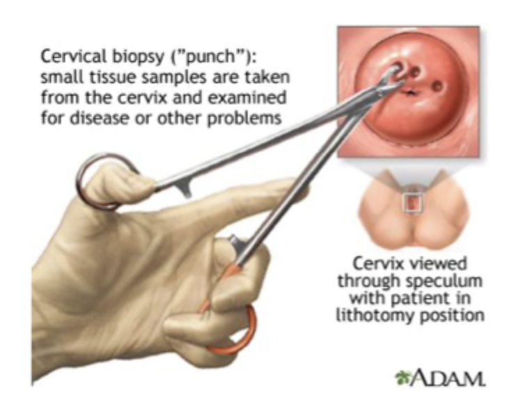
punch method
biopsy method for gynecologic sites after abnormal pap smear → during colposcopy
small, round piece taken out
full thickness of skin + underlying tissue included
can contain skin cells + mucosal layers
cone biopsy
biopsy method for gynecologic sites after abnormal pap smear → during colposcopy
cone of tissue taken out to analyze different layers
done via
LEEP (loop electrosurgical excision procedure) → wire loop heated electric current
cold knife biopsy → scalpel

endocervical curettage (ECC)
biopsy method for gynecologic sites after abnormal pap smear → during colposcopy
scraping lining of cervical canal
deeper into cervix
seeding
when cancer cell stick to medical instruments during biopsy + spread to other biopsy sites
can be prevented by using new needles when going into new sites
head and neck diagnostic methods
palpation
neck nodes → hard, >1cm, nontenderm nonmobile
direct inspection
laryngoscopy
fiberoptic endoscopy
biopsy
FNA
usually looking at small structures
often image-guided to avoid small, delicate structure in HN
staging
requires radiographic imaging
respiratory diagnostic methods
screening
LDCT (low dose CT) → high risk patients
history + physical exam
smoking
occupation history
imaging tests
chest x-ray
tumours >5mm
CT + MRI
tumours close to other structures
brain metastasis
PET
determine if malignant or benign, ie. angiogenesis
lab tests
pulmonary function tests → see if patient can tolerate treatment
CBC → look for bone + liver disease
diagnosis
sputum cytology → non-invasive but cannot determine tumour SITE
bronchoscopy → biopsy w/ US guidance for centrally located tumours
CT-guided FNA → peripheral disease
