PCB30733C Skeletal Muscle LAB
 Call Kai
 Learn
Learn Practice Test
 Spaced Repetition
 Match
Match Flashcards
 Knowt Play
1/107

There's no tags or description
Looks like no tags are added yet.
Name | Mastery | Learn | Test | Matching | Spaced | Call with Kai | Chat |
|---|
No analytics yet
Send a link to your students to track their progress
108 Terms
What are the 3 different types of muscle tissue?
smooth, skeletal, and cardiac
Which type of muscle tissue has intercalated discs?
cardiac
Where is smooth muscle located?
in the walls of most hollow organs, circulatory vessels, and in the digestive tract
Which muscles attach to bones to allow movement of the skeleton?
skeletal muscle
Which muscle type lacks striations?
smooth
Which muscle type is multinucleated?
skeletal (has multiple nuclei per cell)
which muscle type is uninucleated?
smooth
Which muscle type is uninucleated or binucleated?
cardiac
Which muscle types have centrally located nuclei? Which have peripherally located nuclei?
smooth and cardiac - central
-
skeletal - peripheral
Which muscle types are involuntary? Which are voluntary?
smooth & cardiac - involuntary (controlled by ANS)
-
skeletal can be voluntary and involuntary
Is the diaphragm voluntary or involuntary?
both!
the diaphragm is a dome-shaped muscle separating the thoracic and abdominal cavities & is critical for respiration and breathing
-
Typically, the diaphragm contracts & relaxes on its own by the ANS, meaning that it is primarily involuntary. However, you can choose to take voluntary control over the diaphragm if desired. (choosing to hold ur breath)
What is the difference between epimysium, perimysium, and endomysium?
epimysium : surrounds muscle group
-
perimysium : surrounds fascicle
-
endomysium surrounds muscle fiber/cell
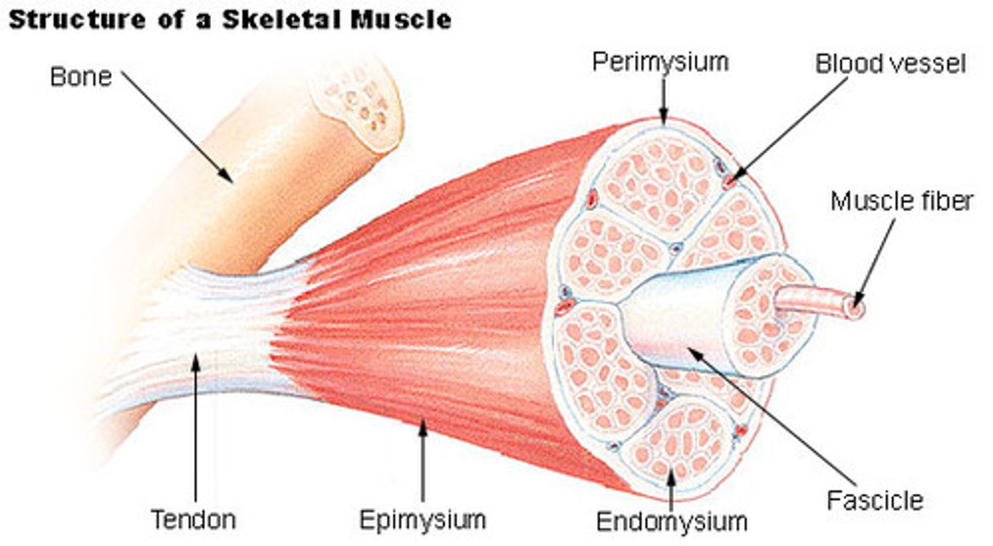
Differentiate between tendons and ligaments.
tendonds attach muscle to bone & ligaments attach bone tobone
What are bundles of skeletal muscle cells called? What surrounds them?
fasicle; perimysium
Flip this card to see pics of skeletal muscle histology :3
see that the skeletal muscle is multinucleated and has peripherally located nuclei !
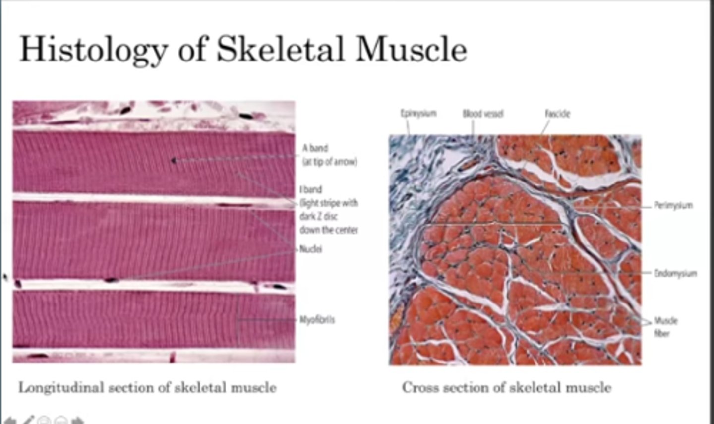
What gives skeletal muscle its striated appearance?
the arrangement of protein filaments
-
it comes from how the myofilaments are arranded within a myofiber
Within each muscle cell/fiber, there are ______________, and within each of those, there are _______________.
Within each muscle cell/fiber, there are myofibrils, within each myofibril, there are myofilaments.
What are myofilaments?
contractile proteins like actin and myosin
Myofibril is composed of repeating filaments called __________________.
sarcomeres
What is known as the functional unit of the mucle?
sarcomere (it is the most basic unit of muscle contraction)
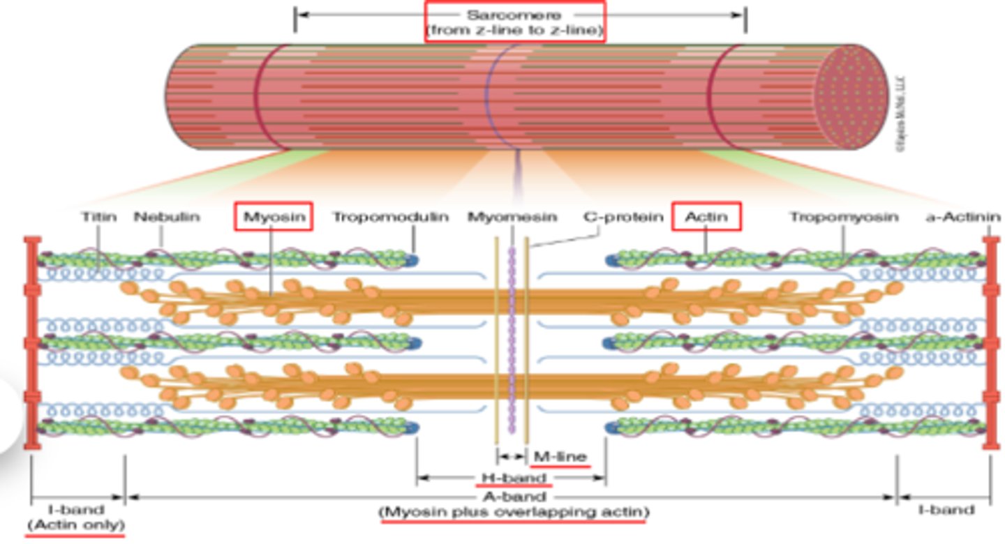
Sarcomeres extend from _______________ to ______________.
Sarcomeres extend from Z-line to Z-line. (this defines the boundaries of one sarcomere)
Actin filaments can also be called what?
thin filaments
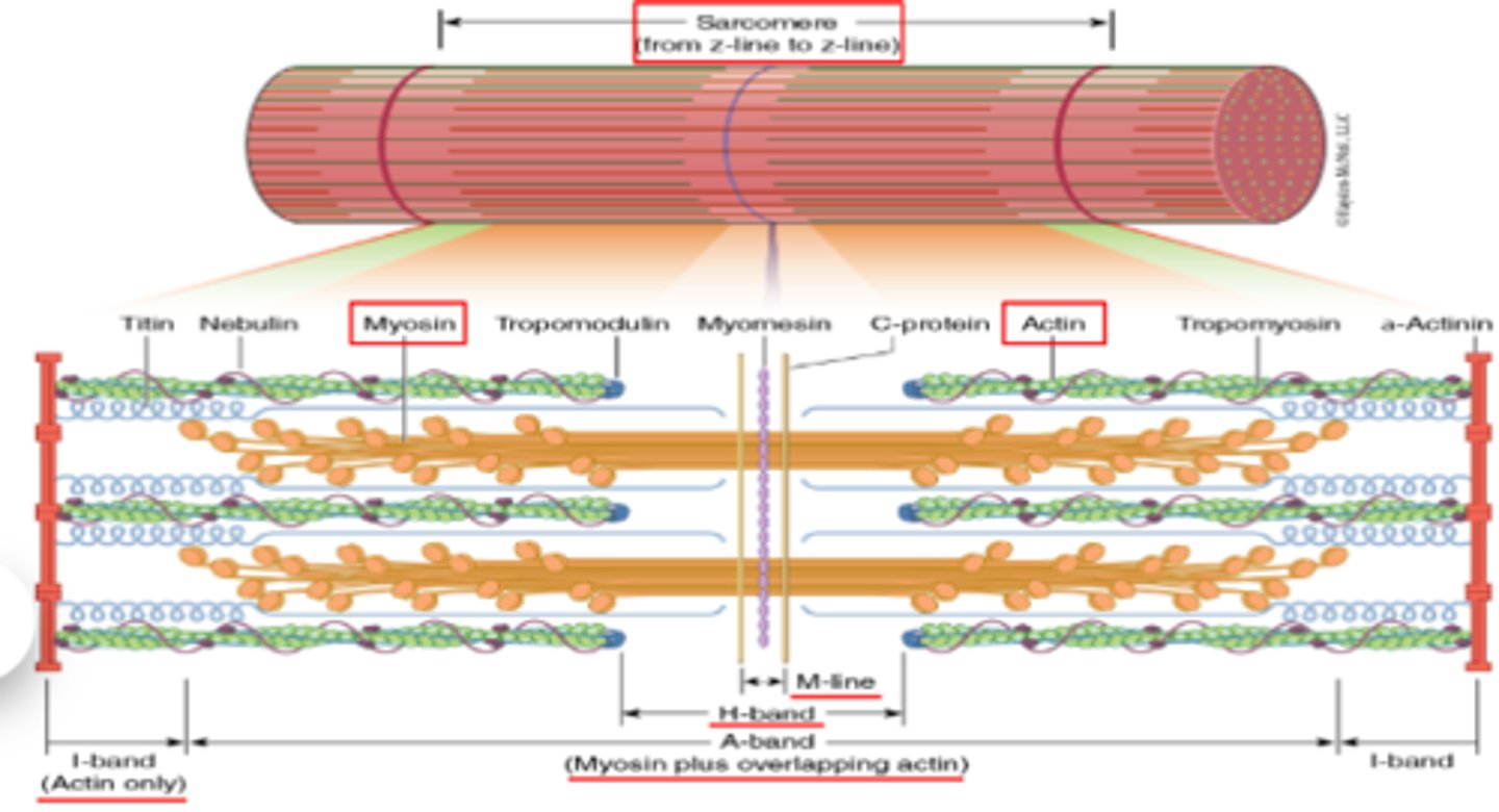
What filament wraps around actin spirally?
tropomyosin
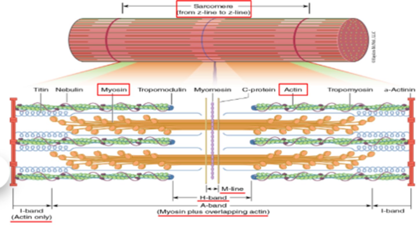
What are the "dots" on the tropomyosin?
troponin
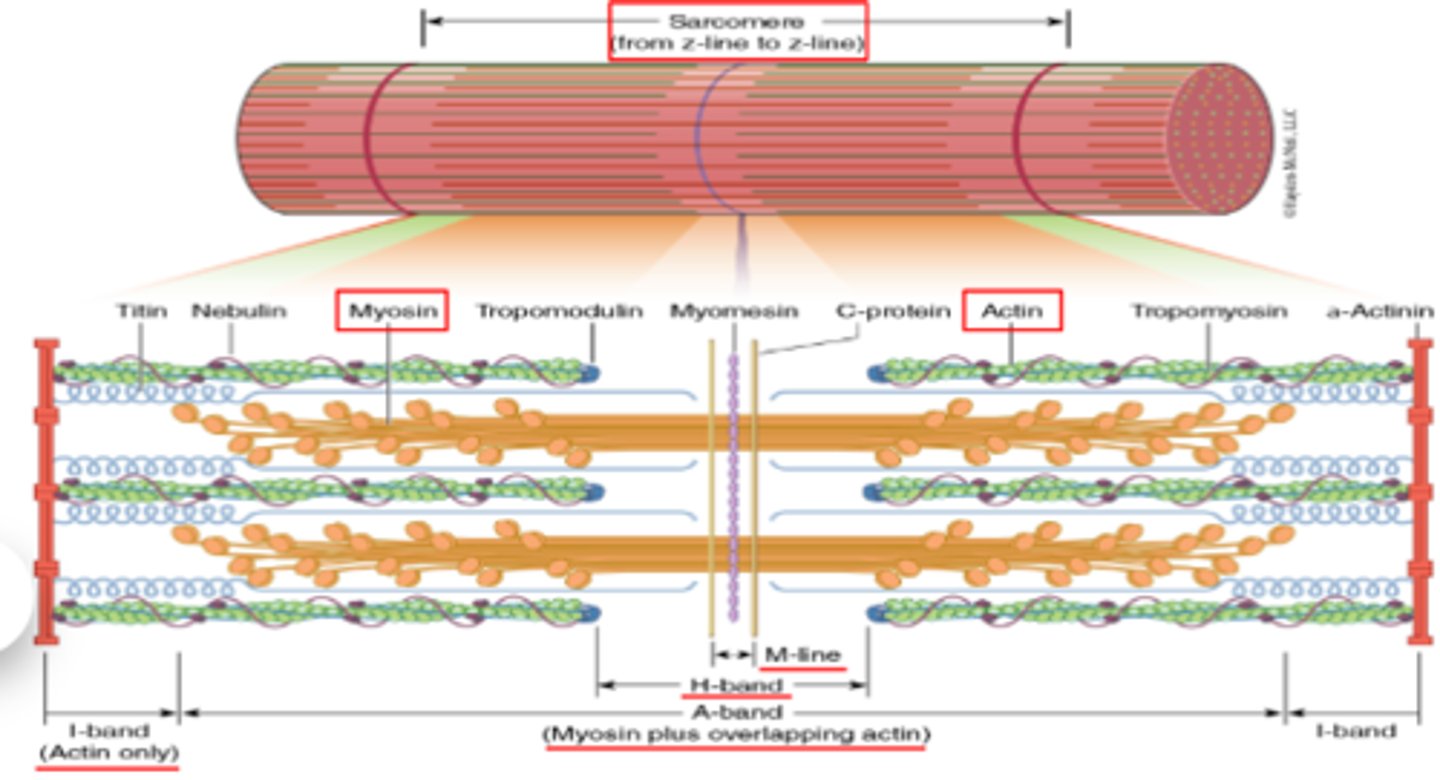
Myosin filaments can also be called what?
think filamenets
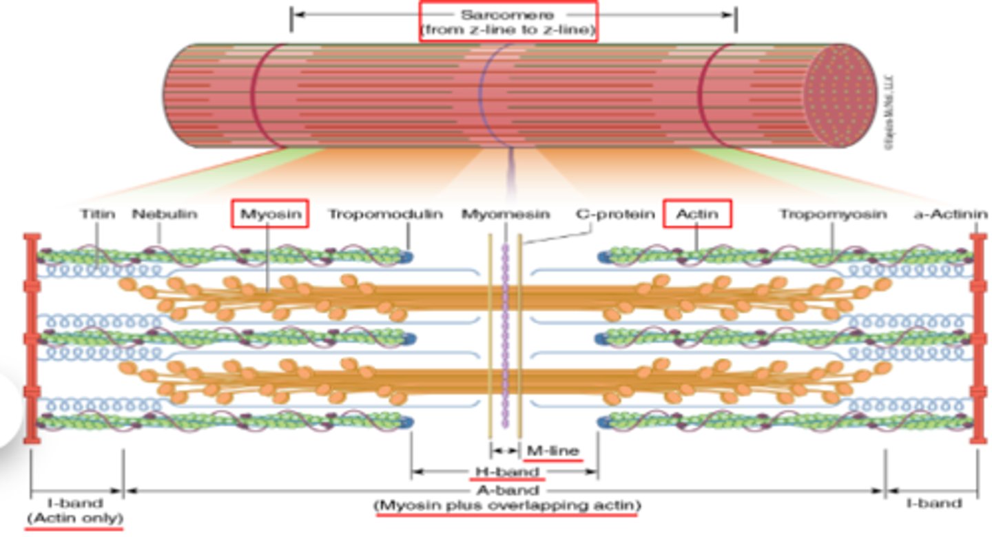
What are the bulbs on myosin filaments?
myosin heads
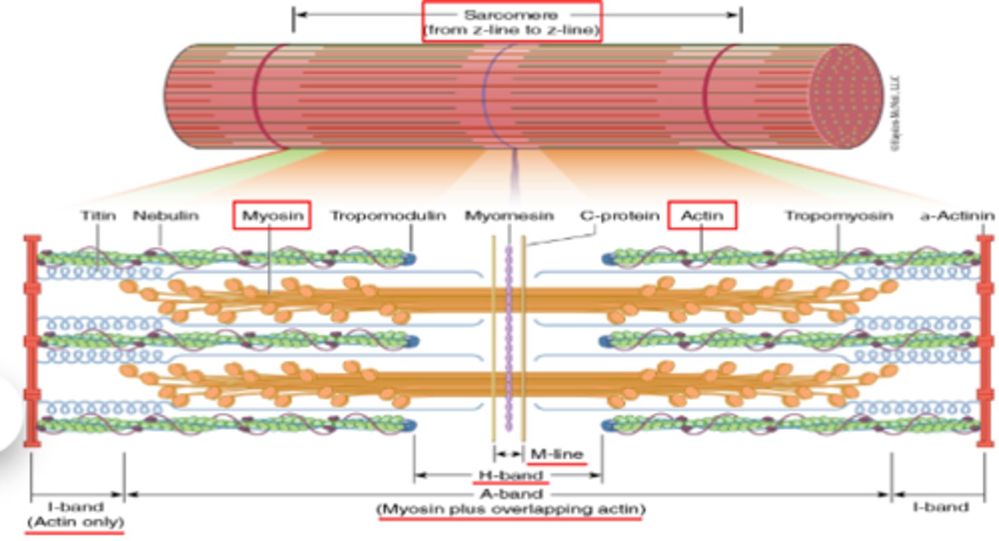
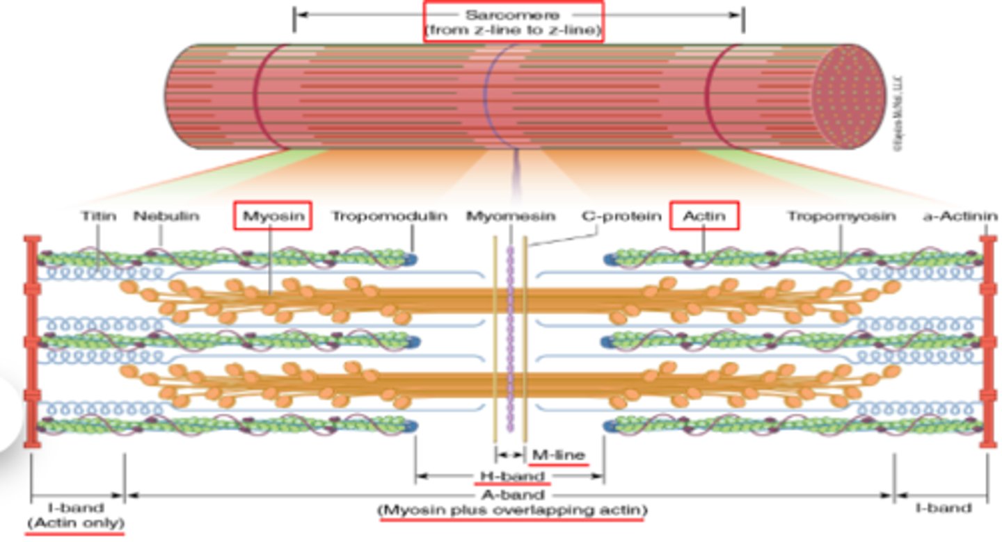
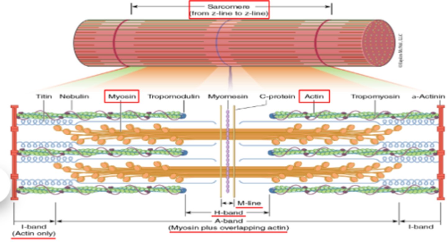
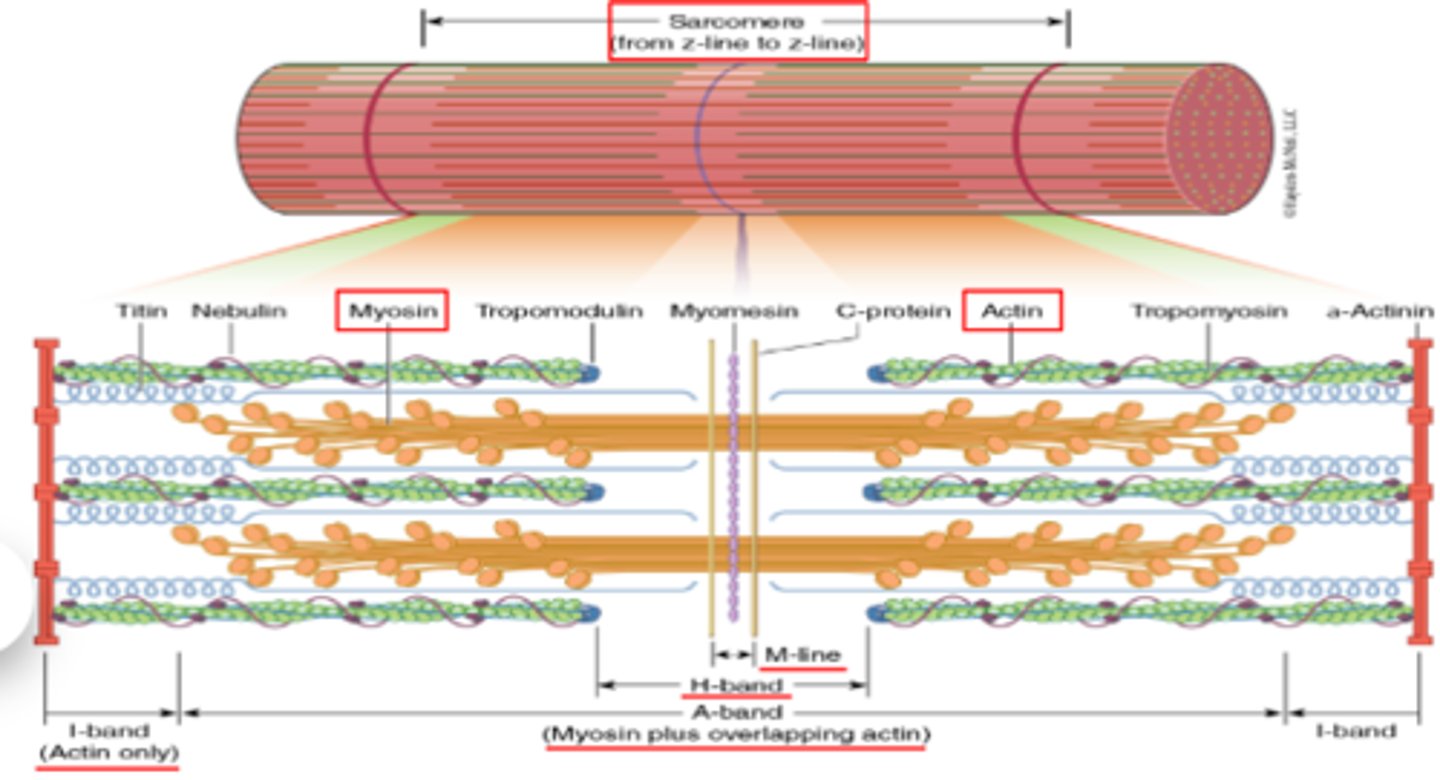
What line is straight down the middle of the sarcomere?
M line (think M for middle)
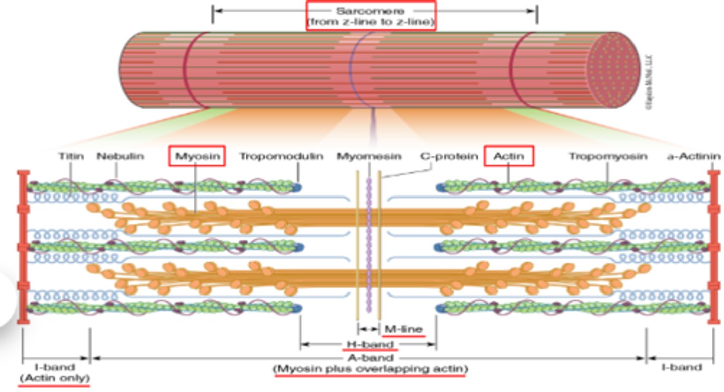
What protein structure composes the M line?
myomesin
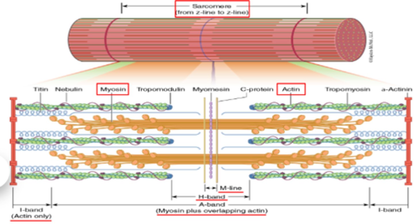
Which region of the sarcomere is composed of actin only?
the I band
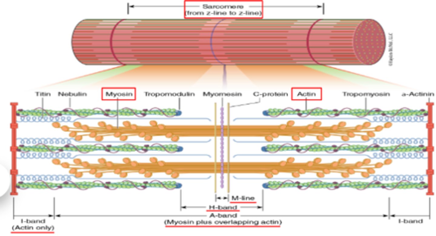
The ____ bands spans the length of the myosin filaments plus overlapping actin.
A band
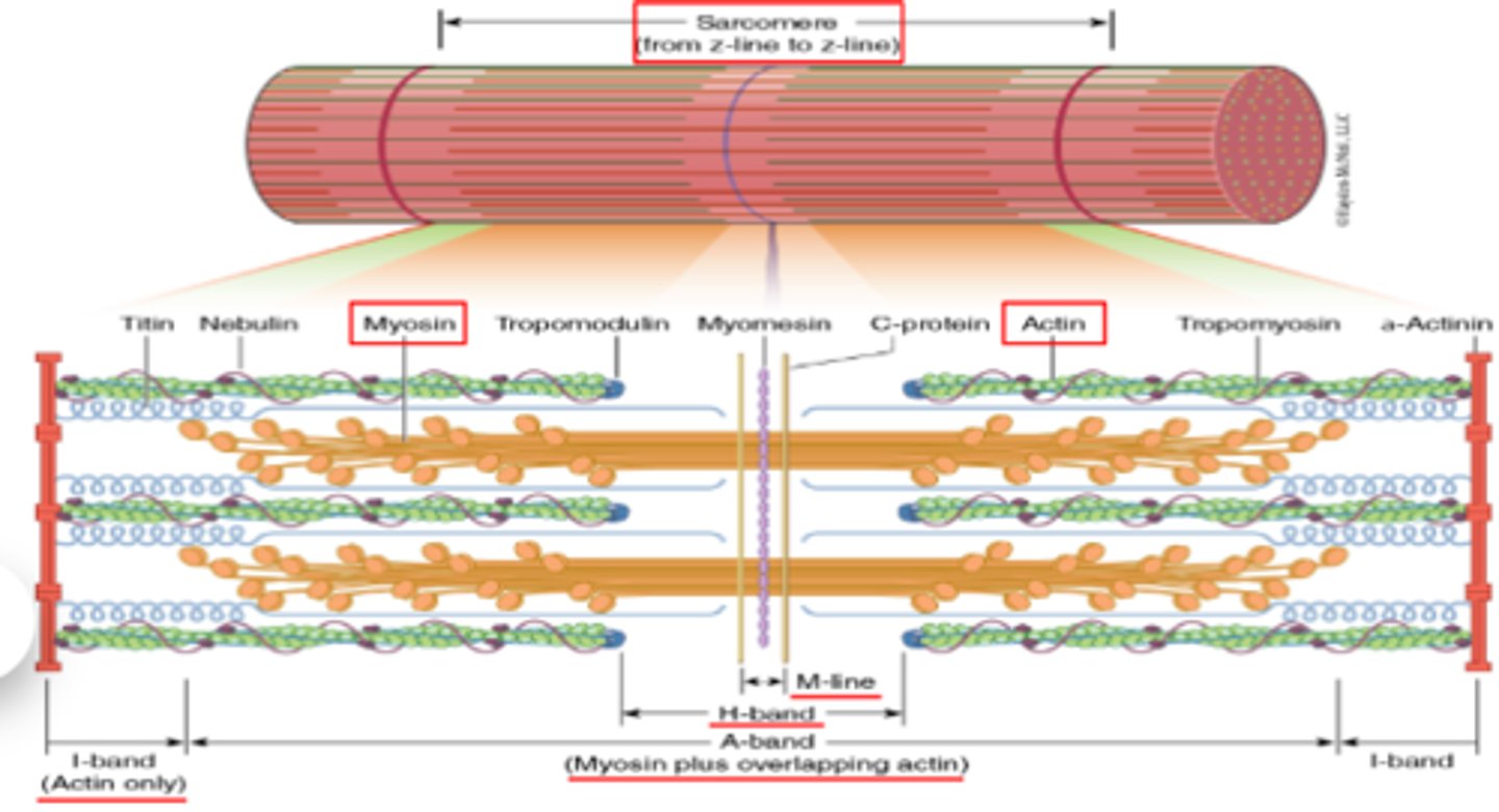
Which band is composed of myosin only?
H band
What is another name for the H band?
H zone
What do myosin heads do when the muscle contracts?
As muscle contracts, myosin heads grab onto the actin, and pull it inward. It will pull the Z lines closer together, which causes the sarcomere as a whole to shorten
T/F : The actin and myosin filaments shorten when muscle contracts
FALSE! They do not structurally change their lengths, they simply just increase their overlap, which makes the sarcomere shorten & contract
dark region of the sarcomere, myosin filaments plus some overlapping actin
A. I band
B. A band
C. H zone
D. M line
E. Z line
A band
Light region of the sarcomere, containing only actin filaments
A. I band
B. A band
C. H zone
D. M line
E. Z line
I band
In the center of each A band is a portion of the myosin with no overlap of actin
A. I band
B. A band
C. H zone
D. M line
E. Z line
H zone
How sarcomeres are divided from each other, found in the center of each I band
A. I band
B. A band
C. H zone
D. M line
E. Z line
Z line
In the center of each A band
A. I band
B. A band
C. H zone
D. M line
E. Z line
M line
What is a muscle plasma membrane called?
sarcolemma
What is the sarcoplasmic reticulum?
smooth endoplasmic reticulum with its interconnecting tubules surrounding each myofibril.
Where are Ca2+ ions stored?
sarcoplasmic reticulum
-
the calcium ions are released when theres a stimulus to make the muscle contract
What are T tubules?
extensions of the sarcolemma (sarcomere plasma membrane) into the interior of the cell. These T tuvules carry the action potential to the entirety of the muscle fiber.
In order for our muscles to contract, the cell membrane has to __________
depolarize
the process that pairs the depolarization of a neuron with the depolarization of a muscle cell.
excitation-contraction coupling
a nerve cell that innervates muscle fibers is called?
motor neuron
What is the spot at which the nerve ending and the muscle fiber meet called?
neuromuscular junction (NMJ)
-
the axon terminnol meets the sarcolemma at the NMJ
What is the first step in excitation-contraction coupling?
action potential is produced in the neuron and it gets propogated down the axon
1. action potential is produced in the neuron and it gets propogated down the axon
2. ?
nerve impulse releases Ach by exocytosis at the axon terminal. then it diffuses across the synaptic cleft of the NMJ. then it binds to receptors on the sarcolemma. this binding induces an impulse that's going to excite our muscle cell.
1. action potential is produced in the neuron and it gets propogated down the axon
2. nerve impulse releases Ach by exocytosis at the axon terminal. then it diffuses across the synaptic cleft of the NMJ. then it binds to receptors on the sarcolemma. this binding induces an impulse that's going to excite our muscle cell.
3.
that impulse is propoated along the sarcolemma down to depolarize the T tubules
1. action potential is produced in the neuron and it gets propogated down the axon
2. nerve impulse releases Ach by exocytosis at the axon terminal. then it diffuses across the synaptic cleft of the NMJ. then it binds to receptors on the sarcolemma. this binding induces an impulse that's going to excite our muscle cell.
3. that impulse is propoated along the sarcolemma down to depolarize the T tubules
4. ?
this triggers Ca2+ channels to open, so there will be an increase in intracellular concentration of calcium within the cytoplasm
-
(in order for a muscle to contract, there must be Ca2+ in the cytoplasm present so its available to bind to troponin)
1. action potential is produced in the neuron and it gets propogated down the axon
2. nerve impulse releases Ach by exocytosis at the axon terminal. then it diffuses across the synaptic cleft of the NMJ. then it binds to receptors on the sarcolemma. this binding induces an impulse that's going to excite our muscle cell.
3. that impulse is propoated along the sarcolemma down to depolarize the T tubules
4. this triggers Ca2+ channels to open, so there will be an increase in intracellular concentration of calcium within the cytoplasm
5. ?
Ca2+ binds to troponin on the thin (actin) filaments
1. action potential is produced in the neuron and it gets propogated down the axon
2. nerve impulse releases Ach by exocytosis at the axon terminal. then it diffuses across the synaptic cleft of the NMJ. then it binds to receptors on the sarcolemma. this binding induces an impulse that's going to excite our muscle cell.
3. that impulse is propoated along the sarcolemma down to depolarize the T tubules
4. this triggers Ca2+ channels to open, so there will be an increase in intracellular concentration of calcium within the cytoplasm
5. Ca2+ binds to troponin on the thin (actin) filaments
6. ?
Tropomyosin moves to allow the interaction of actin and myosin
1. action potential is produced in the neuron and it gets propogated down the axon
2. nerve impulse releases Ach by exocytosis at the axon terminal. then it diffuses across the synaptic cleft of the NMJ. then it binds to receptors on the sarcolemma. this binding induces an impulse that's going to excite our muscle cell.
3. that impulse is propoated along the sarcolemma down to depolarize the T tubules
4. this triggers Ca2+ channels to open, so there will be an increase in intracellular concentration of calcium within the cytoplasm
5. Ca2+ binds to troponin on the thin (actin) filaments
6. Tropomyosin moves to allow the interaction of actin and myosin
7. ?
cross-bridge cycling behins and force is generated
1. action potential is produced in the neuron and it gets propogated down the axon
2. nerve impulse releases Ach by exocytosis at the axon terminal. then it diffuses across the synaptic cleft of the NMJ. then it binds to receptors on the sarcolemma. this binding induces an impulse that's going to excite our muscle cell.
3. that impulse is propoated along the sarcolemma down to depolarize the T tubules
4. this triggers Ca2+ channels to open, so there will be an increase in intracellular concentration of calcium within the cytoplasm
5. Ca2+ binds to troponin on the thin (actin) filaments
6. Tropomyosin moves to allow the interaction of actin and myosin
7. cross-bridge cycling behins and force is generated
8. ?
Ca2+ reaccumulated by the SR and the muscle relaxes
What happens when Ca2+ is low?
tropomyosin blocks the myosin binding site on actin
-
contraction will NOT occur
What happens when Ca2+ is present?
Ca2+ binds to troponin complex
-
conformational change of tropomyosin allows myosin to bind to actin
-
contraction CAN occur
Troponin is a polypeptide complex. What are its 3 subunits? Which one binds Ca2+?
TnC, TnI, TnT
-
TnC binds calcium
Which troponin subunit is a physiological indicator for damage to the heart?
TnI
-
an elevated troponin is often seen in patients who have had a myocardial infarction
Which troponin polypeptide unit helps bind to troponin to tropomyosin>
TnT
(Think Tnt for Troponin2Tropomyosin)
______________ locks the myosin binding sites on actin
tropomyosin
-
the myosin heads have a special spot where they bind on actin so they cannot just readily bind whenever they want to because the tropomyosin is blocking it
actin and myosin have a natural __________ for one another
affinity
-
theyre like magnets, myosin naturally really wants to bind to actin
What happens when calcium binds to troponin?
it causes the troponin to change shape. in doing so, it will prolly tropomyosin away and expose the myosin binding sites
What are the steps of the sliding filament theory?
ATP binds to myosin causing the dissociation of the actin-myosin complex
-
ATP is hydrolyzed, causing myosin heads to return to their resting conformation
-
myosin head binds to actin monomer forming cross bridge
-
P is released. myosin heads change the conformation; the filaments slide past each other
-
ADP is released
What does ATP get broken down into?
ADP & an inorganic phosphate
How does the sarcomere contract?
myosin pulls the actin filament towards the M line, which then pulls the Z lines closer together
How do you get myosin to let go when you cycle back to the beginning of the sliding filament theory?
once another ATP binds to myosin, the length between actin and myosin, the link between the actin and myosin is weakened and the myosin head detaches. the cross bridge breaks between actin and myosin
What is known as the pulling of actin by myosin results in the overall muscle shortening & the generation of force
sliding filament theory
What comprises a motor unit?
a single mootor neuron and the muscle fiber it innervates
Why is a motor unit called that?
because when the single motor neuron fires, all the associated muscle fibers that it innervates, will contract as a unit
T/F : one motor neuron can innervate different numbers of muscle fibers
true!
-
the motor units vary considerably in size
What do the size of motor units tell us?
the kinds of movements that we are able to perform
small motor units are involved with ____________ motor activities.
very precise
-
one nerve will be controlling a very small number of fibers, so it can be very meticulous and precise with that movement
Large motor units are involved with ____________ motor activities.
gross movements
Are eye movements gross or precise?
precise
Is walking/running a gross or precise movement>
gross
is writing a precise or gross movement?
precise
What is recruitment?
the body's way to keep us efficient. it is the process of activating smaller motor units before activating larger ones
-
EXAMPLE:
there is a box on the floor in front of yyou and you have no idea what's in it or how much it weighs. it could be a box of feathers or a box of cement. You are asked to pick it up. If it is a box of feathers and your body does NOT display recruitment, and you jump straight to activating larger motor units, if you lift that box with too much strength or too large of motor units, the box will go flying. Fortunately, that does not happen. The body will recruit just the right amount of motor units to perform or accommodate the action. You start with smaller and call on more if additional strength is needed.
What are the 2 main muscle actions?
isometric and isotonic actions
What is the difference between isometric and isotonic actions?
isometric : muscle contract and length remains; the force generated is insufficient to overcome the load
(EX : if you try to flip a car, your muscles will contract and try to flip it, but overall, will not overcome the load)
(EX : Carrying a box in front of you. the weight of the box is pushing down on you, but your arms are opposing the weight with equal force going upward, so your arms are neither raising nor lowering. they are contracted but they're staying the same length)
-
isotonic : the contraction tension remain constant and the muscle changes in length and moves the load
(EX : picking up a cup of water to bring to your mouth. you have to contract muscle fibers as you move it up to your mouth and then back down to the table & you're changing the length of those fibers, but the tension in the muscle, resulting in the tension from the cup, stays the same.
-
the prefix iso means same and metric refers to measurement/length ; tonic refers to tension/tone, this means it will have the same tension, but you're able to change the length of the muscle fiber because you can overcome the load
What are the two types of ISOTONIC actions?
concentric : muscle contracts and shortens
-
eccentric : muscle contracts and lengthens
is the upward movement of a bicep curl concentric or eccentric?
concentric
-
the fibers contract and shorten when you curl the fibers and pulls the weight upward
Is the downward movement of a bicep curl concnetric or eccentric?
eccentric
-
as youre lowering the weight back down, the muscle has to stay controlled/contracted, otherwise the arm would just go limp and fall straight down, which is not the proper way to do a bicep curl. as you bring the weight back down, you are lengthening the muscle, however, its still contracting to keep that downward movement controlled
the primary mover
A. agonist
B. synergistic
C. antagonistic
A. agonist
muscles that assist the agonist group
A. agonist
B. synergistic
C. antagonistic
B. synergistic
(brachioradialis is a synergistic muscle to the biceps)
muscles that oppose the agonist
A. agonist
B. synergistic
C. antagonistic
C. antagonistic
(triceps are antagonistic to the biceps)
What are the three main types of classifications of skeletal muscle?
Slow twitch (Type I)
-
Fast Twitch (Type IIX & IIA)
Structural and histological features determine the type of fibers based on what 3 things?
size, diameter, and myoglobin
the oxygen binding molecule that facilitates oxygen transport into myocytes
myoglobin
slow twitch muscles is good for what kind of athletes
endurance (distance running)
fast Twitch muscles is good for what kind of athletes
basketball or sprinting
How do slow twitch muscle fibers produce the majority of their energy?
aerobic respiration (means they're going to need a lot of oxygen)
Which type of skeletal muscle?
- myoglobin rich
- contain large numbers of oxidative enzymes
- high mitochondrial volume
- surrounded by more dense capillary networks
- fatigue resistant
- slower maximal shortening velocity (means they do not prodyce large amounts of force; this results in them having a small cross sectional area because large cross sectional areas allow you to produce more force)
- highly efficient
slow twitch - type I
Which type of skeletal muscle?
- small number of mitochondria
- less resistant to fatigue
- rich in glycolytic enzymes
- generate the highest power output
Fast Twitch - Type IIX
-
fatigue easily but have the highest output
-
EX : 100 m sprint athlete
-
think "X" for extreme
Which type of skeletal muscle?
- intermediate fibers
- mixture of Type I and Type IIX
Fast Twitch - Type IIA
-
EX : 800 m sprint athlete
-
Think "A" for all because its a mix of the other types
-
not completely aerobic & will definitely feel some significant fatigue
an increase in muscle fiber diameter due to an increase in myofibril size ; the amount of force that can be generated by a muscle group is proportional to the cross-sectional area of the muscle ; thus, physiological adaptations as a result of strength training are a result an increase in muscle size, not muscle number (I.e hyperplasia)
hypertrophy
result from microscopic injury to the muscle fibers; Delayed Onset Muscle Soreness (DOMS) appears 24-48 hours post microscopic injury ; NOT a result of lactic acid accumulation
muscle soreness
When absolute strength (the total amount of force applied) is compared in untrained men and women, __________ are typically stronger.
men
When youre on your last end of the workout and your muscles are on fire, what is that?
that is lactic acid buildup, but NOT DOMS
T/F : Women exhibit 40% less upper body strength and 33% less lower body strength on average than men. Women have 37-68% the muscle strength of males.
True