topic 12
 Call Kai
 Learn
Learn Practice Test
 Spaced Repetition
 Match
Match Flashcards
 Knowt Play
1/59

There's no tags or description
Looks like no tags are added yet.
Name | Mastery | Learn | Test | Matching | Spaced | Call with Kai | Chat |
|---|
No analytics yet
Send a link to your students to track their progress
60 Terms
Three muscle types
Skeletal, cardiac, smooth
Skeletal muscles:
Where:
Function:
Type of control:
Response:
– Striated muscle attached to bones of skeleton
– Control body movement
– Voluntary control
▪ Responds to somatic innervation, modulated by endocrine control
Cardiac muscle
Where:
Function:
Type of control:
Response:
– Striated muscle found only in heart
– Moves blood through circulatory system
– Involuntary control
▪ Responds to autonomic innervation, spontaneous contraction,
modulated by the endocrine system
Smooth muscle
Where:
Function:
Type of control:
Response:
– Primary muscle of the internal organs and tubes
– Influence movement of material into, out of and within body
– Involuntary control
▪ Responds to autonomic innervation, spontaneous contraction,
modulated by the endocrine system
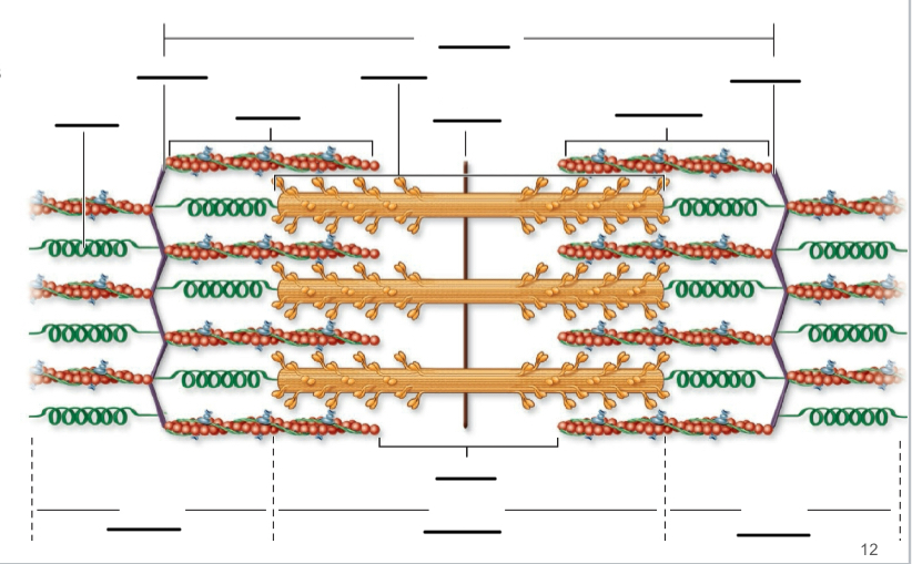
Organization of muscle tissues from largest to smallest
Muscle → fascicle → muscle fiber → myofibril → sarcomere
Organization of a sarcomere: composed of __ and delineated _.
▪ Composed of overlapping thick and thin filaments
▪ Delineated at both ends by z-discs
Sarcomeres =
Myofilaments arranged in repeating units
Thin and thick filaments made up of
Alternating I-bands and A-bands
I bands
▪ Light-appearing regions that contain only thin filaments
▪ Bisected by Z disc
▪ Get smaller when muscle contracts
A band
▪ Dark-appearing region that contains thick filaments and
overlapping thin filaments
▪ Contains H zone and M line
▪ Makes up central region of sarcomere

Identify
Identify

H zone: central portion of A band that:
▪ Only thick filaments present; no thin filament overlap
▪ Disappears with maximal muscle contraction

Identify
Identify

Identify
Identify

M line:
middle of H zone
▪ Protein meshwork structure
▪ Attachment site for thick filaments
Parts of muscle cell (fiber):
Sarcoplasm And has a typical organelles plus contractile proteins

Sarcolemma (plasma membrane):
▪ Has T-tubules (transverse tubules) that extend deep into cell Containing voltage-sensitive calcium channels
Myofibrils are
(hundreds to thousands per cell)
▪ Bundles of myofilaments enclosed in sarcoplasmic reticulum

Sarcoplasmic reticulum
Terminal cisternae: blind sacs of sarcoplasmic reticulum
–Serve as reservoirs for calcium ions
–Two cisternae with T-tubule in between = triad
Sarcoplasmic reticulum Contains
calcium pumps that import calcium
Sarcoplasmic reticulum Contains (extended)
▪ Contains calcium release channels
–Triggered by electrical signal traveling down T-tubule
–Calcium released into sarcoplasm
Myofibrils =
Muscle Fiber Contractile Structures
Myofilamemts
Contractile proteins within myofibrils
Thick filaments consist
Consist of bundles of many myosin protein molecules
Thin filaments
Twisted strands of actin with Myosin binding site where myosin heads attach
Thin filaments Regulatory proteins aren
Tropomyosin and troponin
Accessory proteins are
Titian and nebulin
Titin
– Elastic protein
– Stabilizes the position of the contractile elements
– Elasticity returns stretched muscles to their resting length
• Nebulin
– Inelastic protein
– Aligns actin filaments

Organization of myofibrils
Understand

Neuromuscular junction location and parts
Location where motor neuron innervates muscle
Has the following parts:
–Synaptic knob
–Synaptic cleft
–Motor end plate

Synaptic knob
– Expanded tip of motor neuron axon
Synaptic knob Houses
synaptic vesicles
▪ Small sacs filled with neurotransmitter Ach
Innervation of skeletal fibers Has
Ca2+ pumps in plasma membrane
▪ Establish calcium gradient (more outside the neuron)
Innervation of skeletal fibers Has
Has voltage-gated Ca2+ channels in membrane
▪ Ca2+ flows into cell if channels open (down concern. Gradient)
Motor end plate
Specialized region of sarcolemma with numerous folds
Motor end plate has many
Has many ACh receptors
Motor end plate ACh receptors are
Plasma membrane protein channels
– Opened by binding of ACh
-Allows influx of Na+ (and efflux of K+)
Synaptic cleft
– Narrow fluid-filled space
– Separates synaptic knob from
motor end plate
Synaptic cleft
Acetylcholinesterase here
▪ Enzyme breaks down ACh molecules
Skeletal muscle fibers at rest
Muscle fibers exhibit resting membrane potential (RMP)
– RMP of muscle cell = ~ −90 mV
▪ Established by leak channels and Na+/K+ pumps
(voltage-gated channels are closed
In the Neuromuscular Junction, calcium enters at
The synaptic knob
When Calcium enters at the synaptic knob the nerve signal
travels down axon and opens voltage-gated Ca2+ channels
When voltage-gated Ca2+ channels open
–Ca2+ diffuses into knob
–Ca2+ binds to synaptic vesicles
Neuromuscular junction: excitation of skel. musc. fibers release
• Release of ACh from synaptic knob
–Exocytosis of ACh at
synaptic knob
Neuromuscular junction: excitation of skel. musc. fibers Bind
• Binding of ACh at motor end plate
–Diffuses across cleft
–Binds to receptors
–Excites fiber
Excitation-Contraction Coupling is stimulation
Stimulation of fiber coupled with sliding of filaments
• Synaptic cleft is a
– Narrow fluid-filled space
– Separates synaptic knob from motor end plate
Acetylcholinesterase is housed in _ and is an _.
Housed in synaptic cleft. Enzyme breaks down ACh molecules
Excitation-Contraction Coupling
–Na+ diffuses into cell through voltage-gated channels
▪ Cell depolarizes:
–Becomes less negative (eventually becomes +30 mV)
–Just after Na+ channels open, they close:
▪Voltage-gated K+ channels open
–K+ diffuses out of cell
–Cell repolarizes:
returns to -90mV
AP travels down T-tubules causing
Causes voltage-sensitive calcium channels in T-
tubule membrane to trigger opening of calcium release
channels in SR terminal cisternae
Release of Ca2+ from sarcoplasmic reticulum
–Ca2+ interacts with myofilaments triggering contraction
Slide 42
Slide 44
Slide 45
Sarcomere: Crossbridge Cycling
–Cycling continues as long as Ca2+ and ATP present
–Results in sarcomere shortening as Z discs move closer
together
Sarcomere: Crossbridge Cycling
–Narrowing (or disappearance) of H zone and I band
–Thick and thin filaments remain same length
▪ But slide past each other
Events in Skeletal Muscle Relaxation
1. Termination of nerve signal and ACh release from
motor neuron
2. Closure of Ca2+ channels in sarcoplasmic reticulum
▪ Ca2+ returned to sarcoplasmic reticulum by pumps
3. Troponin returns to original shape
▪ Tropomyosin blockade returns
4. Return of muscle to original position due to its elasticity