RS 1 - Neck and Mediastinum
 Call Kai
 Learn
 Practice Test
 Spaced Repetition
 Match
 Flashcards
 Knowt Play
1/56

There's no tags or description
Looks like no tags are added yet.
57 Terms
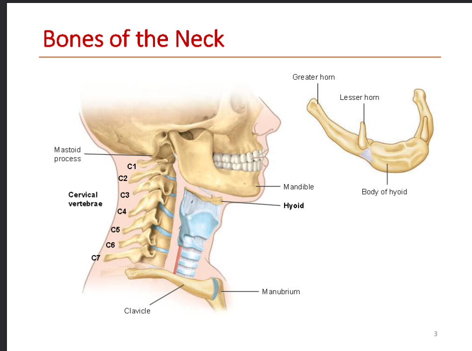
what bones make up the neck
Cervical vertebrae (C1–C7):
Hyoid:
Greater & Lesser horns: These are just the pointy prongs sticking up and back from the main body of the hyoid bone
neighboring bones that frame the neck, including the mandible (lower jawbone), the mastoid process (the bony bump right behind your ear), the clavicle (collarbone), and the manubrium (the top part of your breastbone).
Cervical Vertebrae char
Vertebral foramen: "Foramen" just means "hole." This is the big central tunnel where your spinal cord safely passes through.
Transverse foramen: These are two smaller holes on the left and right sides of the bone. Only neck vertebrae have these, and they act as tunnels for blood vessels going up to your brain.
Bifurcated spinous process: "Bifurcated" means split into two,
Body: The solid, thick drum-shaped front part of the vertebra that bears weight.
Atlas (C1): The very first neck bone that holds up your skull, named after the Greek myth of Atlas holding up the world. It looks like a ring with no solid "body" and lets you nod your head "yes."
Axis (C2): The second neck bone right below the Atlas. It has a special thumb-like peg sticking straight up called the dens (or odontoid process), which acts as a pivot so you can shake your head "no."

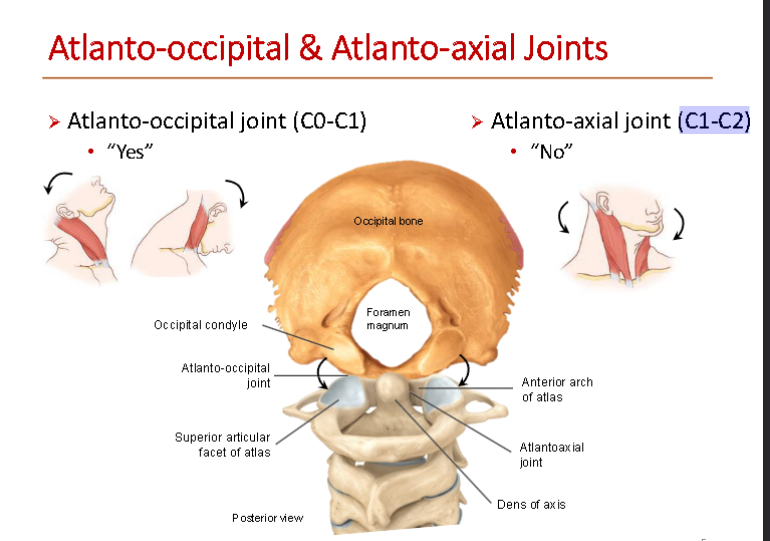
what is the Atlanto-occipital joint
This is the joint between your skull's occipital bone ("C0") and the first neck bone, the Atlas (C1)
allows you to nod ur head yes up and down
what is Atlanto-axial joint
between C1-C2)
allows you to turn ur head left and right “no”
what is the Foramen magnum:
: Literally translates to "big hole." It is the large opening at the bottom of your skull where the brain connects to the spinal cord.
what are Cervical Triangles
divide the side of the neck into two imaginary triangles to make it easier to map out where blood vessels, nerves, and glands are hiding.

what muscles make up the section of the triangles
pink = Anterior Cervical Triangle
(Blue) = Posterior Cervical Triangle
what does the Sternocleidomastoid (SCM do in the cervical trangles?
Divides the neck into anterior and posterior triangles
what is the Platysma Muscle and what is it innervated by?
➢ Very thin muscle of facial expression
➢ Innervated by cervical branch of facial nerve (CN VII)
what is Platysma Muscle main action
Action: tenses skin on the neck, depresses skin on chin
what Muscles are around Cervical Vertebrae
➢ Anterior: longus colli and longus capitis
➢ Lateral: anterior, middle and posterior scalene muscles
➢ Posterior: deep cervical/back muscles
where do Scalene Muscles attach
Transverse processes of cervical vertebrae to 1st and 2nd ribs
Actions of scalene muscles
• Flexion of the neck
• Elevates ribs in forced inspiration
what is the The Scalene Gap?
This is the narrow space sandwiched right between the anterior scalene (AS) and middle scalene (MS) muscles. Two critical structures squeeze right through this gap to reach your arm:
Brachial plexus (green)
Subclavian artery (red)
where does the Phrenic nerve sit?
This nerve sits right on the front (anterior surface) of the anterior scalene muscle

what muscles are attached to the hyoid bone
Muscles attached to the hyoid bone (C3)
Suprahyoid muscles: superior to hyoid
Infrahyoid muscles: inferior to hyoid
what muscles are part of the Suprahyoid Muscles
Mylohyoid
Digastric, anterior belly
Digastric, posterior belly
Stylohyoid
Geniohyoid
main function of suprahyoid muscles?
Elevate hyoid bone during speech and swallowing
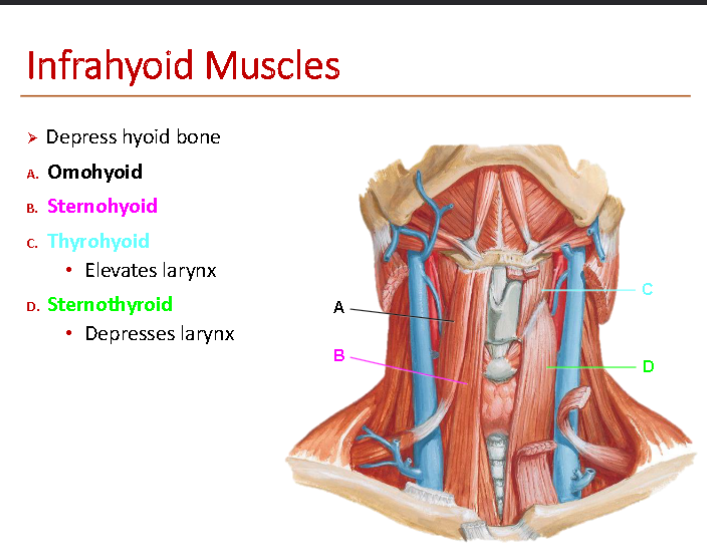
what muscles make up the Infrahyoid Muscles
Omohyoid
Sternohyoid
Thyrohyoid
Sternothyroid
function of Infrahyoid Muscles
Depress hyoid bone
whats another function of Thyrohyoid muscle
Elevates larynx
whats an additional function of Sternothyroid muscle
Depresses larynx
what is superior and inferior attachment for Sternocleidomastoid (SCM) Muscle
Superior attachment - Mastoid process of temporal bone
Inferior attachments - • Manubrium of sternum
• Clavicle
what is scm muscle innervated by and what are its action
Spinal accessory nerve (CN XI)
Lateral flexion with rotation
Flexion of cervical vertebrae
what does Viscera of the Neck mean and what organs are part of this portion of the neck
groups the organs packed into the front of your neck, sitting right behind (posterior to) those flat infrahyoid strap muscles.
Respiratory organs (Breathing)
Alimentary organs
Endocrine organs
which structures are part of respiratory organs
• Larynx
• Trachea
which structures are part of the Alimentary organs
• Pharynx
• Esophagus
what structures are part of the Endocrine organs
• Thyroid gland
• Parathyroid glands
location of Thyroid Gland
➢ Largest endocrine gland
➢ Anterior to trachea at C5-T1 levels
➢ Posterior to infrahyoid muscles
Functions of Thyroid Gland
• Regulates metabolic rate via thyroid hormones (T3 & T4)
• Regulates blood calcium via calcitonin
location of Parathyroid Glands
➢ On posterior aspect of thyroid gland
➢ Usually 4 glands

function of Parathyroid Glands
Regulate calcium and phosphate via parathyroid hormone
Thyroid Vasculature

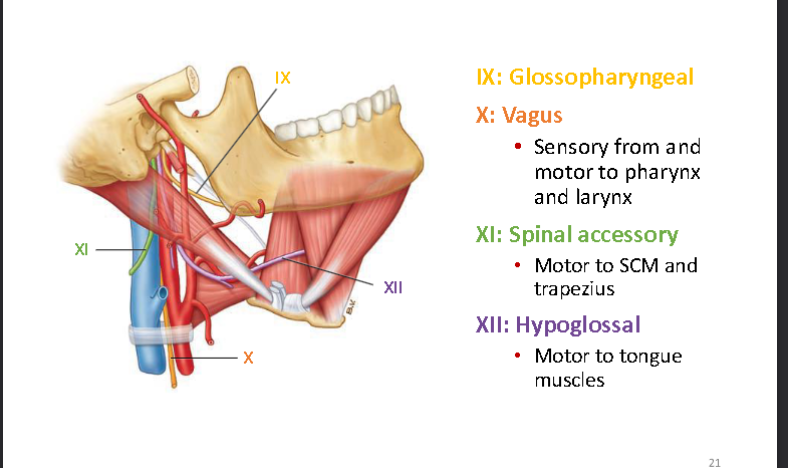
Cranial Nerves in the Neck
what is The sympathetic trunk
is like a vertical "power line" of nerves running up and down the neck. It handles your body's "fight-or-flight" signals (like dilating your pupils, raising your heart rate, or triggering sweat).
Location of sympathetic trunk and function
Between prevertebral layer and carotid sheath
Contains superior, middle, and inferior ganglia
Provides sympathetic innervation for head, neck and heart
what does the Superior Thoracic Aperture contain
Vessels
Nerves
Respiratory organs
Digestive: esophagus
which vessels does it contain
• Common carotid a.
• Subclavian a.
• Brachiocephalic v.
• Thoracic duct
what Nerves does it contain
• Phrenic n.
• Vagus n.
• Recurrent laryngeal n.
• Sympathetic trunk
what Respiratory organs does it contain
• Trachea
• Cervical pleura
what is the Mediastinum, what are its boundaries
“Middle space” between pulmonary cavities
Boundaries:
• Superior thoracic aperture
• Diaphragm
• Pulmonary cavities
• Sternum
• Thoracic vertebrae
what are the Major Organs in the Mediastinum
thymus
great vessels
trachea
esophagus
heart in pericardium
what are different Passages Through the Diaphragm
➢ Caval opening for inferior vena cava (T8 level)
➢ Esophageal hiatus for esophagus and vagus nerves (T10 level)
➢ Aortic hiatus for aorta and thoracic duct (T12 level
what Nerves are in the Mediastinum
Recurrent laryngeal n.
Vagus n
Phrenic n
what is the origin of Phrenic Nerve
cervical plexus (C3,4,5
what is the phrenic nerves pathway
• Enters mediastinum between subclavian artery and vein
• Descends anterior to the roots of the lungs
• Between fibrous pericardium and mediastinal parietal pleura
Motor to diaphragm
where does phrenic nerve gets sensory info from?
• Pericardium
• Parietal pleura (mediastinal,
diaphragmatic)
• Parietal peritoneum (inferior
surface of diaphragm)
what happens in Phrenic Nerve Injury
Normally, when you breathe in (inspiration), both halves of your diaphragm contract and pull downward to make room for your lungs to expand. If one phrenic nerve gets damaged, that half of the diaphragm becomes paralyzed ("denervated") and does the exact opposite of what it should do—called paradoxical movement:
what happens to Denervated side of diaphragm
• Pushed superiorly by abdominal viscera during inspiration
• Pushed inferiorly by positive pressure in thoracic cavity during expiration
whats the pathway of the Vagus Nerve (CN X)
Descends along the common carotid artery
Courses posterior to root of lung and along esophagus
function of vagus nerve x
Parasympathetic to autonomic nerve plexuses
• Cardiac
• Pulmonary
• Esophageal
pathway of Recurrent Laryngeal Nerve
A. Right recurrent laryngeal nerve loops under right subclavian artery
B. Left recurrent laryngeal nerve loops under aortic arch
function of Recurrent Laryngeal Nerve
➢ Motor to most muscles of the larynx
➢ Sensory from inferior larynx
where does Sympathetic Trunk orginate from and what does it contain
Sympathetic output originates from spinal cord T1-L2 (cell bodies of preganglionic neurons)
Sympathetic ganglion contains cell bodies of postganglionic neurons
function of Sympathetic Trunk
Sympathetic trunk connects sympathetic ganglia
• Extends superiorly to the neck and inferiorly to the pelvis
C. Splanchnic nerves contain preganglionic neurons to abd