Lymphatic and Digestive System
 Call Kai
 Learn
Learn Practice Test
 Spaced Repetition
 Match
Match Flashcards
 Knowt Play
1/32

There's no tags or description
Looks like no tags are added yet.
Name | Mastery | Learn | Test | Matching | Spaced | Call with Kai | Chat |
|---|
No analytics yet
Send a link to your students to track their progress
33 Terms
Describe the three functions of the lymphatic system
Fluid balance - collects excess fluid from body tissues and returns it to the blood stream.
Lymph is the term used to describe interstitial fluid once it has entered the lymphatic system. The fluid that forms lymph enters the lymphatic system through tiny lymphatic capillaries which are interlaced among arterioles and venules throughout the body.
Contain valves to ensure one way flow of lymph.
Lymphatic ducts are the final drainage point. They are located at the junction of the jugular and subclavian veins in the neck and drain lymph from lymph vessels into these veins.
lymphatic system is damaged in some way, for example by injury or surgery, interstitial fluid accumulates in the tissue spaces instead of being drained away. This inappropriate accumulation of fluid is referred to as lymphoedema (swelling)
Defence and Immunity - Microorganisms and other foreign substances are filtered from lymph (fluid) by lymph nodes and from blood by the spleen. Immune cells in lymph nodes stand ready to respond to foreign cells or chemicals.
Fat Absorption - from the digestive system. Dietary fats are digested into small molecules (such as fatty acids) and are absorbed by cells of the intestine and then move into the lymphatic system (via lacteals)
Describe the basic functions of the lymph nodes, thymus, tonsil, and spleen
Lymph Nodes:
Small bean-shaped organs located throughout the lymphatic system.
Lymph nodes are especially dense at the cervical, axillary, and inguinal junctions, where lymphatic drainage from the head, arms, and legs converges before returning to the bloodstream.
Made of lymphatic tissue and located along the larger lymph vessels
Lymph that flows through the nodes makes contact with immune cells within the node. These include macrophages, dendritic cells, B and T cells
Thymus:
Bilobed organ located in the space between the sternum and aorta. Connective tissue holds the two lobes closely together
Location where T-cells mature, divide and collect. This allows them to attack pathogens without harming the body cells.
The hormone thymosin is made in the thymus
Tonsils:
Lymphoid nodules located along the inner surface of the pharynx and are important for developing immunity to foreign pathogens that are either inhaled or swallowed.
tonsils are the first line of defence against bacteria and other harmful material and can become enlarged or inflamed due to infection.
Persistent tonsilitis may require surgical removal
Spleen:
Largest Lymphatic organ at approximately 12cm long and is attached to the lateral border of the stomach. The spleen is a fragile organ and appears dark red due to extensive vascularisation (rich blood supply).
Functions include:
Filtering of blood, due to the extensive vascularisation and the presence of macrophages and dendritic cells that remove microbes, debris and worn-out/dying red blood cells.
Immune response to pathogens found in the blood.
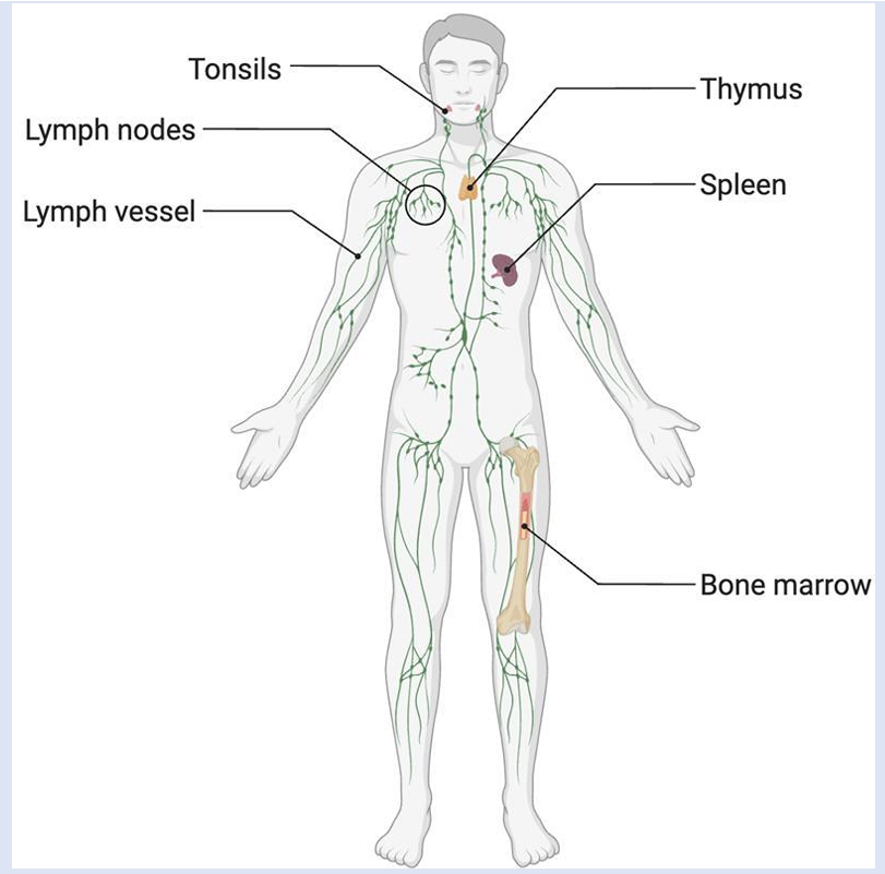
How is lymph transported around the body
Lymph is not actively pumped by the heart, instead it is pushed through vessels by movement in the body and one-way valves to keep lymph moving towards the heart.
Two Mechanisms that do this are:
Skeletal muscle pump. The contraction of skeletal muscle physically squeezes the nearby lymphatic vessel; this compression pushes the lymph forward.
Thoracic pump. Breathing creates pressure changes that ‘pushes’ lymph from the abdomen to the thoracic cavity.
List the 8 major types of lymphatic cells and identify their function
B-Cells
Immune cells that produce antibodies.
An antibody is any of the group of proteins that bind to a specific target on a pathogen, known as an antigen.
An antigen is a chemical marker on the surface of a pathogen that T or B cells can recognise. Once a B cell binds to an antigen, it is activated and becomes a plasma cell.
T-cells
immune cells that mature in the thymus and do not produce antibodies. T cells recognise and attack foreign invaders (antigens)and secrete chemical signals that coordinate the immune response.
Plasma cells
B cells that have differentiated in response to antigen binding. Plasma cells secret large amounts of antibodies.
Natural Killer cells
lymphocytes that are circulating blood cells and contain cytotoxic (cell-killing) granules. They destroy virally infected cells. NK cells are one of the body’s first lines of defence against viruses and certain types of cancer.
Macrophage:
large phagocytic cells that have differentiated from monocytes
Functions include phagocytosis, antigen presentation, chemical secretion, tissue repair and remodelling.
Dendrite Cell:
Mostly found in peripheral tissues (skin and mucus membranes)
Functions include phagocytosis and antigen presentation
Mast Cells:
Do not circulate in the blood, instead mature mast cells are found in the body tissues near blood vessels, the skin, and respiratory tract.
Store inflammatory mediators, including histamine and heparin in large granules.
When exposed to allergens mast cells release the contents of their granules into the immediate vicinity. These mediators cause symptoms of an allergic reaction, including itching, mucus, and inflammation.
Eosinophils:
Anti-parasitic defence - produces toxic proteins such as neurotoxin that paralyses parasites like tapeworm
Regulation of Histamines - secretes enzymes that degrade histamines and other inflammatory chemical to regulation inflammation that could damage tissue.
Basophils:
Motile cells that leave the blood and enter tissues. Secrete chemicals (cytokines) that aid the mobility and action of other leukocytes.
Activate and attract neutrophils and eosinophils.
Describe the three basic kinds of defence involved in the innate immune response
1. Physical and chemical barriers: Prevent pathogens from entering the body or eliminate them before they cause infection.
Physical: Intact skin, mucous membranes, mucus and cilia.
Chemical: Stomach acid, lysozyme in tears and saliva, and antimicrobial substances in skin secretions.
2. Cellular defence: Immune cells recognise, attack and destroy pathogens or infected cells.
Neutrophils and macrophages: Engulf and destroy pathogens through phagocytosis.
Natural killer (NK) cells: Destroy virus-infected and abnormal cells. (Only type of lymphocyte that is part of innate rather than adaptive)
Dendritic cells: Capture antigens and help initiate adaptive immune responses.
3. Inflammatory response and antimicrobial proteins: Chemical mediators coordinate defence, recruit immune cells and limit infection.
Inflammation: Increases blood flow and vascular permeability, allowing immune cells and proteins to reach injured or infected tissues.
Complement proteins: Promote pathogen destruction, opsonisation and inflammation.
Interferons: Help protect cells against viral infection.
Fever: Can support immune activity and inhibit the growth of some pathogens.
Describe the two mechanisms of adaptive immune response
Cell-Mediated Adaptive Immunity:
Driven by T cells
Provides a mechanism for clearing intracellular pathogens (viruses, bacteria, yeasts, protozoans) that are inaccessible to antibody-mediated defences
Acts against parasitic worms, cancer cells, and cells of transplanted tissues and organs
Types of T-cells:
Cytotoxic T-cell (CD8+)
Directly destroy infected or abnormal host cells
Helper T-cell (CD4+)
Recognise antigens displayed by antigen presenting cells and activates B-cells as well as cytotoxic T-cell and macrophages (regulation of the immune system)
Memory T-cell
Long-lived cells descending from cytotoxic T- Cells
Regulatory T-cell
suppress the immune response to prevent excessive reactions and autoimmunity
Humoral (antibody mediated) adaptive Immunity:
Driven by B-cells and antibodies
Humeral refers to antibodies (immunoglobins) dissolved in body fluids. There are five classes of antibodies which are IgM, IgG, IgE, IgD and IgA
Antibodies do not directly destroy cells but mark them for destruction.
Humoral immunity is effective against extracellular viruses, bacteria, yeasts, protozoans, and molecular (noncellular) disease agents such as toxins, venoms, and allergens.
In a mismatched blood transfusion, humoral immunity destroys foreign red blood cells.
Describe the role of chemical messengers and specialised proteins in immune defence
Four Chemical Messengers:
Cytokine - signalling molecule which allows cells to communicate with each other a short distance. Cytokines are secreted into the intercellular space, and the action of the cytokine induces the receiving cell to change its physiology e.g. macrophages release a particular cytokine that cause nearby blood vessels to become leaky
Chemokine - attract cells from a longer distance (chemotaxis). E.g. Macrophages release a chemical that causes neutrophils to leave the blood and move to the affected area.
Interferons are early induced proteins, in that they are produced as they are needed during an immune response. Almost every cell in the body makes interferons.
Cells infected with viruses secrete interferons that travel to adjacent cells and induce them to make antiviral proteins. Thus, even though the initial cell is sacrificed, the surrounding cells are protected.
Complement system is a series of proteins found in the plasma which are made in the liver. Complement proteins becomes more active by infection. Involves multistep cascade that results in a membrane attack complex that punches holes in the plasma membranes of invading microbes leading to their destruction
Describe positive, negative, and clonal selection
After maturation B and T cells enter the blood and migrate to lymph nodes.
Positive selection ensures survival of lymphocytes that react against foreign antigens. These proliferate and form clones
Negative selection eliminates clones of lymphocytes that react against self-antigens
This figure shows this process that occurs in the thymus for T cells. It also occurs for B cells but in the lymph nodes
Clonal selection and activation: once mature, a naïve lymphocyte is activated when it's unique antigen receptor binds to a matching antigen for the first time.
Clonal expansion: the activated lymphocyte proliferates rapidly, producing a clone of many identical cells which are all specific to that same antigen
Cells of the clone differentiate into two functional groups:
effector cells that act immediately against current infection.
Memory cells which persist for a long time and are ready to respond if the same antigen is encountered again
Compare and contrast the roles of MHC class I and II in recognising self- and foreign antigen
Two types of MHC molecules (both are antigen presenting):
Class One - found on the surface of nucleated cells.
Intracellular antigens such as viruses and also cellular proteins are combined with are combined with MHC 1 and transported to the cells surface by a transport vesicle which stimulates a cytotoxic T cell response.
Class Two - found on the surface of antigen presenting cells including B-cells, macrophages, monocytes and dendritic cells.
Extracellular antigens are brought into the cell via endocytosis and combine with MHC class 2 molecules
The vesicle moves to the cell surface. A helper-T cell recognises it and its T-cell receptor binds to the specific antigen.
The APC also needs to give a second co-stimulatory signal before the helper T cell will be activated.
The activated helper T cell secretes cytokines which activate B-cells and antibody production, activate cytotoxic T-cells, and enhance macrophage activity.

Identify the types of antibodies and their actions
Types of Antibodies:
IgA - found in plasma, mucus, tears, milk, saliva, and intestinal secretions. Prevents pathogens adhering to epithelia and penetrating underlying tissues. Provides passive immunity to the newborn through breast milk and colostrum.
IgD - activation of B cells by antigens, surface protein on B cells
IgE - on the surface of basophils and mast cells. Stimulates release of histamine and other inflammatory mediators; immediate hypersensitivity reactions, eosinophil mobilisation in parasitic infections.
IgG - ~80% of circulating blood plasma antibodies; main antibody in secondary immune response; e.g., Anti-D antibodies of the Rh blood group
IgM - 10% of circulating antibodies in plasma. Main antibody in the primary immune response; e.g., anti-A and anti-B agglutinins of the ABO blood group.
Describe the eight functions of the digestive system
Ingestion - introduction of food into the stomach
Mastication - chewing to break down large particles
Propulsion- Deglutition: swallowing, direction of food. Peristalsis: moves material through digestive tract. A wave of circular smooth muscle relaxation moves ahead of the bolus of food or chyme allowing the digestive tract to expand. Then a wave of contraction of the circular smooth muscles behind the bolus of food or chyme propels it through the digestive tract.
Mass Movement - in large intestine. Mixing (segmental contractions) - Secretions introduced into the digestive tract or chyme within the tract begins in one location. Segments of the digestive tract alternate between contraction and relaxation. Material in the intestine is spread out in both directions from the site of introduction. Segmentation separates chyme and then pushes it back together, mixing it and providing time for digestion and absorption
Secretion - lubricate, liquify, digest
Mucus (lubricate, protect from acids), water (liquification), bile (emulsifies fats), Enzymes (chemical digestion)
Digestion - mechanical and chemical
Absorption - movement from tract into circulation or lymph
Elimination - waste products removed from body. The process of elimination of faeces is called defaecation.
Describe the structure of the peritoneum
There are two layers of the peritoneum: visceral (covers organs) and parietal (covers interior surface of body wall).
The membrane covers organs but also provides a flexible scaffold to hold organs in place.
Connective tissue (mesenteries) hold many organs in place while allowing expansion and movement as food passes through.
Retroperitoneal - Certain organs covered by peritoneum on only one surface and are considered behind the peritoneum; e.g., kidneys, pancreas, duodenum
Mesenteries: two layers of peritoneum with thin layer of loose C.T. between. Provide routes by which vessels and nerves pass from body wall to organs
Visceral peritoneum includes multiple large folds that envelope some abdominal organs, holding them to the dorsal surface of body wall. Within the folds are blood vessels, lymphatic vessels, and nerves that innervate the organs.
Identify the structures involved in mastication
Muscles involved: masseter, temporalis, medial and lateral pterygoids.
Elevate mandible: temporalis, masseter, medial pterygoids
Depress mandible: lateral pterygoids
Protraction and lateral and medial excursion: pterygoids and masseter
Retraction- temporalis
Mastication reflex: medulla oblongata, but descending pathways from cerebrum provide conscious control. Controls basic movements Involved in chewing
Describe the function of Salivary Glands
Saliva is 95.5 %water. Remaining 4.5 percent is a mixture of ions, glycoproteins, enzymes, growth factors, and waste products.
Contains the enzyme salivary amylase, catabolises starch into disaccharides maltose and isomaltose. Salivary amylase continues acting until it is inactivated by stomach acids.
Bicarbonate and phosphate ions function as chemical buffers, maintaining saliva at a pH between 6.35 and 6.85.
Salivary mucus lubricates food, facilitating movement in the mouth, bolus formation, and swallowing.
Saliva contains IgA-prevents bacterial infection, and lysozyme, which is antimicrobial.
Parasympathetic nervous signals cause saliva production — sight, smell or thought of food increase production.
Define deglutition and identify the structures involved
Swallowing
Posterior walls of the oropharynx and laryngopharynx contain a group of muscles called the pharyngeal constrictors that contribute to swallowing. Also involves oesophagus.
Three Phases:
Voluntary: bolus of food moved by tongue from oral cavity to pharynx.
Pharyngeal: reflex. Controlled by swallowing centre in medulla oblongata. Soft palate elevates, upper oesophageal sphincter relaxes, elevated pharynx opens the oesophagus, food pushed into oesophagus by pharyngeal constrictors' successive contraction from superior to inferior. Epiglottis is tipped posteriorly due to pressure of the bolus, larynx elevated to prevent food from passing into larynx.
Oesophageal: reflex. Stretching of oesophagus causes enteric NS to initiate peristalsis of muscles in the oesophagus.
Mucosa epithelium protects against erosion from food particles
List the 3 phases of stomach secretion regulation
Cephalic Phase:
The taste or smell of food, tactile sensations of food in the mouth, or even thoughts of food stimulate the medulla oblongata. Parasympathetic action potentials are carried by the vagus nerves to the stomach, where enteric plexus neurons are activated. Stimulates stomach secretory activity ie HCL and pepsin and also the hormones gastrin and histamine.
Gastric Phase
Distention of the stomach activates a parasympathetic reflex. Action potentials are carried by the vagus nerves to the medulla oblongata.
Medulla oblongata stimulates further secretions of the stomach.
Distention also stimulates local reflexes that amplify stomach secretions.
Intestinal Phase:
Chyme in the duodenum with a pH less than 2 or containing lipids inhibits gastric secretions by three mechanisms.
Sensory input to the medulla from the duodenum inhibits the motor input from the medulla to the stomach. Stops secretion of pepsin and HCl.
Local reflexes inhibit gastric secretion
Secretin and cholecystokinin produced by the duodenum decrease gastric secretions in the stomach
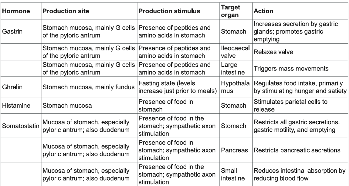
Describe the functions of the four stomach hormones
What are the secretions of the stomach
Chyme: ingested food + stomach secretions
Mucous: produced by surface and neck mucous cells
Viscous and alkaline
Protects from acidic chyme and enzyme pepsin
Irritation of stomach mucosa causes greater mucus
Intrinsic Factor: produced by parietal cells. Binds with vitamin B12 and helps it to be absorbed. B12 necessary for DNA synthesis
HCI: produced by parietal cells
Kills bacteria
Stops carbohydrate digestion by inactivating salivary amylase
Denatures proteins Helps convert pepsinogen to pepsin
Pepsinogen: packaged in zymogen granules released by exocytosis from chief cells. Pepsin catalyses breaking of covalent bonds in proteins
Describe the movements in the stomach and the small intestine
In the Stomach:
a. Peristalsis (mixing and propulsion)
Wave-like contractions of the stomach wall move towards the pylorus (the stomach's outlet).
These contractions push food towards the pyloric sphincter.
b. Churning and retropulsion
Strong contractions in the lower stomach push food towards the pyloric sphincter.
Because the sphincter is mostly closed, much of the food is forced backwards into the stomach.
This mixes food with gastric juice and breaks it into smaller particles, forming a semi-liquid mixture called chyme.
c. Gastric emptying
The pyloric sphincter opens intermittently, allowing small amounts of chyme to enter the duodenum (the first part of the small intestine).
Emptying is regulated by the composition and volume of chyme, with fats and excessive acidity in the duodenum generally slowing gastric emptying.
In the Small Intestine:
a. Segmentation
Circular smooth muscles contract at alternating points along the intestine.
This divides and repeatedly mixes chyme, bringing it into contact with digestive enzymes and the absorptive surface.
Its primary function is mixing, rather than rapid forward movement.
b. Peristalsis
Coordinated waves of contraction propel chyme towards the large intestine.
Circular muscle contracts behind the contents and relaxes ahead of them.
Its primary function is propulsion.
Describe chemical digestion in the stomach
Acidity inactivates salivary amylase and activates lingual lipase-begins breaking down triglycerides into free fatty acids, and mono- and diglycerides.
Breakdown of protein begins through actions of HCl and pepsin.
Only one stomach function necessary to life: the production of intrinsic factor (IF). Intestinal absorption of vitamin B12 is necessary for production of mature red blood cells and normal neurological functioning, cannot occur without IF.
A total gastrectomy (stomach removal)—for stomach cancer, requires vitamin 812 injections.
Contents of the stomach are completely emptied into the duodenum within 2 to 4 hours after a meal.
List the secretions of the small intestine and discuss their function
Fluid primarily composed of water, electrolytes and mucus.
Mucus: Protects against digestive enzymes and stomach acids
Duodenal glands: Produce mucus when stimulated by vagus nerve (parasympathetic), secretin, chemical or tactile irritation of duodenal mucosa
Digestive enzymes of the small intestine are bound to the membranes of the absorptive cells
Disaccharidases: Break down disaccharides to monosaccharides
Peptidases: Hydrolyze peptide bonds
Nucleases: Break down nucleic acids
Endocrine cells: produce regulatory hormones, such as secretin (triggered by acid) and cholecystokinin (triggered by fatty acids and peptides)
List the secretions of the large intestine
Mucus provides protection and parasympathetic NS stimulates goblet cell secretion
Pumps: bacteria produce acid and the following remove acid from the epithelial cells that line the large intestine - Exchange of bicarbonate ions for chloride ions - Exchange of sodium ions for hydrogen ions
Bacterial actions produce gases (flatus) from particular kinds of carbohydrates - found in legumes and in artificial sugars like sorbitol
Bacteria produce vitamin K which is then absorbed
Faeces consists of water, undigested food (cellulose), microorganisms, sloughed- off epithelial cells.
Describe defecation
1. Faeces enter the rectum: Mass movements of the large intestine push faeces into the rectum, stretching its walls.
2. Defecation reflex is initiated: Stretch receptors in the rectal wall detect distension and send nerve signals to the sacral spinal cord.
3. Rectal contractions occur: Parasympathetic nerve signals stimulate contractions of the rectum and sigmoid colon, increasing pressure and pushing faeces towards the anus.
4. Internal anal sphincter relaxes: The internal anal sphincter is composed of smooth muscle and relaxes involuntarily as part of the reflex.
5. External anal sphincter is controlled voluntarily: The external anal sphincter is skeletal muscle. It can be voluntarily relaxed to allow defecation or contracted to delay it.
6. Faeces are expelled: Relaxation of the external sphincter, often assisted by contraction of the abdominal muscles and diaphragm, allows faeces to pass out through the anus.
Describe the blood supply, drainage, and capillary system of the liver
Blood supply: Receives blood from two sources:
Hepatic portal vein: Brings nutrient-rich blood from the gastrointestinal tract and spleen.
Hepatic artery proper: Supplies oxygen-rich blood.
Venous drainage: Blood leaves the liver through the hepatic veins, which drain into the inferior vena cava.
Biliary system: Hepatocytes produce bile, which drains through bile canaliculi into bile ducts. Bile can be stored and concentrated in the gallbladder before being released into the duodenum.
Porta hepatis: The region on the visceral surface through which the hepatic portal vein, hepatic artery proper, hepatic ducts, lymphatic vessels and nerves pass.
Functions of Hepatocytes:
Bile production
Storage
Interconversion of nutrients
Detoxification
Phagocytosis
Synthesis of blood components

Describe the functions of the liver
Bile Production: 600-1000 mL/day. It contains bile salts, bilirubin, cholesterol, fats, fat-soluble hormones, lecithin
Neutralizes and dilutes stomach acid
Bile salts emulsify fats. Most are reabsorbed in the ileum.
Secretin (from the duodenum) stimulates bile secretions, increasing water and bicarbonate ion content of the bile
Storage: Glycogen, fat, vitamins, copper and iron. Hepatic portal blood comes to liver from small intestine.
Nutrient Conversion:
Amino acids to energy producing compounds Hydroxylation of vitamin D.
Vitamin D then travels to kidney where it is hydroxylated again into its active form
Detoxification: Hepatocytes remove ammonia and convert to urea
Phagocytosis: Kupffer cells phagocytise worn-out and dying red and white blood cells, some bacteria
Synthesis of blood components: Albumins, fibrinogen, globulins, heparin, clotting factors
Describe regulation of bile secretion and release by the gallbladder
Gallbladder Structure:
Sac lined with mucosa folded into rugae, inner muscularis, outer serosa
Bile produced constantly in the liver is stored and concentrated
Stimulated by cholecystokinin (from the intestine) and vagal stimulation
Bile exits through cystic duct then into common bile duct
Gallstones: precipitated cholesterol
Can block cystic duct
Can occur because of drastic dieting
Bile:
Bile, including bile salts is produced by liver and stored in gall bladder.
Bile salts are reabsorbed in ileum and stimulate more production of bile.
Secretin from the duodenum also stimulates bile production.
Cholecystokinin stimulates release of bile from gall bladder.

Describe the structure and function of the pancreas
Pancreas both endocrine and exocrine
Head, body and tail Endocrine: pancreatic islets.
Produce insulin, glucagon, and somatostatin.
Exocrine: groups acini (grape-like cluster) form lobules separated by septa. Intercalated ducts lead to intralobular ducts lead to interlobular ducts lead to the pancreatic duct.
Pancreatic duct joins common bile duct and enters duodenum at the hepatopancreatic ampulla controlled by the hepatopancreatic ampullar sphincter

List the pancreatic enzymes and describe their interaction with the duodenal
Enzymatic portion:
Trypsinogen
Chymotrypsinogen
Procarboxypeptidase
Pancreatic amylase
Pancreatic lipases
Deoxyribonucleases and ribonucleases
Enterokinase is attached to the brush border of the duodenal mucosa and activates trypsinogen to trypsin.
Trypsin activates chymotrypsinogen to chymotrypsin.
Trypsin activates procarboxypeptidase to carboxypeptidase.
Trypsin, chymotrypsin, and carboxypeptidase digest proteins: proteolytic.
Pancreatic amylase continues digestion of starch.
Pancreatic lipase digests lipids.
Deoxyribonucleases and ribonucleases digest DNA and ribonucleic acid, respectively.
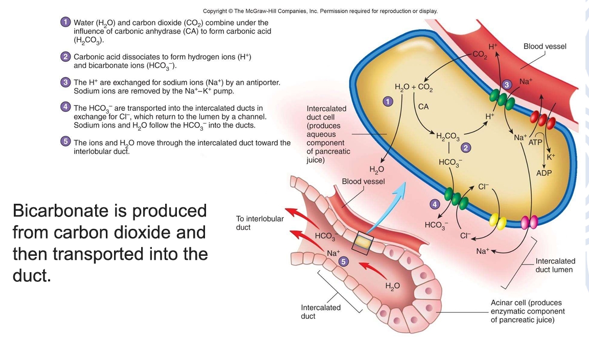
Describe production of bicarbonate in the pancreas
Describe monosaccharide, lipid and amino acid transport
Carbohydrate (monosaccharide) Digestion:
Glucose is transported into cells requiring energy; insulin increases the number of transport proteins
Starch glycogen is broken down into disaccharides (maltose, sucrose, lactose) by enzyme salivary amylase
Disaccharides are broken down into monosaccharides (glucose, fructose, galactose) by enzyme ….
Glucose and galactose are absorbed into blood stream by co-transport with sodium ions.
Fructose is absorbed via facilitated diffusion
Final destination of all sugars is the liver via to the hepatic portal vein
Protein digestion:
Begins in the stomach and ends in the small intestine
Protein is broken down by pepsin into large polypeptides and then small polypeptides and then amino acids which are co-transported with sodium ions into the blood stream with final destination being the liver via the hepatic portal vein.
Lipids:
Include triglycerides, phospholipids, steroids, fat-soluble vitamins
Bile salts surround fatty acid and glycerol to form micelles
Chylomicrons are 90% triglyceride, 5% cholesterol, 4% phospholipid, and 1% protein.
Chylomicrons enter blood stream and travel to adipose tissue. In blood, triglycerides converted back into fatty acids and glycerol where they are transported into the adipose cells, then converted back into triglycerides.
Transport across Intestinal Membrane:
Micelles are converted to chylomicrons and exocytosis into lymph
Summarise the role of VLDL, LDL and HDL in the transport of lipid
All lipids carried in the blood are done so in combination with protein to make it soluble in the blood
Cholesterol: 15% ingested; 85% manufactured in liver and intestinal mucosa
Lipids are lower density than water; proteins are higher density than water
Chylomicrons: 99% lipid and 1% protein (extremely low density); enter lymph
VLDL: 92% lipid, 8% protein
Form in which lipids leave the liver
Triglycerides removed from VLDL and stored in adipose cells.
VLDL has been converted to LDL.
LDL: 75% lipid, 25% protein "bad cholesterol"
Transports cholesterol to cells where taken up by LDL receptors
LDL receptors downregulated once cell's lipid/cholesterol needs are met.
HDL: 55% lipid, 45% protein "good cholesterol"
Transports excess cholesterol from cells to liver
Summarise Glucose Homeostasis
Blood glucose decreases below 80-90mg/dl (fasting)
• liver cells increase catabolism glycogen, amino acids, and glycerol glucose (glycogenolysis and gluconeogenesis)
• Released into blood
Blood glucose level increases
• liver cells increase removal rate of glucose molecules from blood
• Glucose glycogen (short term storage called glycogenesis)
Glucose converted to fat, excreted in urine.
Hormonal Control of Blood Glucose levels:
Insulin - secreted by beta cells of the pancreas to reduce blood glucose levels
Glucagon - secreted by alpha cells of the pancreas to increase blood glucose levels.
Other hormones involved:
Incretins: Gastrointestinal hormones (glucagon-like peptide 1 [GLP-II, gastric inhibitory peptide [GIP]) that affect pancreatic cells; increase insulin release and decrease glucagon release to reduce the blood glucose level
Stress hormones: Adrenaline (acute stress) and Cortisol (chronic stress) to increase blood glucose levels
Growth hormone: Increases the blood glucose level by shifting from carbohydrate to fat catabolism to preserve glucose for use by neurons
Thyroid hormones: Have complex effects on metabolism
![<p><span><strong>Blood glucose decreases below 80-90mg/dl (fasting)</strong></span></p><p>• liver cells increase catabolism glycogen, amino acids, and glycerol glucose (glycogenolysis and gluconeogenesis)</p><p>• Released into blood</p><p><span><strong>Blood glucose level increases</strong></span></p><p>• liver cells increase removal rate of glucose molecules from blood</p><p>• Glucose glycogen (short term storage called glycogenesis)</p><ul><li><p><span>Glucose converted to fat, excreted in urine.</span></p></li></ul><p><span style="background-color: rgb(204, 255, 255);">Hormonal Control of Blood Glucose levels:</span></p><ul><li><p><span>Insulin - secreted by beta cells of the pancreas to reduce blood glucose levels</span></p></li><li><p><span>Glucagon - secreted by alpha cells of the pancreas to increase blood glucose levels.</span></p></li></ul><p><span><strong>Other hormones involved:</strong></span></p><ul><li><p><span><strong>Incretins</strong>: Gastrointestinal hormones (glucagon-like peptide 1 [GLP-II, gastric inhibitory peptide [GIP]) that affect pancreatic cells; increase insulin release and decrease glucagon release to reduce the blood glucose level</span></p></li><li><p><span><strong>Stress hormones</strong>: Adrenaline (acute stress) and Cortisol (chronic stress) to increase blood glucose levels</span></p></li><li><p><span><strong>Growth hormone:</strong> Increases the blood glucose level by shifting from carbohydrate to fat catabolism to preserve glucose for use by neurons</span></p></li><li><p><span><strong>Thyroid hormones:</strong> Have complex effects on metabolism</span></p></li></ul><p></p>](https://assets.knowt.com/user-attachments/bc275334-9041-48ee-81d3-ed05dbeb022d.png)
Discuss the mechanisms for regulating food intake
Hypothalamus plays a part in food intake
Appetite centres in the hypothalamus exert primary control over food intake
• Feeding centre — Orexigenic effects: Factors that trigger appetite
• Satiety centre —Anorexigenic effects: Factors that suppress appetite (anorexia is loss of appetite) These are regulated by a range of hormones that also influence energy expenditure, including:
• Ghrelin —secreted by stomach when it is empty (induces hunger)
• Leptin —secreted by adipose tissue (induces satiety)