NPB 101: Lecture 20 - Cardiovascular Physiology 3: Cardiac Cycle & Regulation
 Call Kai
 Learn
 Practice Test
 Spaced Repetition
 Match
 Flashcards
 Knowt Play
1/21

Earn XP
Description and Tags
Learning Objectives: Trace the cardiac cycle, relating chamber pressues to valve opening and closure, blood flow, and ventricular volume. Interpret a ventricular pressure-volume loop, identifying filling, isovolumetric contraction, ejection, and isovolumetric relaxation. Calculate stroke volume, cardiac output, and ejection fraction from ventricular volumes and heart rate. Predict how changes in preload, afterload, and contractility affect stroke volume and ventricular empyting. Explain how sympathetic and parasympathetic signaling alter pacemaker activity and heart rate. Explain how sympathetic stimulation affects cardiac contraction and relaxation, and integrate changes in heart rate and stroke volume to predict cardiac output.
22 Terms
Cardiac cycle
All the events involved with blood flow through the heart during one heart beat.
Two primary phases:
Systole
Diastole
Systole
The phase of the cardiac cycle when the ventricles contract, pumping out blood into the arteries— sending blood to the lungs through the pulmonary artery and to the rest of the body through the aorta.

Diastole
The relaxation phase of the cardiac cycle when the ventricles relax, allowing for its chambers to refill with blood passed down from the atria.

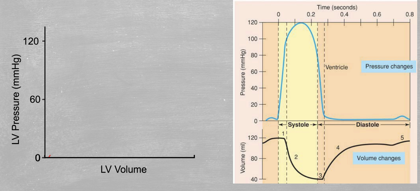
Isovolumetric contraction
First step in Cardiac Cycle Sequence.
Ventricular contraction increases pressure shutting the AV valves
Ejection
Second step in Cardiac Cycle Sequence.
Left ventricle pressure > Aortic pressure
Aortic valve opens
Ventricular volume decreases
Isovolumetric relaxation
Third step in Cardiac Cycle Sequence.
Left ventricle pressure drops below aortic pressure
Aortic valve shuts
Passive filling
Fourth step in Cardiac Cycle Sequence.
Pressure in the ventricles falls below that of the atria
AV valve opens and ventricle fills
Active filling
Fifth step in Cardiac Cycle Sequence.
Atrial contraction delivers the final blood volume to the ventricles
End-diastolic volume (EDV)
Volume of blood in the ventricles at the end of diastole (relaxation/filling)
End-systolic volume (ESV)
The amount of blood remaining in the chamber at the end of systole (contraction/emptying)
Stroke Volume (SV)
The amount of blood pumped out with each contraction.
Equation: SV = EDV - ESV
Regulated by…
Amount of blood coming back to the heart
Preload ~ EDV
How hard it is to push blood out of the heart
Afterload ~ aortic blood pressure
Contractility of the cardiac muscle
Inotropy

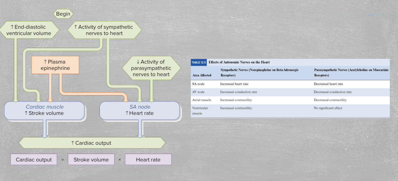
Cardiac Output (CO)
The amount of blood pumped by each ventricle per unit of time
Equation: CO = HR x SV
Factors that affect heart rate or stroke volume will thus affect cardiac output
Heart rate
Cardiac cycles per unit time (beats per minute; 1/ cardiac cycle time).
Regulated by autonomic nervous system
Sympathetic (Fight-or-Flight)
Parasympathetic (Rest-and-digest)
Ejection Fraction (EF)
Fraction of blood pumped from ventricle.
Equation: SV/EDV
Measure of contractility
Pressure Volume Loops
A real-time graphical plot of left (or right) ventricular pressure against ventricular volume during a single complete cardiac cycle (heartbeat).
Ventricular pressure (mmHg)
Ventricular volume (mL)
Preload (Intrinsic)
The degree to which the cardiac muscle cells are stretched before they contract.
Determined by the volume of blood in ventricle when full before contraction (EDV)
Increased preload = increased stroke volume
Related to length tension curve
Operated ascending limb
Afterload (Intrinsic)
The pressure ventricles must produce to open aortic/pulmonary valves.
Anything that increases systemic or pulmonary pressure can increase afterload
Increased afterload decreases Ejection Fraction
Extrinsic Regulation of Heart Contractility
The control of myocardial force generation by factors originating outside the heart.
Two divisions of autonomic nervous system (ANS):
Sympathetic
Parasympathetic

Sympathetic Nervous System
Division of the ANS that drives the heart’s “fight-or-flight” response, increasing cardiac output to meet physical or emotional demands.
Pathway:
Sympathetic cardiac nerves (thoracic spinal nerves) release norepinephrine
Adrenal medulla releases circulating epinephrine via bloodstream
Receptors:
They bind to beta (β₁) adrenergic receptors on the atria and ventricles
Increases Ca2+ release and contraction
Faster Ca2+ re-uptake and relaxation
Effects:
Inotropic effects on contractility
Causes heart rate to increase
Increase in pacemaker AP

Parasympathetic Nervous System
Division of the ANS that acts as the heart’s natural “rest-and-digest” brake petal, slowing down cardiac activity.
Pathway:
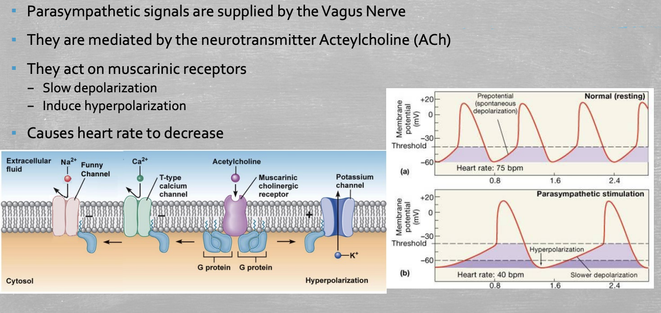
The vagus nerve delivers acetylcholine (ACh) to the heart
Receptors:
ACh binds to muscarinic (M2) receptors
Effects:
Slows depolarization in the heart
Induces hyperpolarization
ACh increases K+ outflow of SA and AV (pacemaker) nodes
Decreases slow Na+ and Ca2+ influx
Slows heart rate
Decrease in pacemaker AP
Autonomic Nervous System
The ANS affects the activity of the pacemaker neurons by affecting the function of individual channels.

Regulation of Cardiac Output