Lower Limb Muscle Attachments and Primary Movement
 Call Kai
 Learn
 Practice Test
 Spaced Repetition
 Match
 Flashcards
 Knowt Play
1/18

Earn XP
Description and Tags
Vocabulary flashcards detailing the anatomical origins, insertions, and primary movements of the muscles of the lower limb.
19 Terms

Psoas Major
Origin:
Transverse process of L1-L5
Vertebral bodies of T12-L4
Lateral surfaces of intervertebral discs
Insertion:
Lesser trochanter
Function:
Hip flexion
External rotation of the thigh
Trunk flexion, lateral flexion of the trunk & postural stability.

Iliacus
Origin:
Iliac fossa of the iliac wing
Insertion:
Lesser trochanter
Function:
Hip flexion
External rotation of the thigh
Trunk flexion/anterior pelvic tilt.

Gluteus Medius
Origin:
Posterior (gluteal) surface of the ilium
Insertion:
Greater trochanter
Function:
Hip abduction (anterior fibres perform hip flexion and internal rotation)
Hip abduction and assist pelvic tilt (Medial fibres)
Hip extension and external rotation (Posterior fibres)

Gluteus Maximus
Origin:
Gluteal surface of the ilium
Posterior superior iliac crest
Sacrum
Coccyx
Insertion:
Iliotibial tract
Gluteal tuberosity
Function: Hip extension.

Adductor Longus
Origin:
Anterior surface of the pubis
Insertion:
Middle third of the linea aspera of the femur
Function:
Hip adduction

Sartorius
Origin:
Anterior superior iliac spine
Insertion:
Medial aspect of the proximal tibia
Function:
Hip flexion
Hip abduction
External rotation.

Vastus Medialis (Quadriceps)
Origin:
Lower, medial, half of the intertrochanteric line
Medial lip of the linea aspera of the femur
insertion:
Via the patella tendon into the tibial tuberosity
Function:
Knee extension

Vastus Intermedius (Quadriceps)
Origin:
The anterior and lateral surfaces of the body of the femur
Insertion:
Via the patella tendon into the tibial tuberosity
Function:
Knee extension

Vastus Lateralis (Quadriceps)
A quadriceps muscle originating from the intertrochanteric line, greater trochanter, gluteal tuberosity, and lateral lip of the linea aspera of the femur; inserting via the patella tendon into the tibial tuberosity; and responsible for knee extension.

Rectus Femoris (Quadriceps)
Origin:
Anterior inferior iliac spine
Insertion:
Via the patella tendon into the tibial tuberosity
Function:
Knee extension
Hip extension (assisting iliopsoas)

Biceps Femoris (Hamstrings)
Origin:
Lower medial ischial tuberosity (long head)
Lateral linea aspera (short head)
Insertion:
Head of the fibula
Lateral tibial condyle
Function:
Knee flexion

Semitendinosus (Hamstrings)
Origin:
Ischial tuberosity
Insertion:
Medial aspect of the proximal tibia
Function:
Knee flexion.

Semimembranosus (Hamstrings)
Origin:
Ischial tuberosity
Insertion:
Medial tibial condyle
Function:
Knee flexion.

Peroneus Longus
Origin:
Head and lateral surface of the fibula
Insertion:
Plantar surface of the medial cuneiform bone
Base of the first metatarsal bone
Function:
Eversion of the foot and plantar flexion.
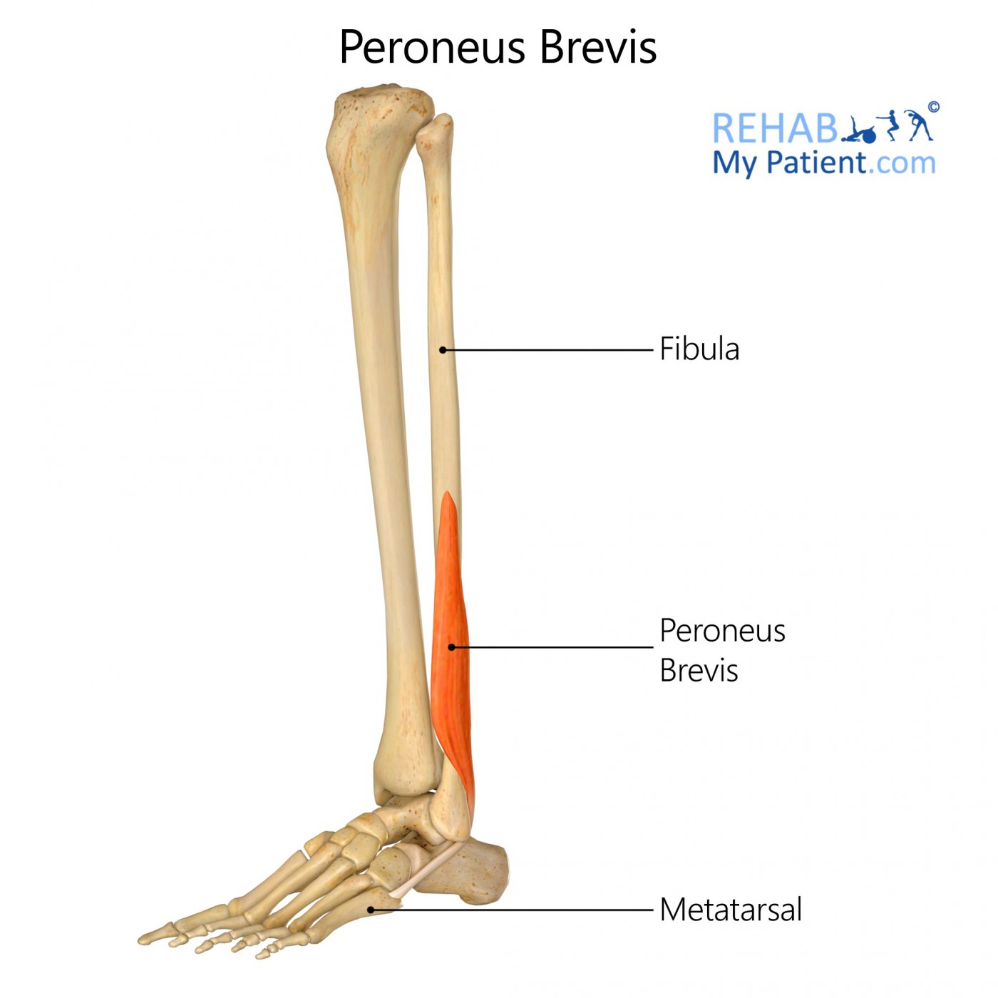
Peroneus Brevis
Origin:
Lateral surface of the fibula
Insertion:
Tuberosity of the fifth metatarsal bone
Function:
Eversion of the foot

Gastrocnemius (Superficial calf muscle)
Origin:
Lateral and medial condyles of the femur
Insertion:
Posterior surface of the calcaneus via the Achilles tendon
Function:
Plantar flexion

Soleus (Calf)
Origin:
Head and upper third of the posterior surface of the fibula
Medial border of the tibia
Soleal line
Tendinous arch spanning between the tibia and fibula
Insertion:
Posterior surface of the calcaneus
Function:
Plantar flexion.

Tibialis Anterior
Origin:
Upper two-thirds of the lateral surface of the tibia
Adjacent interosseous membrane
Insertion:
Medial cuneiform
Base of the first metatarsal bone of the foot
Function:
Dorsi flexion
Foot inversion

Tibialis Posterior
Origin:
Upper two-thirds of the posterior surface of the tibia
Superior two-thirds of the medial posterior surface of the fibula
Insertion:
Tuberosity of the navicular bone
Medial cuneiform
Intermediate cuneiform
Lateral cuneiform
Cuboid
Bases of the 2nd, 3rd, and 4th metatarsals
Function:
Foot inversion
Plantar flexion.