Bio 201 Chapter 6: Bone Tissue
 Call Kai
 Learn
 Practice Test
 Spaced Repetition
 Match
 Flashcards
 Knowt Play
1/57

There's no tags or description
Looks like no tags are added yet.
58 Terms

diaphysis
main section of bone

metaphysis
middle of bone
may contain epiphyseal plate or ossified epiphyseal line
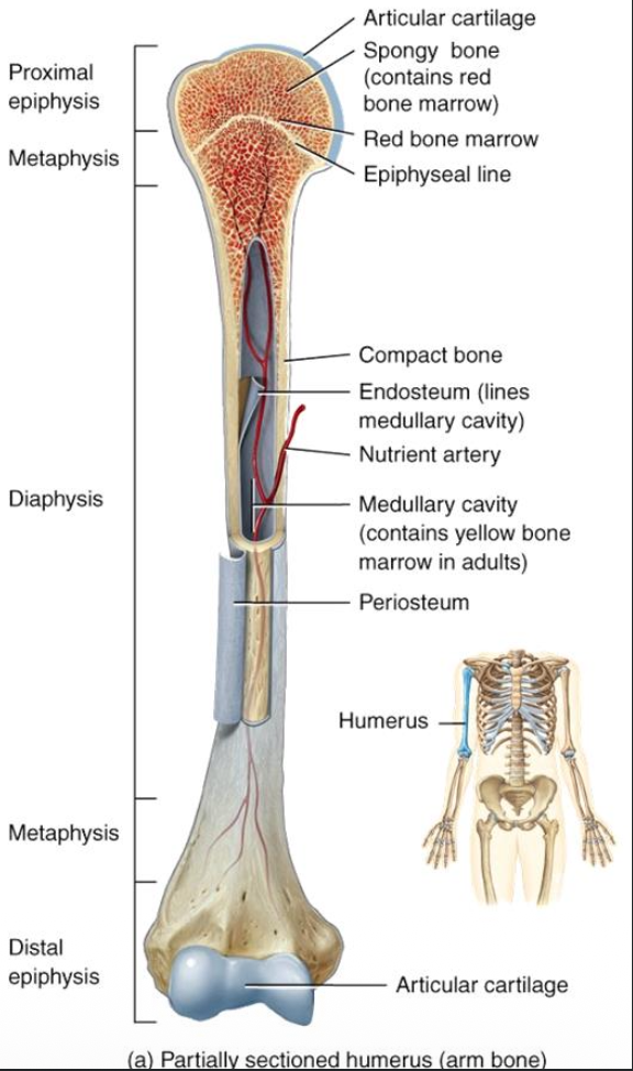
epiphysis
ends of the bone (proximal and distal)
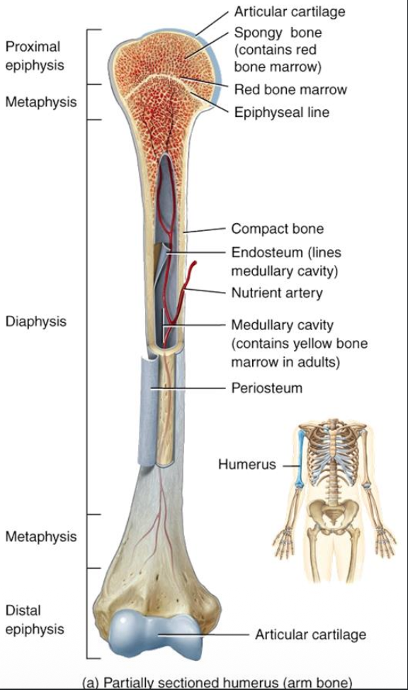
auricular cartilage
contains hyaline cartilage
absorb shock and reduce friction

periosteum
contains dense irregular CT and osteoblasts
help with nourishment and repairs
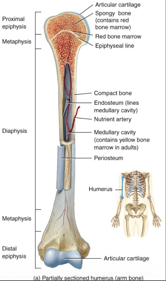
medullary cavity
contains yellow bone marrow and vessels

endosteum
contains osteoblasts and osteoclasts
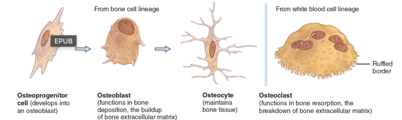
osteoprogenitor cells
bone stem cells
undergo mitosis to become osteoblasts

osteoblasts
help build bone
become osteocytes after secreting enough extracellular matrix

osteocytes
mature bone cells
no cell division
maintain bone structure

osteoclasts
break down bone
help regulate calcium levels

compact bone
protection and support
located beneath periosteum of all bones and diaphysis of long bones

spongy bone
tissue support
located in the epiphysis of long bones
located in interior of bone of flat and irregular bones

osteon
repeating structural unit that makes up compact bone
also known as haversian system
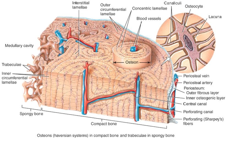
haversian canal
also known as central canal
contains vessels
vertical
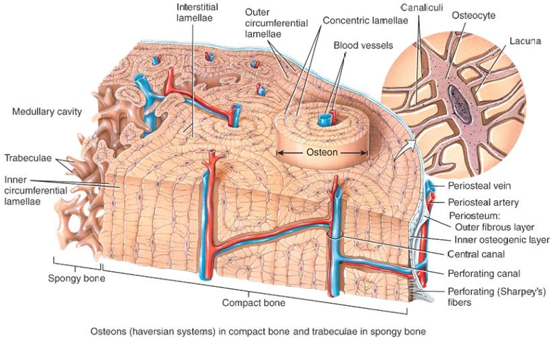
concentric lamellae
circular plates of mineralized extracellular matrix
surround the central canal

lacuna
space between lamellae
contain osteocytes
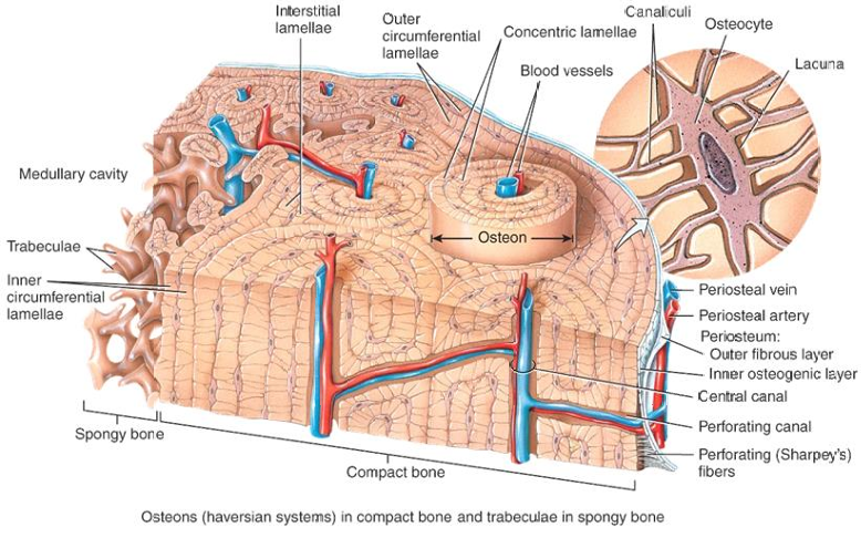
canaliculi
channels that connect lacunae

Volkmann’s canal
all vessels to connect between osteons
horizontal

Sharpey’s fibers
connect to periosteum

trabeculae
layers of lamellae arranged irregularly
present in spongy bone
contain red bone marrow
hips, ribs, sternum, vertebra, proximal ends of humerus and femur

periosteal arteries and veins
with nerves
enter diaphysis via interosteonic canals

nutrient arteries and veins
supply the compact and spongy bone
in medullary cavity, supplies bone to the epiphyseal line

metaphyseal arteries and veins
supply the marrow and epiphysis
stage 1 of bone formation
in utero
stage 2 of bone formation
growth until adulthood
stage 3 of bone formation
remodeling throughout lifetime
stage 4 of bone formation
repair when damagec
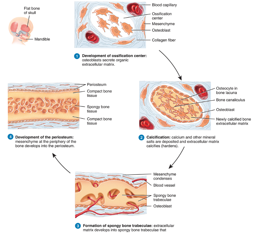
intramembraneous ossification
produces flat bone from mesenchyme
1: develop ossification center: mesenchyme → osteogenic → osteoblast → osteocyte
2: calcification: ECM calcifies due to Ca+ and minerals
3: trabeculae form: due to ECM calcifying and coming together
4: periosteum forms
5: compact bone replaces some trabeculae

endochondral ossification
create long bone form hyaline cartilage
1: cartilage model grows: chondroblast → chondrocytes, mitosis occurs and growth starts from the inside out
2: primary ossification center develops: bone replaces cartilage of the diaphysis, follows steps of intramembranous bone formation
3: medullary cavity develops: breakdown of newly formed spongy bone leaves an empty cavity in shaft
4: secondary ossification center develops: bone replaces cartilage, but of the epiphysis
5: formation of the articular cartilage and epiphyseal plate

interstitial growth
lengthwise
occurs on epiphyseal side of epiphyseal plate (4 zones)
replacement of cartilage on diaphyseal side by endochondral ossification
beginning of adulthood; epiphyseal plate → epiphyseal line

resting cartilage
no longer functioning in bone growth/anchor to epiphysis
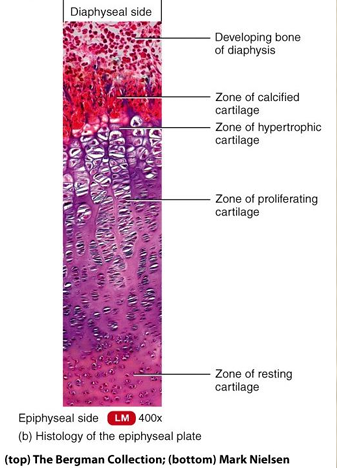
proliferating cartilage
chondrocytes undergoing mitosis, make extracellular matrix

hypertrophic chondrocytes
mature chondrocytes

calcified cartilage
dead calcified chondrocytes

appositional growth
growth in width
1: create groove (periosteum helps)
2: grooves fold together
3: osteoblast inside the canal creates lamellae
4: osteon is created

bone remodeling
replacing old bone with new bone

resorption
osteoclasts release enzymes and acids that break down bone
minerals can be recycled via bloodstream, especially phosphorous and calcium

deposition
osteoclasts leave and osteoblasts help rebuild broken-down bone
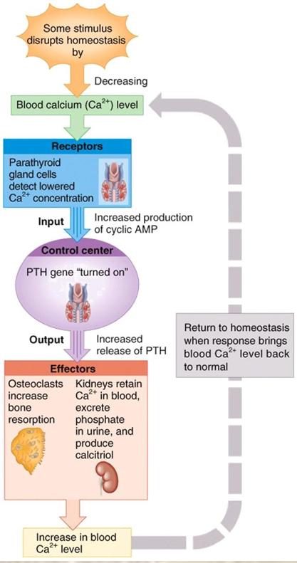
decreasing blood calcium level
2: parathyroid gland releases parathyroid hormone
3: osteoclasts break down bone and kidneys retain calcium
4: blood calcium increased

increasing blood calcium level
2: thyroid gland releases calcitonin
3: osteoblasts rebuild bone and deposit calcium into bone
4: blood calcium decreased
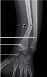
greenstick
partial; only occurs in children
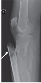
open
also known as compound; broken ends of bone protrude through skin

comminuted
bone is splintered or crushed

impacted
one end of fractured bone is driven into the other

spiral
occurs when one end of a fractured bone is rotated
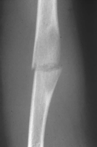
transverse
closed or complete fracture horizontal across long axis of bone

oblique
closed or complete fracture diagonally along bone

displaced
bone fragments move out of normal alignment

reactive phase
formation of fracture hematoma (6-8 hours)
clot forms around fracture due to disruption of vessels
inflammation occurs
osteocytes die and osteoclasts/macrophages remove dead material

reparative phase stage 1
form fibrocartilaginous callus (3 weeks)
fibroblasts produce collagen
chondroblasts produce cartilage
reparative phase stage 2
form bony callus (3-4 months)
formation of bone
fibrocartilage replaced
flat bones
protect internal organs such as heart, lungs, and brain
ex. cranial bones, ribs, scapulae, sternum
long bones
support the weight of the body, facilitate movement
ex. humerus, ulna, radius, femur, tibia and fibula, metacarpals and metatarsals, phalanges
short bones
cube-shaped components of wrists and ankles
ex. carpals, tarsals
irregular bones
vary in shape and structure
ex. vertebrae, pelvic bones
collagen
organic component of bone
provides bone with tensile strength and gives it the ability to withstand stretching
calcium hydroxyapatite
inorganic component of bone
provides bone with compressional strength and gives it the ability to withstand compressive forces and provides hardness to the bone