Anatomy and Physiology Lab Comprehensive Review
 Call Kai
 Learn
Learn Practice Test
 Spaced Repetition
 Match
Match Flashcards
 Knowt Play
1/33

Earn XP
Description and Tags
Comprehensive vocabulary flashcards covering anatomical planes, directional terms, tissues, integumentary structures, axial and appendicular skeletal anatomy, joints, and muscular organization.
Name | Mastery | Learn | Test | Matching | Spaced | Call with Kai | Chat |
|---|
No analytics yet
Send a link to your students to track their progress
34 Terms
Parietal Serous Membrane
The outer serous membrane layer that lines the inner walls of body cavities, including the parietal pleura, parietal pericardium, and parietal peritoneum.
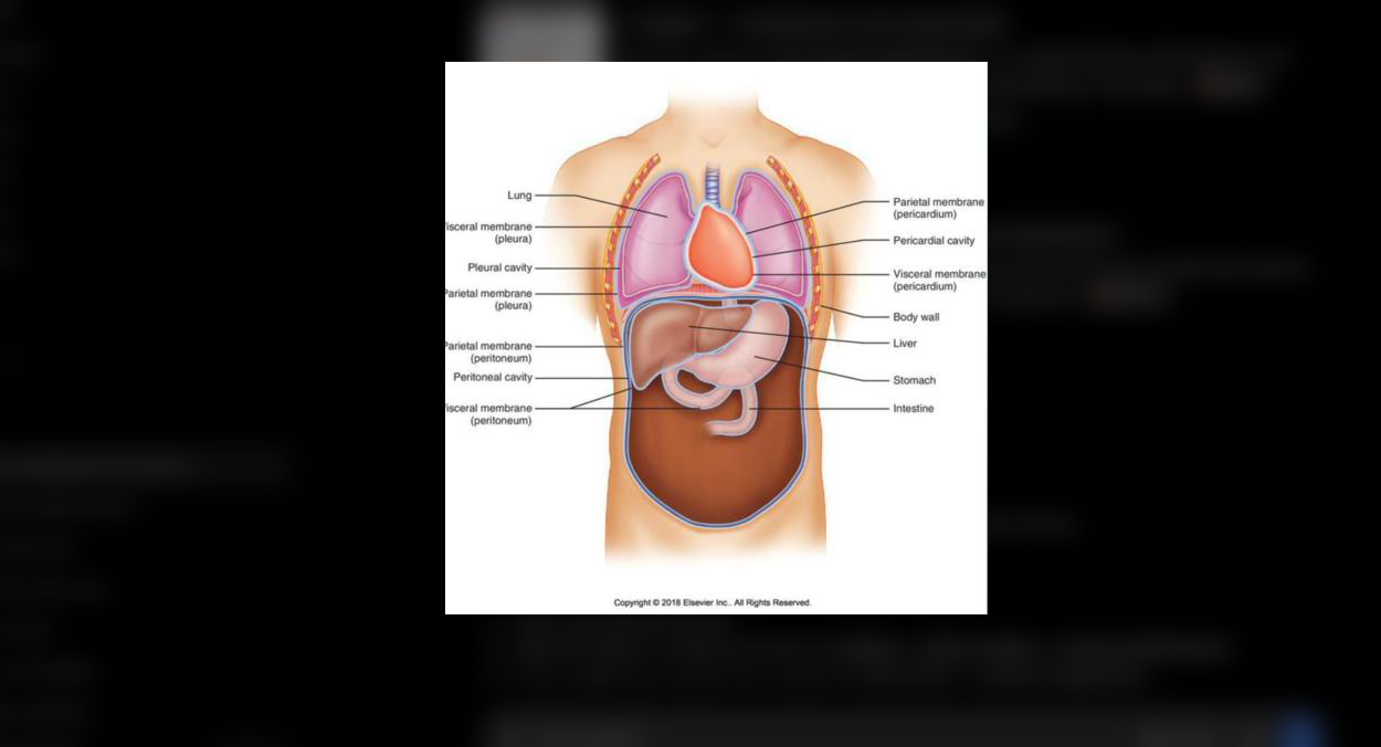
Visceral Serous Membrane
The inner serous membrane layer that directly covers the external surface of organs within body cavities, including the visceral pleura, visceral pericardium, and visceral peritoneum.

Coronal (frontal) Plane
An anatomical plane oriented vertically that divides the body or an organ into anterior (front) and posterior (back) portions.

Midsagittal Plane
A vertical plane passing directly through the midline that divides the body or an organ into equal right and left halves.

Stratum Basale
The deepest epidermal layer consisting of a single row of actively dividing stem cells that continuously produce new keratinocytes.

Stratum Corneum
The most superficial layer of the epidermis, composed of multiple layers of flattened, dead, anucleate, fully keratinized cells.

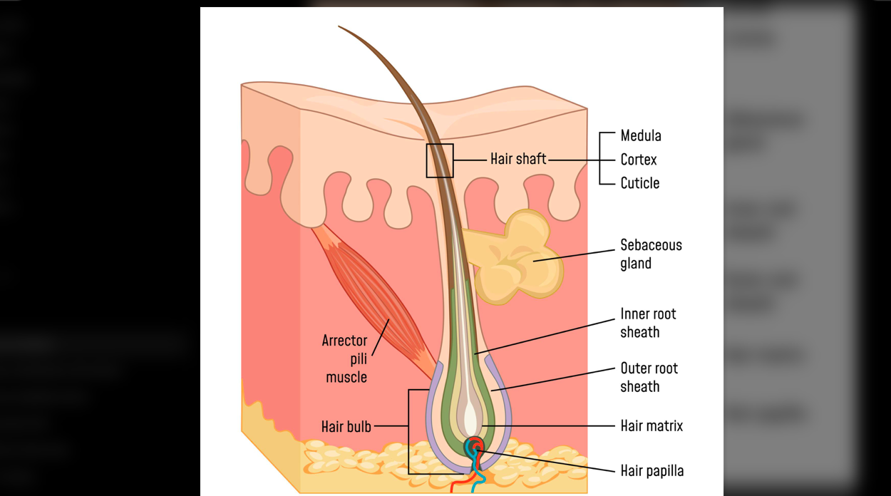
Arrector Pili Muscle
A bundle of smooth muscle attached to a hair follicle that contracts to pull the hair shaft upright, producing goosebumps.
Lamellar Corpuscle
A layered cutaneous mechanoreceptor located deep in the dermis or hypodermis that senses deep pressure and high-frequency vibration.

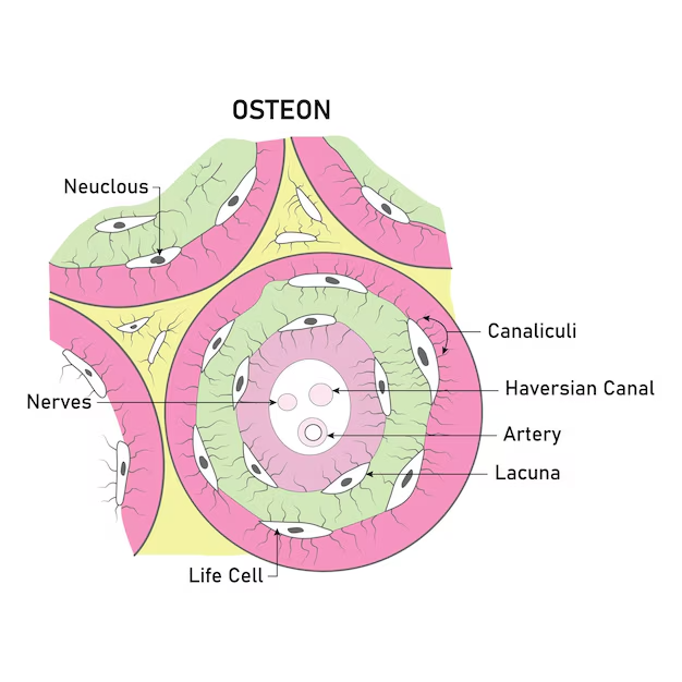
Osteon
The fundamental microscopic structural and functional unit of compact bone, consisting of concentric lamellae surrounding a central canal.

Canaliculi
Microscopic hairlike canals connecting adjacent lacunae and the central canal, facilitating nutrient exchange and communication between osteocytes.
Epiphyseal Plate
A hyaline cartilage plate situated in the metaphysis at each end of a growing long bone that provides the site for longitudinal bone growth.

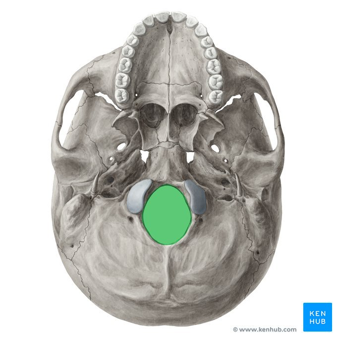
Foramen Magnum
A large opening in the base of the occipital bone through which the medulla oblongata and spinal cord exit the cranium.
Sella Turcica
A prominent saddle-shaped depression in the superior surface of the sphenoid bone that cradles the pituitary gland.
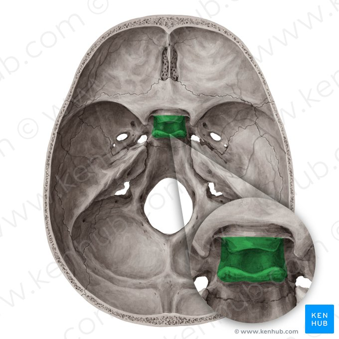
Crista Galli
A sharp upward triangular projection of the ethmoid bone that serves as an anterior attachment point for the falx cerebri.

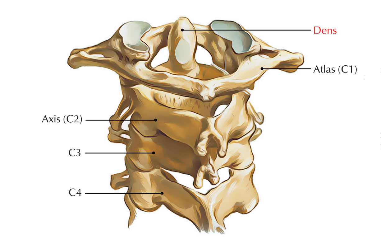
Dens
A prominent tooth-like vertical process projecting superiorly from the body of the axis (C2) vertebra, articulating with the atlas (C1) to allow head rotation.
Glenoid Cavity
A shallow articular depression on the lateral angle of the scapula that receives the head of the humerus to form the glenohumeral (shoulder) joint.

Deltoid Tuberosity
A roughened, triangular elevation located on the lateral aspect of the humeral diaphysis that serves as the insertion point for the deltoid muscle.

Olecranon Process
A large, thick, curved bony projection at the proximal end of the ulna that forms the point of the elbow and lodges in the olecranon fossa of the humerus.

Acetabulum
A deep, concave, cup-like socket on the lateral surface of the coxa formed by the union of the ilium, ischium, and pubis, articulating with the head of the femur.

Obturator Foramen
A large opening in the coxa bounded by the pubis and ischium, transmitting obturator vessels and nerves.

Medial Malleolus
The prominent downward bony projection located at the distal medial end of the tibia that forms the medial bulge of the ankle.

Lateral Malleolus
The prominent downward bony process at the distal end of the fibula that forms the outer lateral bulge of the ankle.

Scaphoid, Lunate, Triquetrum, Pisiform, Trapezium, Trapezoid, Capitate, Hamate.
Sally Left The Party To Take Cathy Home
Synarthroses
Joints categorized by functional classification as completely immovable, providing maximum mechanical stability.
Amphiarthroses
Joints categorized by functional classification as slightly movable, balancing mobility with structural support.
Diarthroses
Joints categorized by functional classification as freely movable, encompassing all synovial joints.
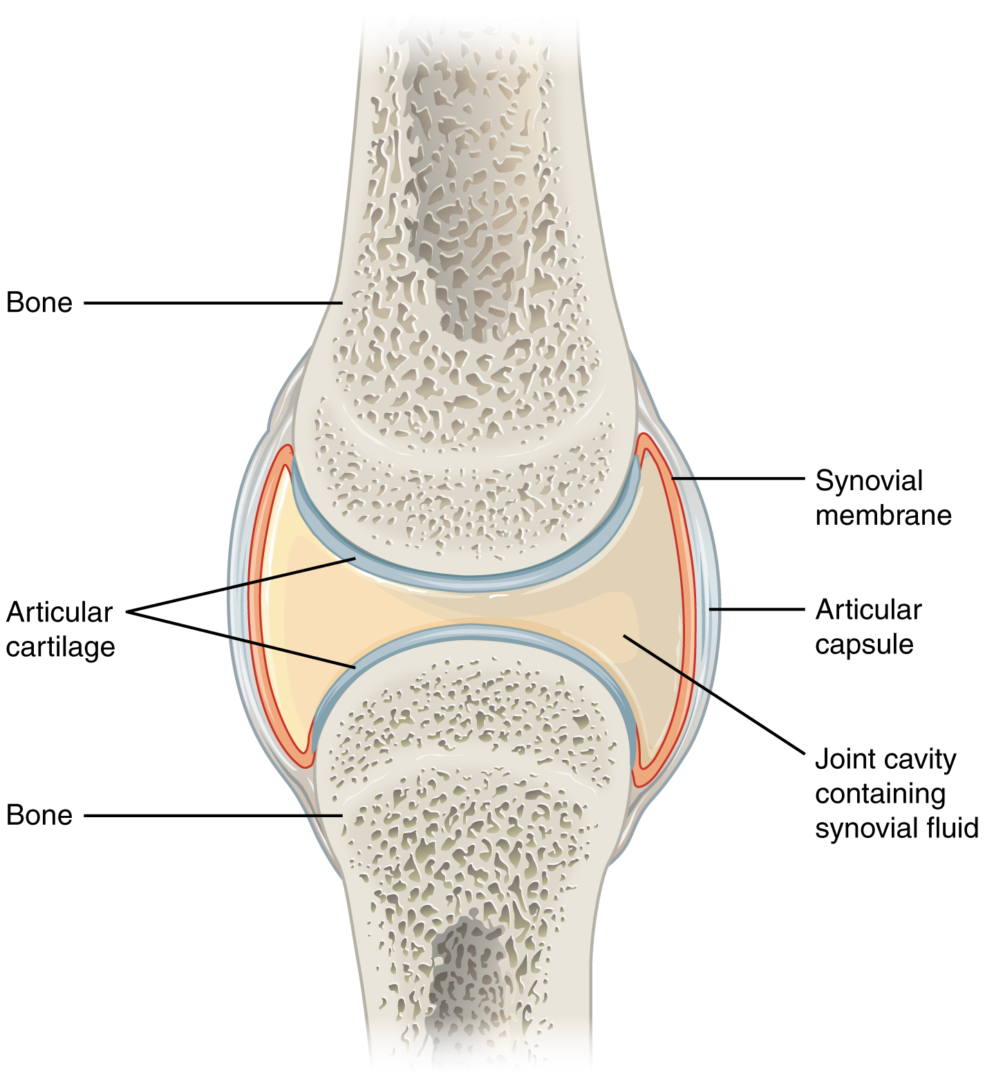
Synovial Membrane
The inner vascular layer of the articular capsule of a synovial joint that secretes synovial fluid to lubricate and nourish articular cartilage.
Anterior Cruciate Ligament
An intracapsular ligament of the knee that connects the anterior intercondylar area of the tibia to the lateral femoral condyle, preventing anterior translation of the tibia.

Epimysium
An external sheath of dense irregular connective tissue surrounding an entire skeletal muscle.

Perimysium
A layer of fibrous connective tissue that envelops and bundles individual skeletal muscle fibers into discrete fascicles.

Endomysium
A delicate sheath of loose connective tissue that surrounds each individual skeletal muscle fiber.

Sarcolemma
The specialized plasma membrane of a muscle fiber that envelops the sarcoplasm and propagates electrical action potentials into T tubules.

Motor End Plate
The specialized, highly folded region of the muscle fiber sarcolemma situated beneath the synaptic cleft that contains acetylcholine receptors.

Sarcomere
The basic contractile repeating functional unit of a myofibril, defined anatomically as the segment spanning between two adjacent Z discs.
