Week 10 - Nucleic Acid Chem + Techniques
 Call Kai
 Learn
 Practice Test
 Spaced Repetition
 Match
 Flashcards
 Knowt Play
1/23

There's no tags or description
Looks like no tags are added yet.
24 Terms
Ribonucleotides
structural units of RNA = ribonucleoside 5′-monophosphates.
Ribonucleotide nomenclature, symbols, and corresponding nucleosides:
Adenylate (adenosine 5′-monophosphate) | Symbols: A, AMP | Nucleoside: Adenosine.
Guanylate (guanosine 5′-monophosphate) | Symbols: G, GMP | Nucleoside: Guanosine.
Uridylate (uridine 5′-monophosphate) | Symbols: U, UMP | Nucleoside: Uridine.
Cytidylate (cytidine 5′-monophosphate) | Symbols: C, CMP | Nucleoside: Cytidine.
RNAs Types + Functions
Major cellular RNA classes:
Ribosomal RNAs (rRNAs): Components of ribosomes required for protein synthesis.
Messenger RNAs (mRNAs): Intermediaries carrying genetic information from one or a few genes to a ribosome for protein synthesis.
Transfer RNAs (tRNAs): Adapter molecules that translate information in mRNA into a specific sequence of amino acids.
Additional specialized types of RNA produced in cells:
snRNA: Functions in various nuclear processes (e.g., splicing).
snoRNA: Facilitates chemical modification of RNAs.
miRNA: Regulates gene expression.
siRNA: Silences gene expression.
lncRNA: Regulates gene expression.

RNA Primary Structure
mRNA is always single-stranded + right-handed helical conformation.
Base-stacking interactions are strongest between two purines.
Can base pair with complementary regions of DNA or RNA.
Paired strands run antiparallel.

RNA Secondary Structure
Complementary RNA strands is an A-form right-handed double helix.
Breaks caused by mismatched or unmatched bases —> in bulges or internal loops.
Internal loops form between palindromic sequences.
Extensive base-paired helical segments form in RNAs.
Hairpins are the most common type of secondary structure.

RNA (3D) Tertiary Structure
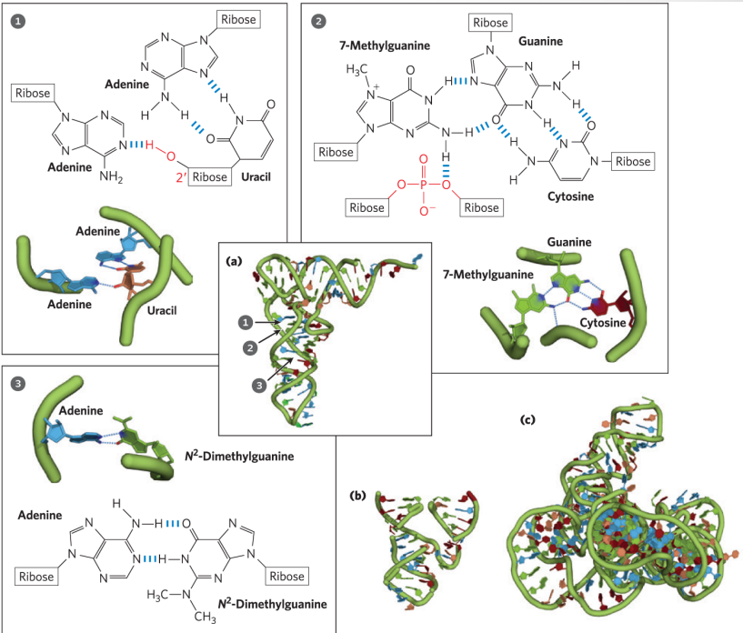
Multiple secondary structures and unusual base-pairing forms create the 3D structure of rRNA, mRNA, and tRNA.
Messenger RNA (mRNA)
synthesized by DNA transcription.
mRNA sequence is complementary and antiparallel to the template strand of DNA.
Functions as the blueprint for protein synthesis (translation) by encoding amino acid sequences.

Ribosomal RNA (rRNA)
Integral components of ribosomes —> serve as cellular machinery for protein synthesis.
In translation, the genetic codes of mRNA are converted into functional proteins.
Highly conserved across species, underscoring their fundamental role in translation.
Many antibiotics target bacterial ribosomes, disrupting protein synthesis (tetracyclin)
Transfer RNA (tRNA)
tRNA has a distinctive cloverleaf-like structure containing 4 critical regions:
Amino acid arm.
D arm.
Anticodon arm.
T or TψC arm.
Amino acid arm: Carries a specific amino acid esterified by its carboxyl group to the 2′-OH or 3′-OH group of the A residue at the 3′ end of the tRNA.
Anticodon arm: Contains the anticodon sequence.
D arm: Helps in the proper folding and stabilization of the tRNA structure.
T or TψC arm: Contains thymine (T) or pseudouridine (Ψ), involved in binding tRNA to the ribosome during protein synthesis.

The Wobble Hypothesis
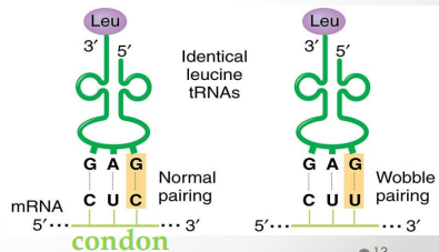
Proposes that the first base of the anticodon (5′ → 3′) pairs loosely with the corresponding codon base.
Loose pairing permits rapid dissociation of the tRNA from its codon during protein synthesis.
Rules of the Wobble Hypothesis:
First base U can recognize A or G.
First base G can recognize U or C.
First base I (inosine) can recognize U, C, or A.
Genetic Information Flow

DNA Denaturation + Heat
Denaturation: Defined as the unwinding of the double helix into two single strands.
Induced by pH extremes or high temperatures.
Process disrupts hydrogen bonds and base-stacking interactions between strands.
Annealing: Rewinding of separated complementary strands when temperature or pH is returned to physiological ranges (occurs in 2 steps).
Denaturation temperature (tm): The temperature at which 50% of double-stranded DNA is converted to single strands.
(tm) increases proportionally with higher(G triple bond C) base pair content.

DNA Partial Denaturation + UV Absorbance Effects
Partially denatured regions within DNA form bubbles, which are typically rich in A=T base pairs.
Hypochromic effect: Observed decrease in UV light absorption when complementary strands pair into a double helix.
Hyperchromic effect: Observed increase in UV light absorption when double-stranded nucleic acid undergoes denaturation.
260 nm = transition from double-stranded to single-stranded DNA.

RNA Denaturation
RNA duplexes exhibit higher heat denaturation stability than DNA duplexes.
RNA-DNA hybrid duplexes generally possess intermediate stability.
DNA Non-Enzymatic Transformations
Definition: Spontaneous chemical modifications occurring in DNA without enzyme catalysis.
Common Types of Transformations:
Mutation
Deamination (removal of an exocyclic amino group)
Depurination (loss of a purine base)
Pyrimidine dimers (cross-linking caused by UV light)
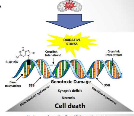
Oxidation (damage from reactive oxygen species)
Methylation (\(-\text{CH}_3\))
Causes: Exposure to chemical reagents, radiation, or reactive oxygen species (ROS) produced during cellular metabolism.
Mutations + Alkylating Agents
Mutations: Structural alterations in DNA producing permanent changes in encoded genetic information.
Strongly linked to aging processes and carcinogenesis.
Alkylating agents: Modified nucleotides and chemical agents that add alkyl groups
Deamination + Depurination
Deamination: Spontaneous loss of exocyclic amino groups from bases.
Depurination: Hydrolysis of the N-beta-glycosyl bond connecting the base to the pentose sugar.
Pyramidine Dimers
Pyrimidine dimers: Covalent bond formation between adjacent pyrimidine bases (T or C) caused by UV radiation.
Forms Thymine-Thymine, Cytosine-Cytosine, or Thymine-Cytosine dimers.
Structural types include Cyclobutane dimers or 6-4 photoproducts.
Abnormal bonds introduce structural kinks that distort the DNA backbone, causing replication errors.

Oxidation
Reactive Oxygen Species (ROS): Metabolic byproducts including hydrogen peroxide, hydroxyl radicals, and superoxide radicals.
Guanine most susceptible to oxidative damage, lowest one-electron reduction potential among DNA bases
Cellular organisms maintain elaborate defence systems against oxidative stress.
Enzymatic Transformations: Methylation
Adenine (A) and Cytosine (C) residues undergo enzymatic methylation (CH3) more frequently than Guanine (G) or Thymine (T).
DNA methylases transfer methyl groups using S-adenosylmethionine as a methyl donor.
In eukaryotic genomes, 5% of cytidine residues are methylated.
Biological functions: DNA replication, gene expression regulation, development, cell differentiation, and epigenetic control.

Chemical Synthesis of DNA (Phosphonamidite Method)
Phosphonamidite method: Automated chemical approach for oligonucleotide synthesis.
Nucleotides are added sequentially in the 3' —> 5' direction.
Solid-Phase Synthesis: Performed on a solid support matrix, allowing rapid separation of the growing DNA chain from reaction byproducts.
Four-Step Reaction Cycle:
Detritylation
Activation and coupling
Capping
Oxidation
Features high yield, precision, and efficiency for producing custom DNA sequences.

Polymerase Chain Reaction (PCR)
PCR: In vitro method used to exponentially amplify targeted DNA segments.
Relies on DNA polymerases to synthesize new strands from dNTPs using a DNA template.
DNA polymerases attach new nucleotides to the 3' ends of preexisting oligonucleotides called primers.
Three Core Thermal Steps (1 Cycle):
Denaturation (95°C): Separates template strands.
Annealing (55°C): Synthetic primers bind to template strands.
Extension (72°C): Thermostable Taq DNA polymerase synthesizes new strands in the 5' —> 3' direction.
Capable of detecting and amplifying a single DNA molecule.
Key Applications:
Amplification of rare DNA from ancient samples (40,000+ years old).
Tracing evolutionary relationships.
Forensic investigations.
Asymptomatic detection of viral infections and oncogenic mutations.
Prenatal genetic disease diagnosis.
DNA Sequencing
Sanger Sequencing:
Known as dideoxy chain-termination sequencing.
Uses modified dideoxynucleoside triphosphates (ddNTPs) lacking a 3'-OH group to terminate strand extension.
Fragments are separated by size using agarose gel electrophoresis.
Automated DNA Sequencing:
Each of the four ddNTPs is tagged with a distinct fluorescent dye.
Uses capillary gel electrophoresis paired with laser detection to record sequence chromatograms.
Next-Generation Sequencing (NGS):
Illumina NGS: Delivers 300 bp read lengths, 300 Gb per run, with high accuracy.
Nanopore Sequencing: Reads up to 4 Mb, 48 Gb per run, with reasonable accuracy.