Lab 7: Spinal Cord, Brain Stem, Peripheral Nervous System, and Special Sense
 Call Kai
 Learn
 Practice Test
 Spaced Repetition
 Match
 Flashcards
 Knowt Play
1/8

Earn XP
Description and Tags
Fall 2026 Professor Rana Khankan
9 Terms
What are the four different divisions of the spinal cord?
Cervical (C1-C7), Thoracic (T1-T12), Lumbar (L1-L5), and Sacral (S1-S5)

Know the anatomical views to help with vertabrae labelling.
Anterior = Ventral & Posterior = Dorsal

Very helpful image for identifying dorsal vs ventral locations of vertebrae + artery courses
Once again, relying on anatomical position helps greatly when identifying specific locations of vertebrae


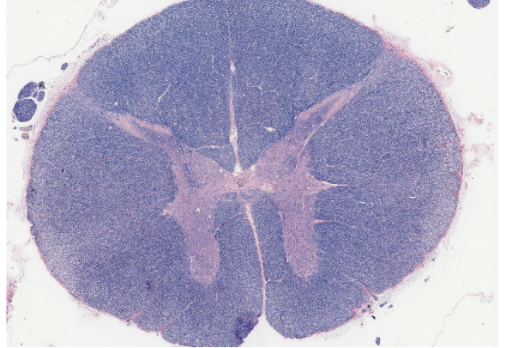
Know the staining of gray and white mater for lumbar vertabrae
Gray mater = Stains pink (butterfly shape) & White mater = Stains dark purple
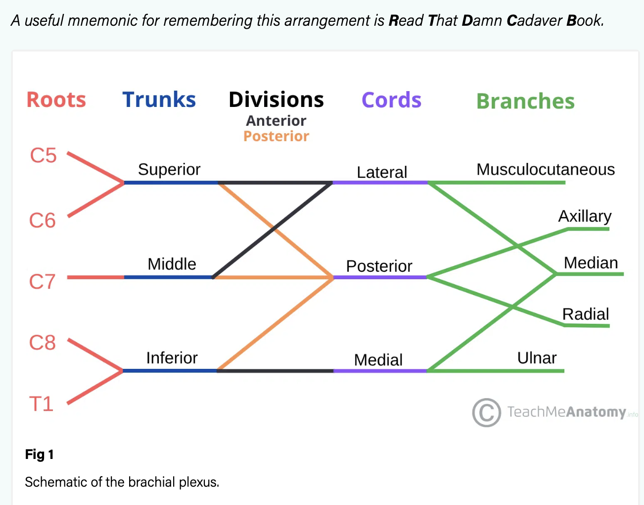
Visual image displaying the major components of the plexus “READ THAT DAMN CADAVER BOOK!”
Roots, trunks, divisions, chords, and radial nerve

Cadaver image of Axillary Nerve
Branches out to the lateral (→) side of shoulder. It’s behind your median nerve.

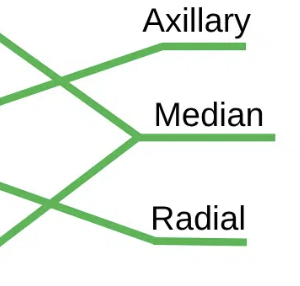
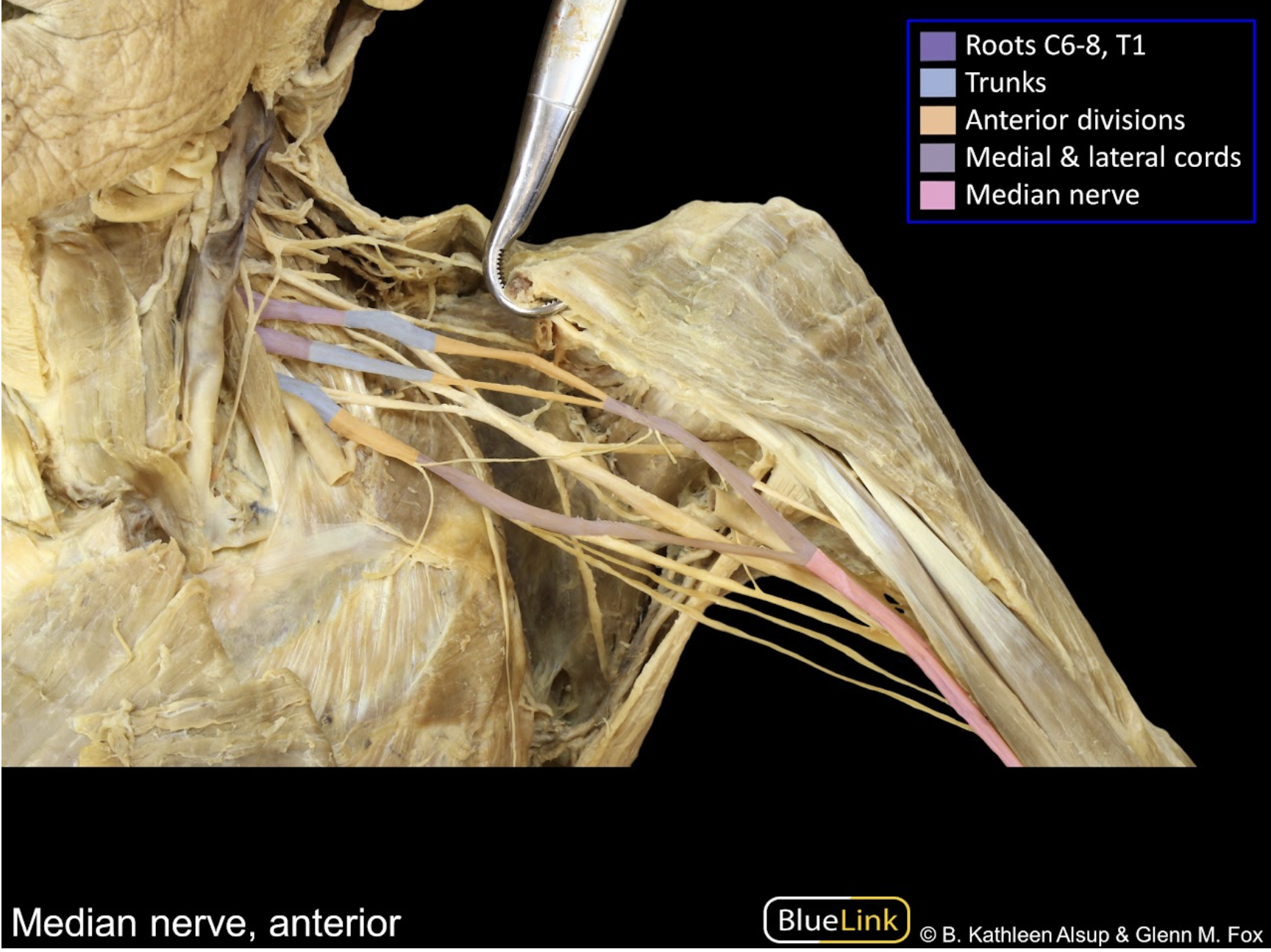
Cadaver image of median nerve
Branches from medial/lateral cords. Nerve falls above/on top of the axillary nerve. The medial nerve is the center portion of letter M: /|\
Cadaver image of the ulnar nerve
Very thin. located on the top layers of nerves (medially)
