ch 12 nervous system and tissue
 Call Kai
 Learn
 Practice Test
 Spaced Repetition
 Match
 Flashcards
 Knowt Play
1/60

There's no tags or description
Looks like no tags are added yet.
61 Terms
sensory input
information gatered by sensory receptors—external (senses) and internal (bladder full) changes
integration
interpretation process of sensory input—-spinal cord (bridge between sensory input and motor output)
motor input
activation of effector organs (muscles and glands) produces response
ex: “i might be thirsty” → gets water bottle
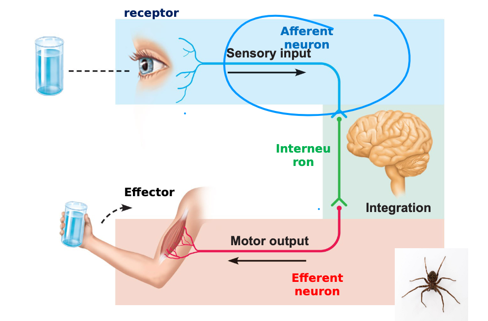
afferent neuron
sensory information — INFORMATION IN

interneuron
nerve cell in the CNS that connects other neurons and processing information
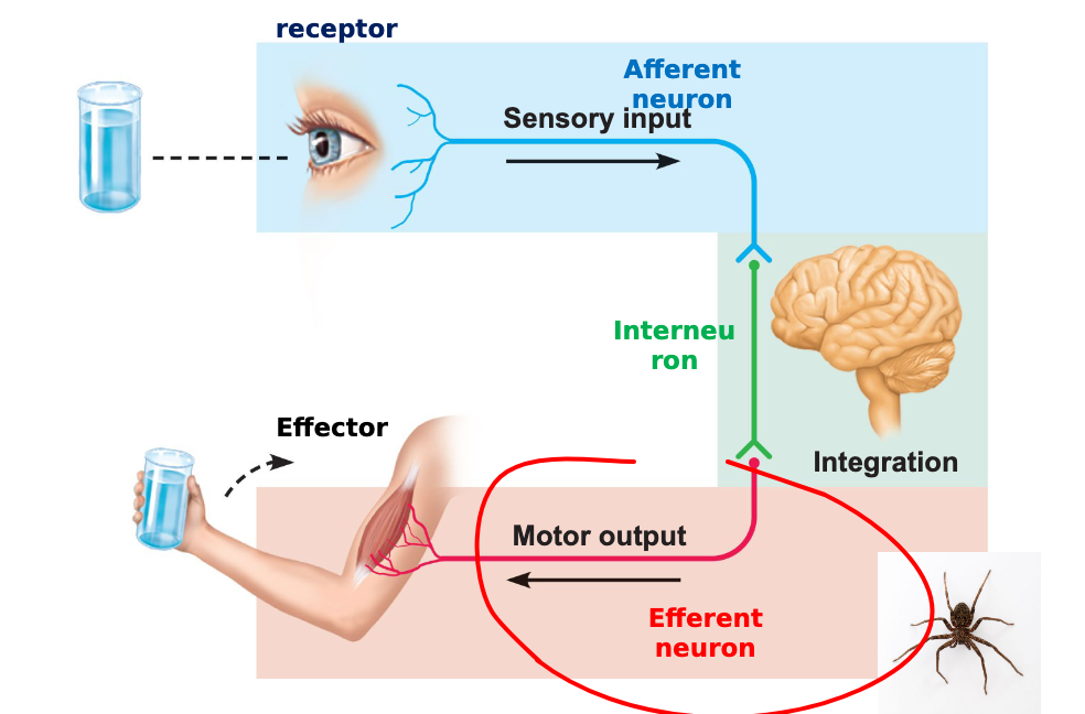
efferent neuron
reaction — INFORMATION OUT
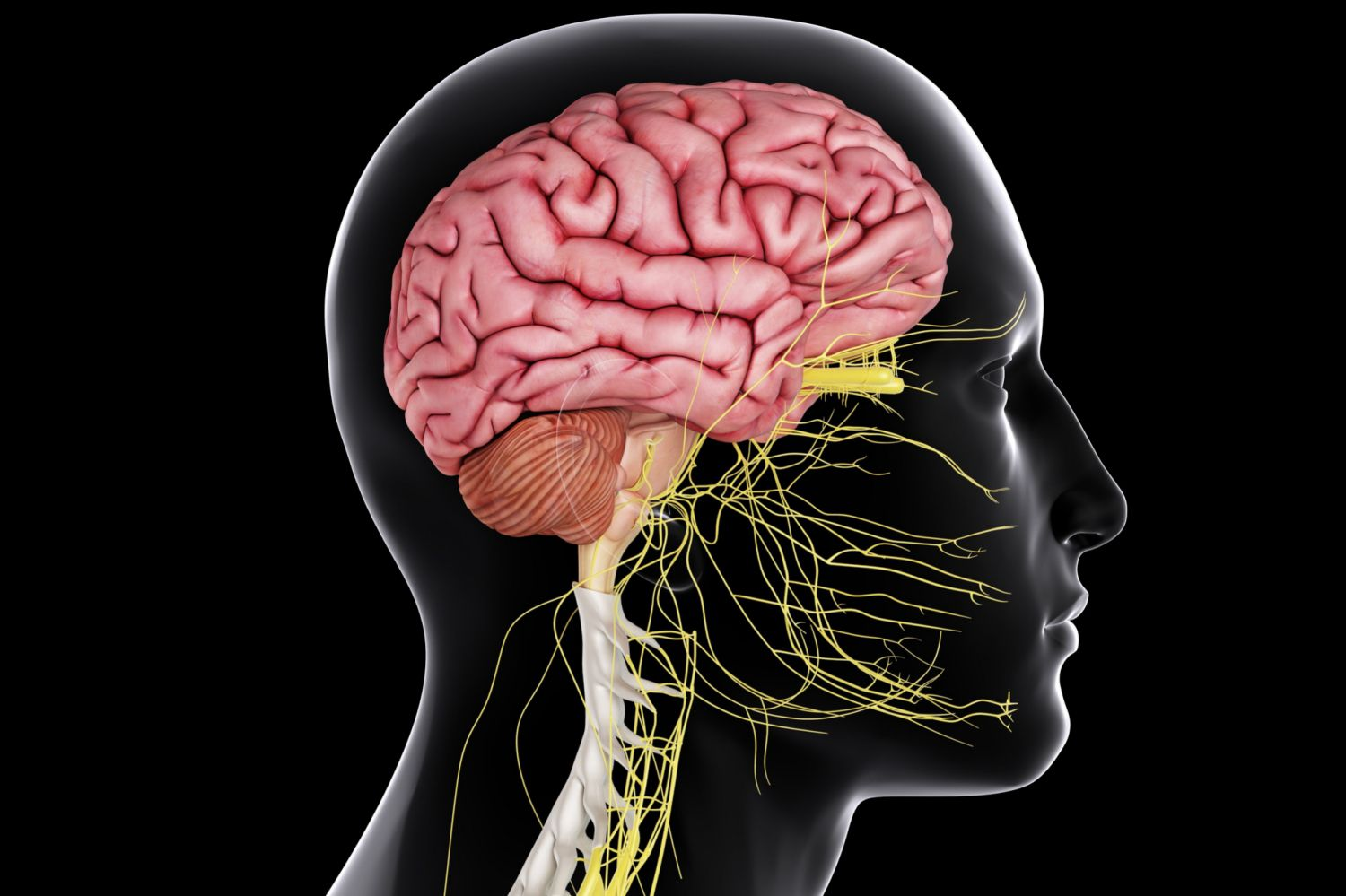
central nervous system (CNS)
brain and spinal cord
command center

peripheral nervous system (PNS)
paired spinal and cranial nerves carry msgs to and from the CNS
two directions (sensory and motor division)
INFORMATION GOING INTO THE BRAIN (spinal and cranial nerves)
sensory (afferent) division
TO THE BRAIN — UNCONSIOUS
somatic afferent fibers
viseral afferent fibers
somatic afferent fibers
carrying incoming signals convey impulses from skin, skeletal muscles and joints into the CNS
viseral afferent fibers
convey impusles from internal organs, blood vessels and glands into the CNS
motor (efferent) division
AWAY FROM THE BRAIN—transmits impulses from the CNS to effector organs (muscles & glands)
somatic nervous system
autonomic nverous system


somatic (voluntary) nervous system
CONSCIOUS control of skeletal muscles
ex: smiling, making leg move, activating skeletal muscle

autonomic (involuntary) nervous system
regulates smooth, cardiac muscles and glands
two functional subdivdisions:
sympathetic
parasympathetic

sympathetic division
fight or flight

parasympathetic division
rest, read and digest
2 principal cell types
1) neurons
2) neuroglia

neurons
excitable cells that transmit electrical → main cells that SENDS the msg going into the somantic/motor division
100 BILLION NEURONS IN OUR BRAIN
neuroglia
glial cells — supporting cells
astrocytes (CNS)
microgliail (CNS)
ependymal (CNS)
oligodendrocytes (CNS)
schwann cells (PNS)
nuclei…
…clusters of cell bodies in CNS
ganglia…
…clusters of cell bodies in PNS
tracts…
…a highway bundle of nerve fibers in the CNS
nerves…
…a highway bundle of nerve fibres in the PNS

astrocytes
star like
support (MANY ROLES) — most abundant cell
helps form blood brain barrier
helps block certain things going into the brain

microglial
defensive (phagocytes—protects/detect dmged or infections)
immune cells of CNS

ependymal
produces and moves CSF (spinal fluid)
surrounds brain and spinal cord — epithelial lining

oligodendrocytes
CNS — produces myelin, insulating axons

schwann cells
PNS — produces myelin, insulating axons
vital to regenerate damaged PN fibers
nerves do NOT regenerate if its completely cut off
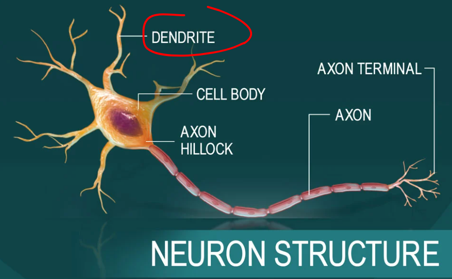
dendrites
receptive regions → getting/sending info into the brain

cell body/soma
biosynthetic center and receptive region — surounds nucleus (rough ER)
biosynthetic
center of a neuron

rough ER
makes neurotransmitters — proteins

axon
impulse generating and conducting region

axon hillock
where impulse STARTS—area where axon rises

node of ranvier
small and periodic gap in the insulating myelin sheath

axon terminals
distal terminations of branches of an axon — arrival of axon potential (synpases) — LEADS INTO THE TARGET CELL

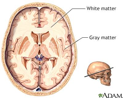
white matters
dense collections of myelinated fibers (tracts)
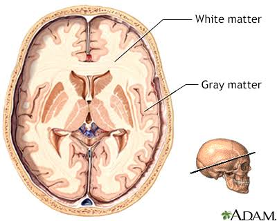

gray matters
mostly neuron cells bodies and unmyelinated fibers

unipolar
one projection — SENSORY NEURONS
multipolar
multi projections — MOTOR NEURONS AND INTERNEURONS

bipolar
two projection — ONLY NEURONS OF THE EYE
very rare
the ionic concentrations of a cell at rest is…
…increase concentration of K inside the cell
inside of cell is more NEGATIVE when resting

resting membrane potential
potential difference across the membrane of a resting cell — no charge
-70 mV in neurons → inside of a membrane is negatively charged relative to outside
outside: Na+, inside K+


when a cell is at rest…
…potassium (K+) inside and sodium (Na-) outside

membrane potential
concentrations of iosn across membrane CHANGES
permeability of membrane to ions changes
sending/receiving signals
2 types of signals:
graded potentials
action potentials
graded potentials
local signals (dendrites)
short lived
depolarizations (EXCITE) or hyperpolarizaton (INHIBIT)
graded potential spreads as local currents — sodium gates allows the cell to be negative/positive
inhibitory potentials
bad angel prevents action potential
excitatory potentials
good angel triggers action potential

action potential
brief reversal of membrane potential with total amp of 100 mV
occurs in muscle cells and axons of neurons
doesnt decrease in magnitude over distance
long distance neural communcation (MAIN FORM)
NO STOPPING A MSG UNLESS NERVE IS SEVERED
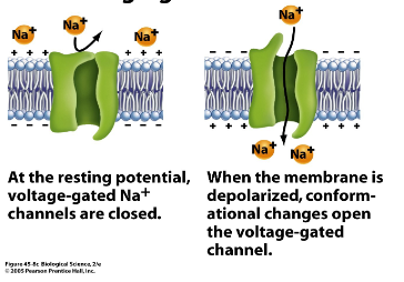
resting state
leakge channels for Na+ and K+ are open

depolarization
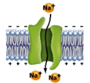
depolarizing local currents open voltage-gated Na+ channels → Na+ ENTERS
OUTSIDE TURNS NEGATIVE—-INSIDE TURNS POSITIVE

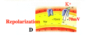
repolarization
slow voltage sensitive K-gates open → K+ LEAVES
K+ exits the cell and internal negativity is restored
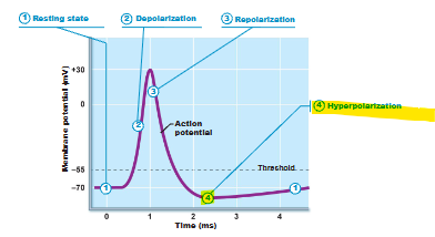
hyperpolarization
some K+ channels remain open allowing excessive K+ efflux

strong stimuli causes AP to occur…
…more frequently (# of impulses per second)
CNS determines stimulus intensity…
…by the frequency of impulses (higher frequency means stronger stimulus)
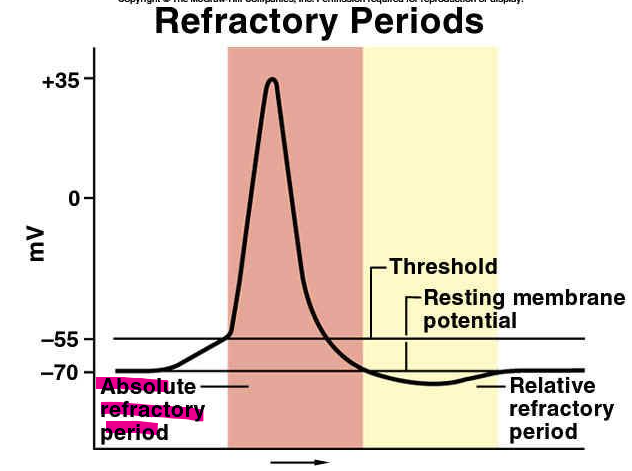
absolute refractory period
time from the opening of the Na+ channels until the resetting of the channels
another AP cannot fire during the period
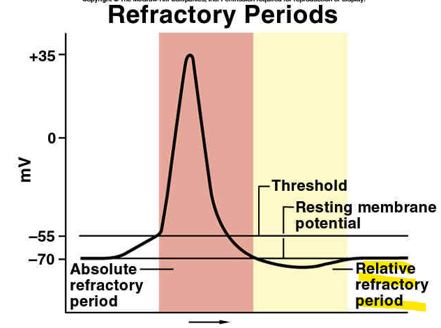
relative refractory period
repolarization is occuring
threshold for AP generation is elevated
exceptionally strong stimulus may generate an AP
axonal loss in mulitple sclerosis…
…destruction of myelin sheaths in the CNS become nonfunctional
chemical synapse
mediates informatino transfer from one neuron…
to another or an effector cell (NMJ)
release of a neurotransmitter
2 parts:
axon terminal of the presynaptic neuron
receptor region on the postsynaptic neuron
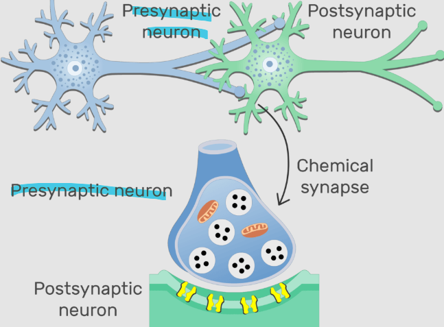
presynaptic neuron
sends a signal toward a synapse — axon

postsynpatic neuron
receives that signal across the synapse — dendrite