6 - Bones
 Call Kai
 Learn
Learn Practice Test
 Spaced Repetition
 Match
Match Flashcards
 Knowt Play
1/98

There's no tags or description
Looks like no tags are added yet.
Name | Mastery | Learn | Test | Matching | Spaced | Call with Kai | Chat |
|---|
No analytics yet
Send a link to your students to track their progress
99 Terms
What are the Five Primary Functions of the Skeletal System
1. Support
2. Storage
3. Blood Cell Production
4. Protection
5. Leverage
The Skeletal System Includes:
Bones and associated Cartilages
Ligaments and other connective tissues
What are Bone (Osseous) Tissue?
Dense, supportive connective tissue
What do Bone (Osseous) Tissue contain?
specialized cells
What do Bone (Osseous) Tissue produce?
solid matrix of calcium salt deposits
What do Bone (Osseous) Tissue form around?
collagen fibers
Organic components of bone (about one-third of bone mass):
Cells
Collagen fibers
Ground substance
Inorganic components of matrix:
Primarily hydroxyapatite
Calcium phosphate and calcium hydroxide (Ca10(PO4)6(OH)2)
Cells of Bone
Osteoprogenitor cells
Osteoblasts
Osteocytes
Osteoclasts
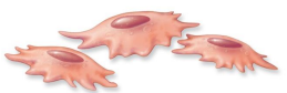
Osteoprogenitor cells
Mesenchymal stem cells in endosteum and periosteum

Osteoblasts
secrete osteoid

Osteocytes
maintain matrix and detect mechanical stress on a bone

Osteoclasts
Large, multinuclear cells that secrete hydrochloric acid and enzymes that dissolve matrix

Compact bone
Solid and relatively dense, and are the external surfaces of long and flat bones

Spongy bone
contain trabeculae and are the internal surface of bones

What is an Osteon?
the basic unit of compact bone

What are Osteocytes are arranged in?
concentric lamellae

Osteocytes arranged around
a central canal containing blood vessels
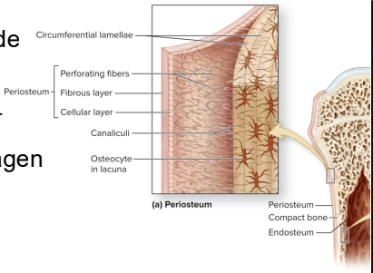
Circumferential Lamellae
form around long bone and bind osteons together

Periosteum are formed on
the outside of Circumferential Lamellae except joint capsules

Periosteum layers
fibrous and cellular layer

Perforating fibers - collagen
bone
periosteum
joint capsule
tendon/ligament

Endosteum
incomplete cellular layer

What does the endosteum line?
medullary cavity and central canal

Endosteum covers the
trabeculae of spongy bone

Endosteum contain what kind of cells?
Osteoblasts
Osteoprogenitor cells
Osteoclasts
Typical adult has how many bones?
206 (>270 to <200)
What are Bones classified by?
Shape
Internal tissue organization
Bone markings (surface features; marks)
Six Bone Shapes
1. Sutural bones
2. Irregular bones
3. Short bones
4. Flat bones
5. Long bones
6. Sesamoid bones

Structure of a Long Bone
Diaphysis
Epiphysis
Metaphysis

Diaphysis
shaft of the bone
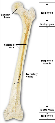
What do Diaphysis contain?
compact bone
medullary cavity
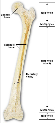
What are Epiphysis?
ends of long bone
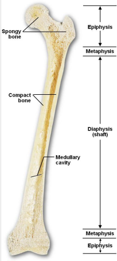
Epiphysis
Mostly spongy bone
Surrounded by compact bone

What are Metaphysis?
Where diaphysis and epiphysis meet
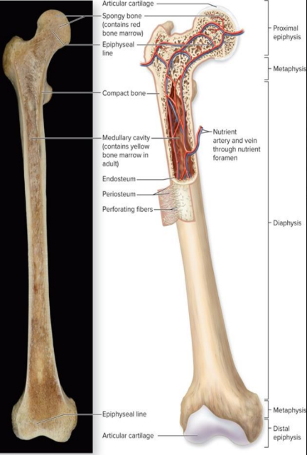
Spongy Bone
No osteons
open network of trabeculae

Where is red bone marrow made?
Spongy Bone

Medullary Cavity contain
yellow marrow
Human bones grow until about
age 25
What is Osteogenesis?
Bone formation
What is Calcification?
process of depositing calcium salts
When does calcification occur?
during bone ossification and in other tissues
What is Ossification?
process of replacing other tissues with bone
What are the main forms of ossification?
Endochondral ossification
Intramembranous ossification
Endochondral Ossification
Ossifies bones that originates as cartilage
Intramembranous ossification
makes flat bones
Growth in length at epiphyseal plate: Epiphyseal to Diaphyseal:
1. Zone of resting cartilage
2. Zone of proliferation
3. Zone of hypertrophy
4. Zone of calcification
5. Ossified bone
Summary of bone development
1. Cartilage model forms
2. Bone collar forms
3. 1° ossification center
4. Medullary cavity
5. 2° ossification center
6. Growth continues

What is Appositional Growth?
growth in width
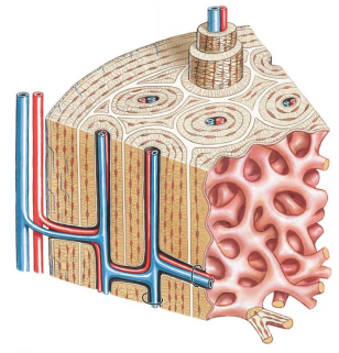
Appositional Growth thickens and strengthens
long bone
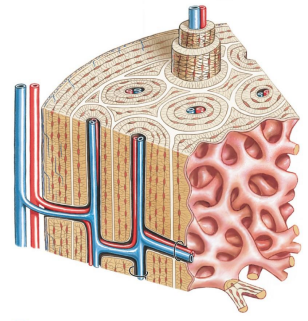
Appositional Growth adds more layers of
circumferential lamellae
Bones stop growing around
your early 20’s

Epiphyseal Lines form when
long bone stops growing
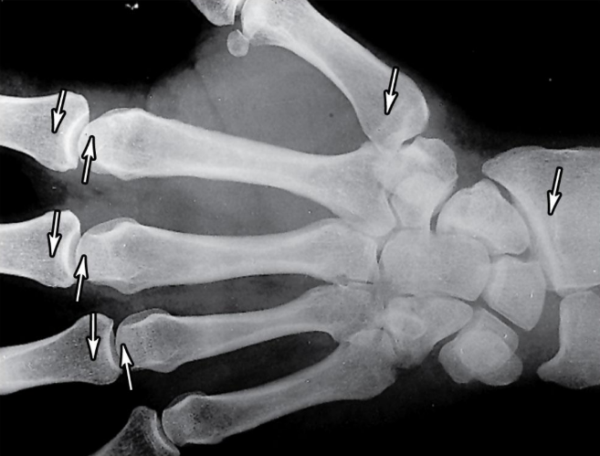
When long bone stops growing, after puberty:
Epiphyseal cartilage ossifies which is visible on X-ray as epiphyseal line

Intramembranous Ossification (dermal ossification)
Flat bones of the skull
Mesenchymal tissue model

Three main steps to Intramembranous Ossification (dermal ossification)
Mesenchymal cells differentiate to osteoblasts
Osteoblasts begin ossification
Spongy bone formation – remodeled

Blood Supply of Mature Bones
Nutrient Artery and Vein
Metaphyseal Vessels
Periosteal Vessels

Nutrient Artery and Vein
Supply the epiphyseal cartilage
A single pair of large blood vessels
Enter the diaphysis through the nutrient foramen
Femur has more than one pair

Metaphyseal Vessels
Where bone growth occurs

Where bone growth occurs
Blood to superficial osteons
Secondary ossification centers during development
Process of Bone Remodeling the adult skeleton:
Maintains itself
Replaces mineral reserves
Continually recycles and renews bone matrix
Involves osteocytes, osteoblasts, and osteoclasts
Effects of Exercise on Bone
Remodeling allows bones to acclimate
Stressed bones become stronger/denser
Broken bones, osteoporosis, bedridden
Stressed bones become
stronger/denser
Bone Degeneration affects:
Up to one third of bone mass
Normal Bone Growth and Maintenance
Need good Nutrition and Hormones
Nutrition
Dietary source of Calcium and Phosphate
Drink Milk, Eat Bones!
Hormones – Calcitriol
Synthesized in the kidneys
What does Calcitriol require?
Vitamin D3
Where is Vitamin D3 obtained from?
the sun
Other vitamins that are important:
Vitamin C
Vitamin A
Vitamins K and B12
Vitamin C
collagen synthesis/stimulation of osteoblast differentiation
Vitamin A
stimulates osteoblast activity
Vitamins K and B12
help synthesize bone proteins
Other hormones informant for bone development and growth
Growth hormone and thyroxine
Estrogens and androgens
Calcitonin and parathyroid hormone
Growth hormone and thyroxine
determine how large bones grow, secreted by pituitary gland
Hypersecretion of GH leads to
gigantism
Hyposecretion of GH leads to
pituitary dwarfism
Estrogens and androgens
sex differences in height
Estrogens and androgens
Calcium homeostasis; how much calcium is in the blood
Calcium Homeostasis
Bones store calcium and other minerals
Calcium ions are vital to:
Membranes
Neurons
Muscle cells, especially heart cells
Fractures
Cracks or breaks in bones
Fractures are repaired in four steps
1. Formation of clot
2. Formation of callus
3. Ossification of callus
4. Remodeling

Clot formation
Fibrous network
Bone cells in the area die
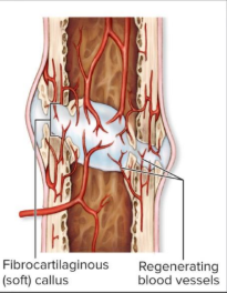
Calluses stabilize the break
Cells of the endosteum and periosteum
Fibrocartilage callus (soft)

Ossification of
cartilage callus

Remodeled to original look
osteoblasts and osteoclasts
up to a year
little to no evidence

Effects of Aging on the Skeletal System
Reduction in collagen and calcium
Osteopenia begins between ages 30 and 40
The epiphyses, vertebrae, and jaws are most affected
Reduction in collagen and calcium
bones become less dense
Osteopenia begins between ages 30 and 40
Bones become thinner and weaker with age
Women lose 8% of bone mass per decade, men 3%
The epiphyses, vertebrae, and jaws are most affected
fragile limbs
Reduction in height
Tooth loss
Osteoporosis
Severe bone loss
menopause
Osteoporosis Over age 45, occurs in ______ of women
29%
Osteoporosis Over age 45, occurs in ______ of men
18%

Identify 1
Articular cartilage

Identify 2
spongy bone

Identify 3
Medullary cavity

Identify 4
Endosteum

Identify 5
Periosteum