Biology chapter 5 & 9
 Call Kai
 Learn
Learn Practice Test
 Spaced Repetition
 Match
Match Flashcards
 Knowt Play
1/30

There's no tags or description
Looks like no tags are added yet.
Name | Mastery | Learn | Test | Matching | Spaced | Call with Kai | Chat |
|---|
No analytics yet
Send a link to your students to track their progress
31 Terms
What is membrane made of? why?
Phospholipid molecules.
Hydrophilic head (polar)
Hydrophobic tails (non polar)
Head towards water, tail away from water
The phospholipid bilayer is fluid and flexible - lipids can move within the membrane, and membranes can bend and form vesicles
proteins
sterols
carbohydrates

Sterols
Inside membrane with polar and nonpolar ends.
Cholesterol - major sterol in animal cells, inserted between phospholipids to regulate fluidity of membrane with temperature change.
Influences how tightly phospholipids pack

Proteins
Transport proteins - move ions, water, solutes
Receptor proteins - bind signals → responses
Recognition proteins - identify self vs foreign
Adhesion proteins - cells attach to one another
Integral membrane proteins - around the bilayer, transport molecules and act as receptors:
Hydrophobic regions interact with lipid tails.
Hydrophilic regions face aqueous environments or create channels.
Peripheral proteins - attach to membrane/integral proteins (support signaling, structure, or enzyme activity)

Carbohydrates
Found on the outer membrane surface
Glycolipids = lipid + carbohydrate.
Glycoproteins = protein + carbohydrate.
In animal cells, these can form the glycocalyx
Main function: cell recognition, cell to cell communication, attachment aid
The Fluid Mosaic Model (Singer & Nicolson, 1972)
membrane as a fluid phospholipid bilayer with molecules that can move
laterally
Membrane proteins - within or attached to the bilayer, creating a "mosaic" appearance
Cholesterol - regulates membrane fluidity and stability in animal cells.
Carbohydrates - attached to lipids and proteins. Cell recognition, signaling, and adhesion.
Membrane - dynamic, allowing transport, communication, membrane repair, and vesicle formation.
The model explains how membranes are selectively permeable while remaining flexible and adaptable.
Misconceptions:
Everything moves at the same rate
Bags holding things
Proteins are loose
Why fluidity is important?
Proteins need correct lipid environment to function
Fusion, vesicle formation, and signaling depend on fluidity
Too rigid: transport and protein movement can be impaired.
Too fluid: permeability and stability can be disrupted

Factors Affecting Fluidity
Temperature
Fatty acids
Cholesterol

Selective Permeability
What crosses depends on the properties of the molecule and the presence of membrane transport proteins.
Pass:
Small Nonpolar Molecules
Limited Pass:
Small Uncharged Polar Molecules
Do not Pass:
Large Polar Molecules
Charged Ions

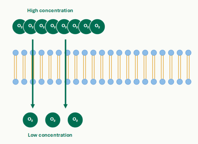
Simple diffusion
Movement Down a Concentration Gradient
Higher concentration → lower concentration
No transport protein
No ATP
Small, nonpolar, lipid-soluble molecules
O₂, CO₂, N₂, steroid hormones, and other lipid-soluble molecules
Facilitated diffusion
Passive transport through proteins
Down concentration gradient or electrochemical gradient
No ATP
Requires transport protein
Used by water, ions, sugars, amino acids, and other polar molecules
Channel Proteins: Fast, Selective Passageways
Hydrophilic pores through the bilayer
Aquaporins - water channels
Ion channels - specific ions to move down electrochemical gradients
Many ion channels are gated: open, closed, or intermediate states.
Carrier Proteins
Binds solute on one side
binding → conformational change
solute is released on opposite side
Carriers can become saturated when all binding sites are occupied

Active transport
Primary: Moving Against a Gradient Requires Energy
Lower concentration → Higher concentration
Requires ATP (directly or indirectly)
Maintains ion gradients and membrane potential
Supports nutrient uptake, waste removal, pH regulation, and cell signaling
Secondary: Symport and Antiport
ion moving down its gradient drives another solute uphill
Symport: both substances move in the same direction
Antiport: substances move in opposite directions
Energy comes indirectly from ATP used to build the ion gradient

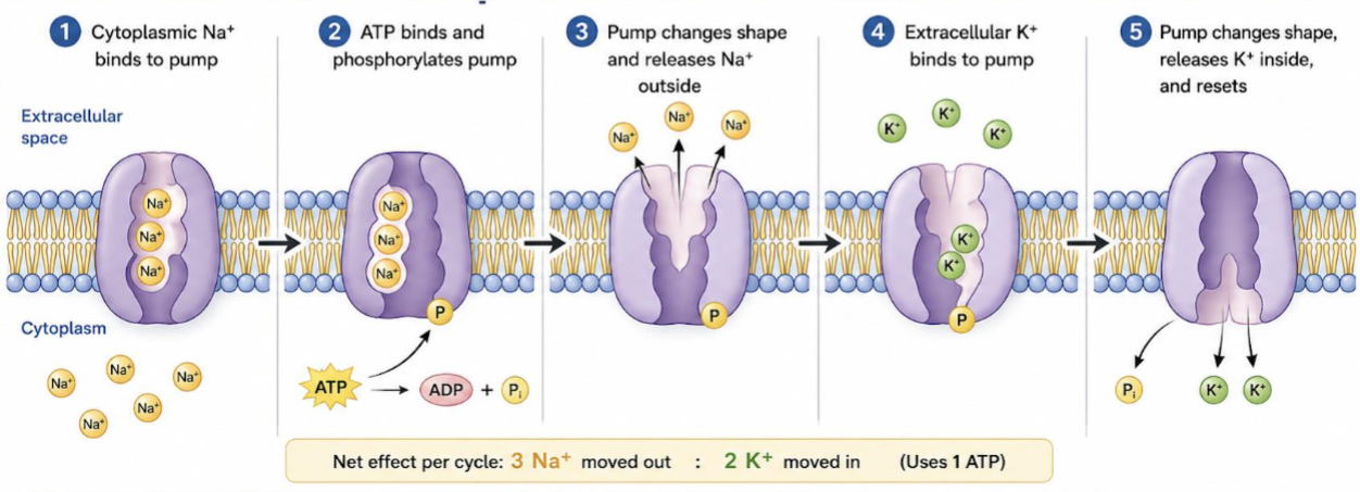
Active transport: Na⁺/K⁺ Pump
Builds an Electrochemical Gradient
Found in the plasma membrane of animal cells
ATP → pump 3 Na⁺ out and 2 K⁺ in per cycle
creates ion gradient (contributes to membrane potential)
Provides stored energy for secondary active transport
Membrane Potential: Electrical Energy Across a Membrane
▪ Unequal ion distributions create a voltage across the membrane.
▪ The inside of many cells is relatively negative compared with the outside.
▪ Membrane potential is essential for neurons, muscle cells, and transport.
▪ Electrochemical gradients combine concentration and electrical forces.

Vesicle Transport
• Large molecules move by membrane vesicles.
• Exocytosis - exports material and adds membrane to the plasma membrane.
• Endocytosis - imports material and removes membrane from the plasma membrane.
• Both processes require energy and involve membrane remodeling.
Exocytosis
Export by Vesicle Fusion
Vesicles from Golgi complex
Vesicles from inside fuses with the membrane
Cargo released outside
Vesicle membrane becomes part of the plasma membrane

Endocytosis
Import by Vesicle Formation
Bulk - non specific import
Receptor Mediated - selective import
Target molecule binds specific receptors
receptors cluster in pits reinforced by clathrin
The pit pinches off to become a vesicle
Cargo may be digested, receptors can be recycled
Phagocytosis - Engulfing Large Particles
Cells extend membrane lobes around a large particle or cell.
The enclosed particle forms a large vesicle called a phagosome.
Vesicles can fuse with lysosomes for digestion.
Important in immune defense and feeding by some protists.

Signal transduction
Many signals cannot cross the membrane directly
Receptor receives the signal outside or inside
Transduction relays and amplifies the signal inside the cell
Response changes cell activity ( cell activity, such as secretion, transport, gene expression, or movement)

Reception
Receptors Recognize Specific Signals
Ligands - signal molecules that bind receptors
Membrane receptors bind hydrophilic signals outside the cell
Binding changes receptor shape/activity
Specific receptors allow cells to respond only to certain signals

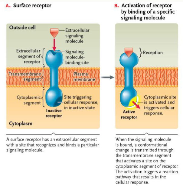
Surface Receptors
Transmembrane Proteins
Polar signals can’t cross the hydrophobic core
Surface receptors bind signals outside
Binding changes receptor shape
Cytoplasmic side initiates an internal response pathway
G-Protein-Coupled Receptors
▪ Seven transmembrane segments.
▪ Signal binding activates a G protein associated with GPCR
▪ G proteins GDP-bound (inactive) → GTP-bound (active) states.
▪ Activated G proteins regulate activate a signal relay through second messenger.

Receptor Tyrosine Kinases
Ligand binding promotes receptor dimerization.
Kinase domains phosphorylate tyrosine’s on the partner receptor.
Phosphorylated sites recruit signaling proteins.
RTKs often regulate cell growth, division, and differentiation

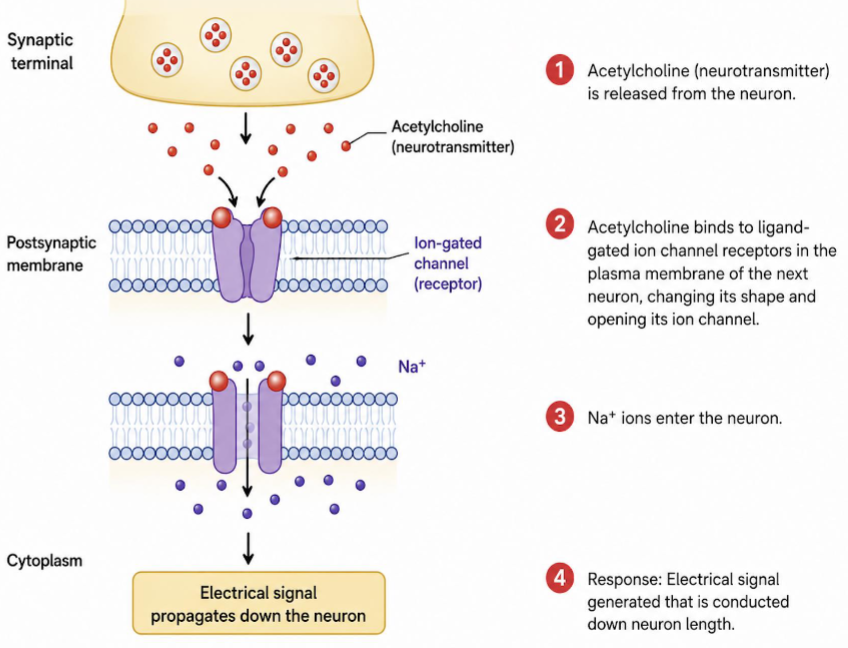
Ligand-Gated Ion Channels
Ligand binding changes channel conformation.
The channel opens or closes.
Ion flow changes membrane potential or cellular activity.
These receptors are important in synaptic signaling
Internal Receptors
Nonpolar signals such as steroid hormones can cross the membrane
They bind intracellular receptors
Activated receptor complexes regulate gene expression
Responses are often slower but longer-lasting than ion channel responses

Transduction: Relay, Amplify, and Integrate
Signals are often amplified: one activated receptor can activate many intracellular molecules.

Signal Amplification
One receptor can activate many molecules downstream
Enzyme cascades amplify the response
More catalytic steps → greater amplification
Amplification helps cells respond to very low signal concentrations

Signal amplification second messenger
cAMP is a 2nd messenger
Adenylyl cyclase converts ATP to cAMP.
cAMP diffuses through the cytoplasm, activates protein kinases.
Phosphodiesterase breaks cAMP down to help turn the signal off.

Signal transduction Pathway: GPCR Pathway
▪ Ligand binds to GPCR, which activates G protein
▪ G protein activates an effector enzyme → produces second messengers → activate protein kinases to phosphorylate target proteins

Signal transduction Pathway: Ras/MAP Kinase Pathway
▪ Activated RTKs → activate Ras (a small G protein)
▪ Ras initiates a kinase cascade, activating multiple kinases that lead to changes in gene expression and cell proliferation
▪ Linking external signal to membrane signaling to cell proliferation decisions

Response: Changing Cell Behavior
▪ Responses may be rapid, such as opening ion channels or triggering secretion.
▪ Responses may be slow, such as changing gene expression.
▪ Signals can regulate membrane traffic, including exocytosis and endocytosis.
▪ Cells integrate multiple signals before committing to a response.

Off Switches
Signals must be turned off to avoid excessive responses.
Ligands can be degraded or removed.
Receptors can be endocytosed and degraded or recycled.
Second messengers can be broken down.
G proteins inactivate themselves by hydrolyzing GTP to GDP

Cross-Talk and Integration
Cells often receive many signals at the same time and pathway can converge on shared targets.
Integration allows flexible, context-dependent responses
