B and T cell tolerance
 Call Kai
 Learn
 Practice Test
 Spaced Repetition
 Match
 Flashcards
 Knowt Play
1/5

There's no tags or description
Looks like no tags are added yet.
6 Terms
What are the two checkpoints of tolerance, and why are both needed?
The first checkpoint is central tolerance, which happens where lymphocytes are generated: the thymus for T cells and the bone marrow for B cells. Here the goal is to eliminate self-reactive cells before they leave.
The second checkpoint is peripheral tolerance, which happens in peripheral organs such as the spleen, lymph nodes and tissues. It is needed because the central checkpoint is not perfect: some self-reactive T or B cells slip through to the periphery, so further control mechanisms are required there.
How does the thymus use positive and negative selection to control self-reactive T cells?
During T cell development in the thymus, the TCR specificity is generated through many random rearrangements, so some T cells will recognize self-antigens. Positive selection happens in the thymic cortex, where T cells interact with cortical epithelial cells. T cells that bind self-peptide-MHC with adequate affinity receive survival signals and are "trained" and eligible for function, becoming CD4 or CD8 single-positive. Negative selection is the test that follows, where T cells are exposed to self-antigens (regulated by the transcription factor AIRE) presented via MHC. If they bind too strongly, they fail the exam and undergo deletion by apoptosis. A third outcome: cells that bind with medium strength can escape deletion and become thymic regulatory T cells (tTregs).
What are the four peripheral mechanisms that control self-reactive T cells that escaped the thymus?
Self-reactive conventional T cells that reach the periphery can be controlled in four ways.
(1) Ignorance: the self-antigen is present in too low an amount or with too low affinity to activate the TCR. T cells are separated from the antigens that can activate them (e.g., by the blood–brain barrier).
(2) Deletion:
Death by Neglect: T cells die when they are outcompeted for essential growth factors (like IL-2) by surrounding activated cells.
Chronic TCR Stimulation: Because self-antigens are never cleared, repetitive TCR signaling constantly upregulates Fas/FasL, driving AICD to remove persistent autoreactive T cells.
High-Affinity Deletion: Self-antigens that bind to the TCR too strongly directly trigger intracellular apoptotic pathways to eliminate dangerous clones.
AICD (Fas/FasL): Activated T cells express both Fas and FasL; their interaction triggers cell suicide (apoptosis). Thsi is how apoptosis works.
(3) Inhibition:
1. Lack of Co-stimulation (Signal 2 Deficiency) Anergy
When antigen presentation occurs without co-stimulatory signals, T cells enter a state of functional unresponsiveness termed anergy. In this state, the T cell cannot produce IL-2 and fails to achieve full activation.
2. Upregulation of Inhibitory Receptors
To actively block T cell activation, inhibitory co-stimulatory molecules such as CTLA-4 and PD-1 are expressed. CTLA-4 outcompetes CD28 for B7 binding, while PD-1 delivers inhibitory signals that shut down downstream TCR pathways.
Anergy and inhibition are commonly triggered by chronic antigenic stimulation (e.g., persistent exposure to self-antigens or high systemic doses of soluble protein antigens) or presentation by non-activated, resting antigen-presenting cells (APCs).
(4) Suppression: the cell is converted into a regulatory T cell by the cytokine environment. The overall aim is to keep the self-reactive T cell from becoming activated.

How are peripherally induced FoxP3+ Tregs generated, and how do they suppress the immune response?
When naive T cells in the body encounter self-antigens, the surrounding chemical environment can transform them into induced regulatory T cells (iTregs).
The main trigger for this change is TGF-β, a signaling protein made by surrounding tissue and epithelial cells. TGF-β turns on the master gene FoxP3, which officially converts the cell into a regulatory T cell. At the same time, the T cell needs IL-2 to survive and multiply. Another signal, IL-10, plays a smaller supporting role during this conversion—its main job comes later, when the Treg uses it as a tool to calm down inflammation.
Once converted, these induced Tregs work to stop autoimmune attacks using four main suppression mechanisms:
(1) blocking costimulation (indirect suppression) through inhibitory molecules. Normally, T cell activation needs a second signal: CD28 on the T cell binds CD80/CD86 on the antigen-presenting cell. Induced Tregs disrupt this by using inhibitory molecules such as CTLA-4 (and PD-1 binding PD-L1) to take the place of CD28 binding. Because this activating signal is blocked, no IL-2 is produced, so the T cell cannot survive or proliferate. When this inhibitory interaction happens between the T cell and a dendritic cell, the dendritic cell also produces metabolites (IDO) that have an inhibitory effect on effector T cells. Together, these effects create a tolerogenic, anti-inflammatory environment.
(2) converting extracellular ATP into anti-inflammatory adenosine using their cell surface enzymes which then it binds receptors on other cells and induces anti-inflammatory reactions, which dampens inflammation.
(3) sequestering IL-2 with their high CD25 expression, so nearby T cells lack survival signals
(4) directly killing effector T cells through perforin and granzyme granules that trigger apoptosis.
What are Tr1 cells, and how do they suppress inflammation?
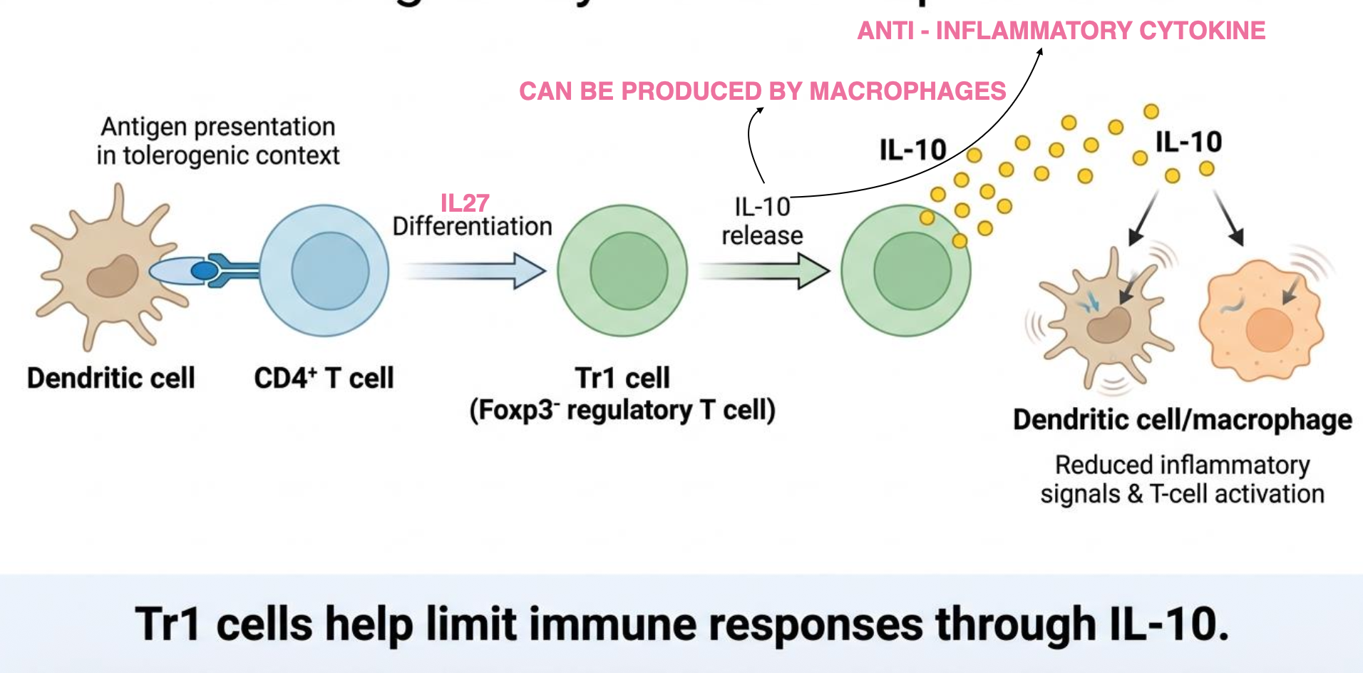
Tr1 cells are a type of regulatory T cells that are FoxP3⁻. They develop in the periphery during chronic antigen exposure, especially in an IL-10-rich environment, and their development is further promoted by IL-27. They mainly suppress immune responses by secreting high levels of IL-10, an anti-inflammatory cytokine. IL-10 inhibits APC function and reduces the activation, proliferation, and survival of pro-inflammatory effector cells, such as Th1 cells. This helps control the immune response and prevent excessive tissue inflammation.
How do B cells achieve central and peripheral tolerance?
B cells develop in the bone marrow, so central tolerance happens there, and peripheral tolerance happens in the spleen. In the bone marrow, a B cell that binds a self-antigen strongly first attempts receptor editing, changing its BCR to a non-self-reactive one. If receptor editing does not work, the cell undergoes deletion by apoptosis. Some B cells become anergic (unresponsive) when the antigen is present in low amounts. In the periphery, the same logic applies: strong self-antigen binding causes deletion; weak binding or low antigen leads to anergy or ignorance; and if naive B cells that recognize self-antigen require co-stimulation from helper T cells. If they encounter anergic T cells (or receive no T-cell help), they fail to receive survival signals (CD40/CD40L interaction and cytokines), inducing B-cell anergy and subsequent apoptosis due to growth factor starvation.
