Great Vessels
 Call Kai
 Learn
Learn Practice Test
 Spaced Repetition
 Match
Match Flashcards
 Knowt Play
1/37

Earn XP
Description and Tags
URR study guide
Name | Mastery | Learn | Test | Matching | Spaced | Call with Kai | Chat |
|---|
No analytics yet
Send a link to your students to track their progress
38 Terms
What are the 3 vessel wall layers?
tunica intima (inner layer)
tunica media (middle, muscular layer)
thicker in arteries
thinner in veins for flexibility of the lumen size due to varied flow volumes
tunica externa (outer layer)

Common iliac arteries
where are they located?
what part of the body do they supply?
doppler characteristic?
at the aortic bifurcation - umbilicus level
supplies blood to legs and pelvis
high resistance waveform - triphasic
aortic segments (what are the parts of the aorta)
ascending aorta
aortic arch
descending thoracic aorta
descending abdominal aorta

Great vessels (image)

Indications to scan the aorta
abdominal pain
pulsatile mass
aneurysm seen on x-ray
follow up aneurysm
trauma
What lab value can indicate active bleeding in the body, or that an aneurysm is leaking?
low hematocrit
How do you obtain a longitudinal view of the aorta and IVC in the same image?
position patient in decubitus position
use coronal approach to angle through the abdomen to visualize the aorta and IVC at the same time
Average Normal Aortic Measurements
Proximal = 2.0 - 2.6 cm
Mid = 1.6 - 2.4 cm
Distal = 1.1 - 2.0 cm
Iliacs = 0.6 - 1.4 cm
Doppler characteristics of the Aorta
high resistance
clean spectral window
biphasic waveform above renal arteries
due to low resistance branches to organs
triphasic waveform below renal arteries
supplies lower extremities
occlusion can change resistance and pulsitility of waveform
aneurysms can show increased turbulence
aortic flow above the renal arteries
biphasic
low resistance branches to organs
most organs have low resistance vascular beds that require continuous antegrade flow

aortic flow below the renal arteries
triphasic
supplies the lower extremities and pelvic
the muscular tissues of the lower extremities have high resistance vascular beds
normal diastolic flow will occur in 2 phases: diastolic flow reversal followed by a small amount of antegrade flow.

Aortic ectasia
occurs when the aorta does not taper in size distally
size of aorta remains constant from proximal to distal
can be a precursor to aneurysm formation
AAA - abdominal aortic aneurysm
what is it? measurements?
caused by what?
who’s at risk?
focal dilation of the aorta
greater than 3 cm
increase in diameter greater than 50% between 2 adjacent segments
commonly caused by atherosclerotic disease
high risk for males, fam hx, smoking, CHTN
Types of aneursyms
(what’s the most common?
and most common location?)
saccular
fusiform
berry (a saccular cerebral aneurysm)
mycotic
most common = fusiform below the renal arteries

Aneurysm above the renal arteries requires immediate surgical intervention.
True or False? Why?
True
the aorta segment above the renal arteries supplies vital organs
aneurysm below renal arteries
most common location for fusiform aneursyms
surgical intervention required if diameter is > 5.5 cm
What AP measurement indicates aneurysm in the common iliac artery?
greater than 1.5 cm
Follow up exams after AAA identified
annual follow up on AAA = 4 - 4.4 cm diameter
semiannual follow up on AAA > 4.5 cm
complications of aortic aneurysm
rupture (most common)
decreased flow to lower extremities
blue toe syndrome with thrombus accumulation and embolization
affects renal circulation and systemic blood pressure, if suprarenal or juxtarenal in location
Large AAA can compress the IVC, causing reduced flow toward the heart and pedal edema
Which 2 structures guarantee a complete evaluation of the abdominal aorta?
celic axis (complete evaluation of the proximal abdominal segment)
iliac arteries (complete evaluation of the infrarenal abdominal segment)
What to do if you see an aneurysm?
measure true lumen size if thrombus is present
measure length of affected segment
document AAA location related to renal arteries
turn on color
turbulence seen with color and PW
Yin Yang Sign = swirling of blood in the body of the aneurysm
most common splanchic artery aneurysm
splenic artery
endovascular aortic repair
percutaneous stent or graft placement through groin
if leak is present, native sac increases in size
blue toe syndrome
embolic material lodges into the digital arteries of the toes leading to cyanosis of the distal tissues
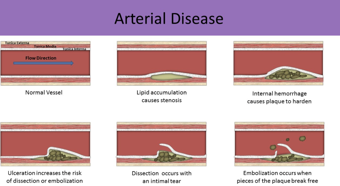
Dissection
how does it happen?
Increased risk of what?
most common where?
What can occur in the false lumen?
intimal layer tears and allows flow between the intima and media layers into a blind pocket (false lumen)
causes weakened vessel wall
risk of vessel rupture
most common at aortic arch
thrombosis can occur in false lumen → stenosis/occlusion and ischemia distal to dissection
Marfan syndrome
what is it?
how do patients with this syndrome appear like?
associated with?
genetic disorder that affects the connective tissue of the heart, vessels and bones
tall, thin, long extremities & fingers
associated with aortic dissection, aneurysm, valve insufficiency
aortic dissection (image)

aortic dissection (image)

aortic dissection (image)
Aortic rupture
measurement?
symptoms?
what can rupture cause?
high risk if aneurysm > 7 cm in diameter
back pain, hypotension
hypovolemic shock due to sudden blood loss
pseudoaneurysm
what is it?
caused by?
what does it look like?
how is it treated?
blood escapes all 3 vessel layers into surrounding tissues and is encapsulated within the tissues
caused by trauma, invasive procedures
round sac of blood connected to the vessel by a neck/stalk
treated with compression of the stalk or thrombin injection
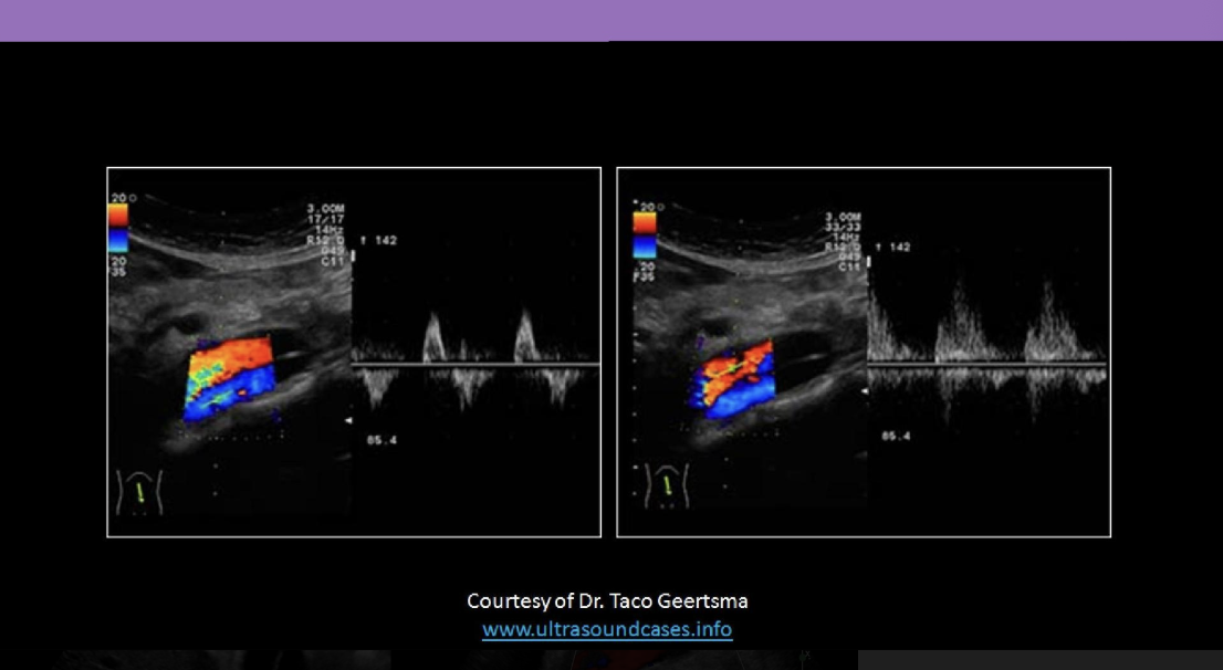
pseudoaneurysm sonographic appearance
yin/yang sign, turbulent flow
high resistance to-fro flow in stalk; low resistance to-fro flow in body
rounded, anechoic structure adjacent to main artery
color demonstrates connection between artery and structure
depth is measured to the pseudo-aneurysm for needle selection with thrombin injection

AORTIC STENOSIS
most commonly caused by?
can be related to what?
flow pattern
proximal to the stenosis?
distal to stenosis?
what happens to velocity at the stenosis?
What happens if the stenosis is suprarenal?
caused commonly by atherosclerosis
also can be related to AAA thrombosis, arteritis
higher resistance proximal to stenosis
increased velocity at stenosis
dampened waveform (tardus parvus) distal to stenosis
if stenosis is suprarenal → causes renal ischemia → systemic HTN
Leriche Syndrome
caused by?
what part of the body does it affect?
symptoms?
flow pattern?
related to?
AKA aortoiliac occlusive disease
occlusion at the abdominal aorta above the bifurcation of the CIA
bilateral symptoms and flow changes in the lower extremities
symptoms:
fatigue, intermittent claudication with ischemic pain, absent or diminished femoral pulses, pallor, coldness
low resistance, post-stenotic flow
related to erectile dysfunction (bc internal iliac arteries supply blood to the male genitalia)
retroperitoneal fibrosis
what is it?
where does it occur?
cause related to?
what can happen to the IVC and gonadal veins?
AKA ormond disease
idiopathic overgrowth of fibrous tissue around and atherosclerotic aorta
at the level of aortic bifurcation and inferior to the pelvis
can be related to drugs, infection, malignancy or cancer therapy
may compress IVC → bilateral pedal edema
may compress gonadal veins → scrotal swelling

What is the normal AP diameter of the aorta?
What measurement indicates an aneurysm?
normal = under 3 cm
Aneurysm = > 3 cm in AP diameter
How should the aorta measure from proximal to distal?
At what level of the aorta should the largest diameter be measured?
normal diameter of aorta should decrease from superior to inferior
greatest diameter is at the level of the celiac axis
How do you accurately measure the AP diameter of the aorta?
measure from outer to outer wall