HTN Renal Disease
 Call Kai
 Learn
Learn Practice Test
 Spaced Repetition
 Match
Match Flashcards
 Knowt Play
1/34

There's no tags or description
Looks like no tags are added yet.
Name | Mastery | Learn | Test | Matching | Spaced | Call with Kai | Chat |
|---|
No analytics yet
Send a link to your students to track their progress
35 Terms
How kidney disease causes hypertension
Loss of nephrons → impaired sodium excretion + RAAS activation → increased blood pressure.

How hypertension causes kidney disease
Chronic high BP → arteriolar hyalinosis, ischemia, glomerulosclerosis, tubular atrophy, interstitial fibrosis.


Histologic finding: arterionephrosclerosis
Hyaline arteriosclerosis, medial thickening, narrowed lumens, global/focal glomerulosclerosis, tubular atrophy.

Histologic finding: hypertensive emergency
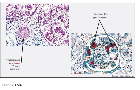
Endothelial swelling, fibrinoid necrosis, RBC fragments in vessel walls, thrombi, “onion-skin” hyperplastic arteriolitis.


Histologic finding: thrombotic microangiopathy
Schistocytes lodged in vessel walls, endothelial swelling, thrombosis occluding arterioles and glomeruli.
Physical exam clues for hypertensive emergency
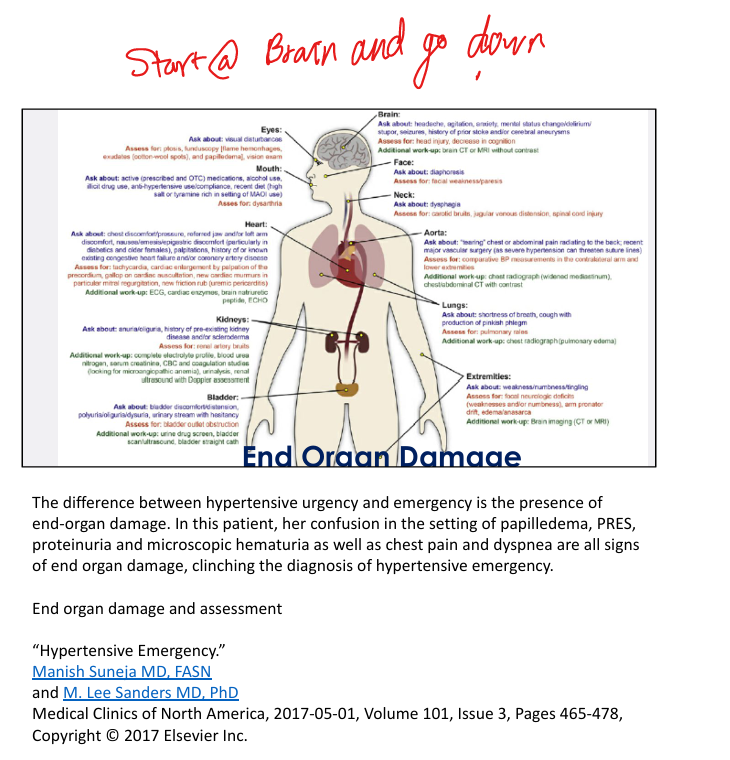
Papilledema, confusion, focal neurologic deficits, pulmonary edema, chest pain, oliguria.

Laboratory clues for hypertensive emergency
Proteinuria, hematuria, elevated creatinine, MAHA (schistocytes), thrombocytopenia.


End-organ damage: brain
Headache, confusion, seizures, PRES, stroke.

End-organ damage: eyes
Papilledema, flame hemorrhages, cotton-wool spots.

End-organ damage: heart
Chest pain, MI, arrhythmia, acute heart failure, pulmonary edema.

End-organ damage: kidneys
Rising creatinine, oliguria/anuria, proteinuria, hematuria.
RAAS activation in hypertensive emergency
Afferent arteriolar injury → ischemia → macula densa senses low flow → massive renin release → worsens BP.

Pathogenesis of hypertensive emergency
Extreme BP → endothelial injury → fibrinoid necrosis → thrombosis → ischemia → RAAS activation.

Definition of hypertensive emergency + tx
BP ≥180/120 with evidence of end-organ damage. Tx: Antihypertensives like labetolol, nicardipine, sodium nitroprusside

Renal artery stenosis: clinical clues
Resistant HTN, abdominal bruit, acute rise in creatinine after ACEi/ARB, flash pulmonary edema.


Renal artery stenosis: renin/aldosterone pattern
High renin + high aldosterone.

Fibromuscular dysplasia features
Young women, distal renal artery involvement, “string-of-beads,” responds well to revascularization.


Atherosclerotic renal artery stenosis features
Older adults, proximal renal artery plaque, associated with PVD/CAD, treated medically.

APOL1 nephropathy clinical pattern
Strong family historyoften black patients), progressive CKD, heavy proteinuria, seen almost exclusively in Black patients.

APOL1 nephropathy histology
Collapsing glomerulopathy, segmental/global sclerosis, prominent podocyte injury.


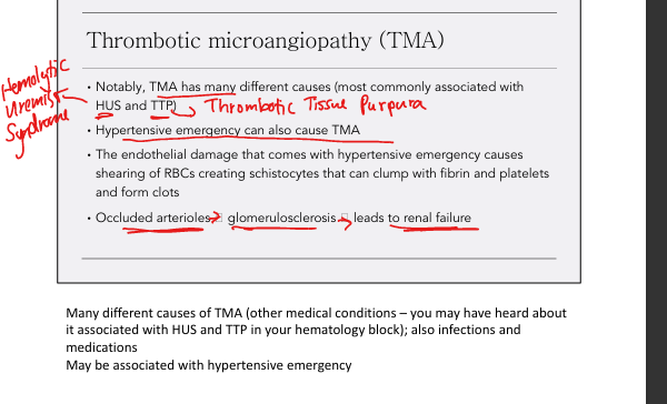
Thrombotic Microangiopathy (TMA) causes relevant to hypertension
Hypertensive emergency, HUS (Shiga toxin), TTP, drugs (cocaine, chemo).

HUS distinguishing features
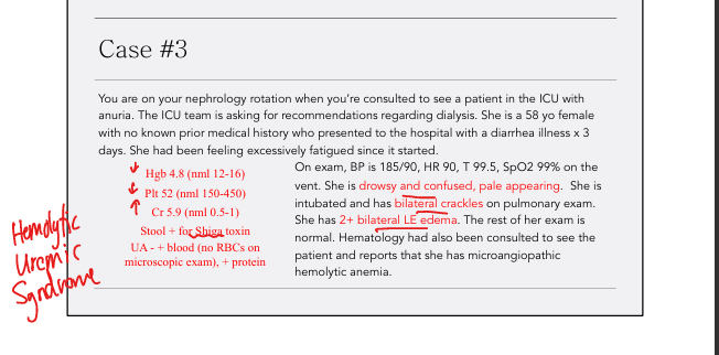
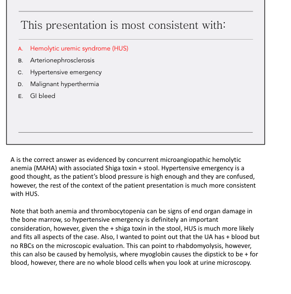
MAHA(microangiopathic hemolytic anemia) + thrombocytopenia + AKI + Shiga toxin–positive stool.
Why hypertensive emergency causes Microangiopathic Hemolytic Anemia (MAHA)
Endothelial injury → RBC shearing → schistocytes → thrombosis → ischemia.

IV medications for hypertensive emergency
Labetalol, nicardipine, clevidipine, sodium nitroprusside.
Why BP must be lowered gradually in hypertensive emergency
Sudden drops impair autoregulation → worsen ischemia in brain, heart, kidneys.
Why CKD worsens hypertension
Reduced nephron mass → impaired sodium excretion → volume expansion → RAAS activation.
Why hypertension worsens CKD
Chronic pressure → vascular thickening → ischemia → glomerulosclerosis → progressive renal failure.
Why ACEi/ARB worsen creatinine in bilateral Renal Artery Stenosis
Efferent dilation → drop in GFR when renal perfusion already low.


Lab findings suggesting Thrombocytic Microangiopathy (TMA)
Schistocytes, thrombocytopenia, elevated LDH, low haptoglobin, AKI.


UA findings in HUS
Hemoglobin-positive dipstick with no RBCs (hemolysis), proteinuria.

Why renal biopsy is avoided in TMA
Severe HTN + thrombocytopenia → high bleeding risk.
TX for 1 sided Renal Artery Stenosis vs 2 sided (bilateral) RAS
1 Sided: use RAAS blockage (ACEi/ARBs)
2 Sided (bilateral: use diuretics, as RAAS block would cause renal failure as they are reliant on RAAS)

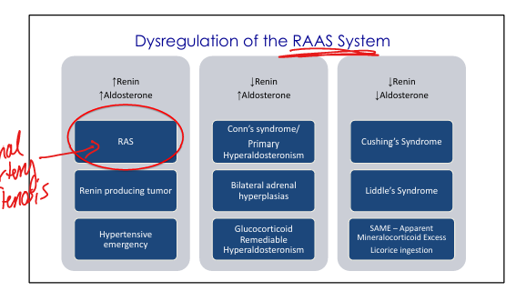
High Renin, High Aldosterone Differential
Renal artery stenosis, renin producing tumor, hypertensive emergency

Low renin, High aldosterone Differential
Primary hyperaldosteronism, bilateral adrenal hyperplasias, Glucocorticoid Remediable Hyperaldosteronism

Low renin, Low aldosterone Differential
Cushing sydrome, Liddle’s syndrome, Syndrome of Apparent Mineralcorticoid Excess (SAME)