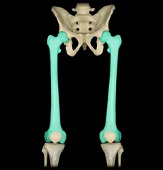
Rad 101 femur hip and pelvis
 Call Kai
 Learn
 Practice Test
 Spaced Repetition
 Match
 Flashcards
 Knowt Play
1/187

There's no tags or description
Looks like no tags are added yet.
188 Terms
Anatomical position, feet straight up and down
How is the femur rotated?
Anthropoid
winder anterior and posterior than right to left male pelvis
gynecoid
Round pelvis female
platypelloid
wider right to left than anterior and posterior pelvis female
Ischial tuberosity
what is C pointing at

Greater trochanter
what is F pointing at

how much do you flex the knee for a lateral femur?
45 degrees
How much do you rotate the legs inward for AP Pelvis, Proximal AP femur and AP unilateral hip
15-20 degrees
CR angle for danelius miller method?
CR perpendicular to the femoral neck and the IR going in at 45 degrees in the crotch.
CR angle for clements nakayama method
Angle CR 15-20 degrees down into femoral neck posteriorly. Angle the IR 15 degrees from vertical.
Angles for outlet pelvis CR?
20-35 degrees cephalad for males
30-45 degrees cephalad for females
CR 1-2 inches lower than the greater trochanter
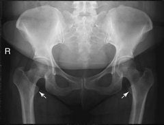
Femur externally rotated, lesser trochanter fully visible
How is the femur rotated?
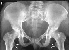
15-20° Internal Rotation, lesser trochanter barely visible (true AP)
How is the femur rotated?

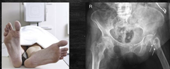
Affected femur placed mediolaterally, unaffected side placed in anatomical position
How would the feet lie to determine a hip fracture?
Find the ASIS (S1-2), go 1-2in medially, then move 3-4in down to neck of femur.
How do you find the the neck of the femur?

Pelvis (os coxae) (Innominate bones)
Serves as the base of the trunk, Forms connection between vertebral column & lower limbs, Consists of 4 bones

Lumbosacral joint
pertaining to the joint between L5 and the sacrum
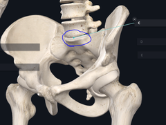
Sacrum
5 fused vertebrae

Sacroiliac joint
the joint between the sacrum and the ilium
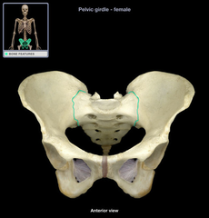
Coccyx
tailbone
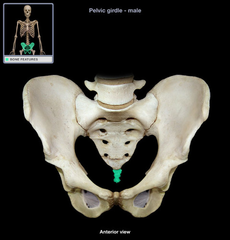
Ilium
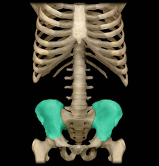
Body of Illium

Ala or wing of Illium
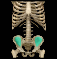
Iliac crests
Portion of innominate bones that define the most superior, bilateral aspect of the pelvic cavity. They are palpable external landmarks that aid in evaluating the pelvis.

Anterior Superior Iliac Spine (ASIS)
The attachment for the inguinal ligament. Creates a noticeable bump on the two coxa.

Posterior Superior Iliac Spine (PSIS)
Posterior projection of the iliac crest and serves as an attachment for the posterior sacroiliac ligaments

Anterior Inferior Iliac Spine (AIIS)

Posterior Inferior Iliac Spine (PIIS)

Ischium
the curved bone forming the base of each half of the pelvis.
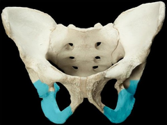
body of ischium

Ramus of ischium
joins the inferior ramus of the pubis anteriorly
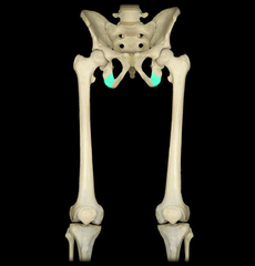
Upper body of ischium

Lower body of ischium

ischial spine
located superior to the ischial tuberosity and projects medially into the pelvic cavity

Greater sciatic notch
allows blood vessels and the large sciatic nerve to pass from the pelvis posteriorly into the thigh

Lesser sciatic notch
inferior to ischial spine

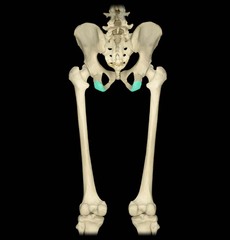
Ischial tuberosities
strongest part of hip bone, what we sit on
Pubis (Pubic bone)
most anterior portion of the hip bone
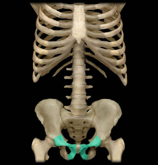
Body of pubis
adjacent to the pubic symphysis

Superior ramus
Extends anteriorly & medially from body of pubis

Symphysis pubis
a cartilaginous joint that is the point of fusion for two pubic bones

Inferior ramus
Pass down & posterior from symphysis pubis to meet the ramus of ischium

Obturator foramen
opening in hip bone formed by the pubic and ischial rami
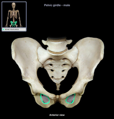
Acetabulum
hip socket, accepts head of femur to form hip joint

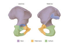
ilium
ischium
pubis
What 3 bones make up the acetabulum?
Brim of pelvis
Divides pelvic into two cavities, defined by superior portion of symphysis pubis anteriorly and the superior prominent part of the sacrum posteriorly

Greater or False pelvis
The general area above or superior to the oblique plane through the pelvic brim, the alae,(wings) of the ilia (ilium pleural) form the lateral and posterior limits of the false pelvis while the abdominal muscles of the anterior wall define the anterior limits

Lesser or True pelvis
Area inferior to the plane through the pelvic brim, A cavity surrounded by bony structures, Size and shape of the true pelvis is of greatest importance during the birth process

Inlet (superior aperture)
Oblique plane defined by the brim of the pelvis

Outlet (inferior aperture)
Defined by the two ischial tuberosities & the tip of the coccyx

The line between the two Ischial tuberosities and the coccyx
What forms the triangularly shaped outlet?

Cavity of the pelvis
Area between the inlet & outlet, During the birth process the baby travels through the inlet, cavity, & outlet of the true pelvis
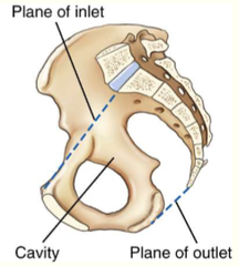
Head travels through the pelvic inlet, then through the cavity, and lastly the outlet
During routine birth, how does the baby travel? (what does it pass through)
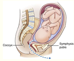
Ilium
Ischium
Pubis
Sacrum
Coccyx
What bones form the pelvic ring?

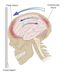
Contrecoup injury
Injury on the pole opposite the site of impact, typically seen in the pelvis or skull where the bones are tightly fixed together.
Gynecoid
Round

Platypelloid
Wider right to left than anterior to posterior

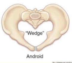
Android
Heart shaped
Anthropoid
Wider anterior to posterior than right to left

General shape
Angle of pubic arch
Ischial Spines
What's the difference between male and female pelvis?

Wider, Ilia are more flared, shallower front to back, round (gynecoid) and wider left to right rather than anterior to posterior (platypelloid)
What is the shape of a female pelvis?

Narrower & Deeper (Anthropoid), Less flared with heart-shaped pelvic inlet (android)
What is the shape of a male pelvis?

Male, less than 90°
Based off the angle of the pubis, is this a male or female pelvis? Why?

Female, obtuse angle greater than 90°
Based off the angle of the pubis, is this a male or female pelvis? Why?
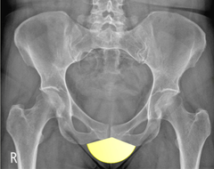
Male, more protrusion of ischial spine into inlet
Based off the protrusion of the ischial spines, is this a male or female pelvis? Why?

Female, less protrusion of ischial spine into inlet
Based off the protrusion of the ischial spines, is this a male or female pelvis? Why?

Avulsion fractures of the pelvis
These fractures cause extreme pain and are difficult to diagnose if not imaged properly. Fractures occur in adolescent athletes who experience sudden, forceful, or unbalanced contraction of the tendinous and muscular attachments. The force of tendons and muscles sliding over the tuberosities, ASIS, AIIS, superior corner of the symphysis pubis, and iliac crest may cause avulsion fractures.
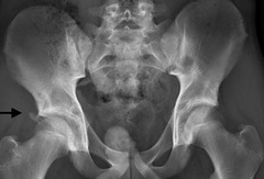
RIGHT Anterior Inferior Iliac Spine (AIIS)
1?
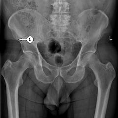
RIGHT Anterior Superior Iliac Spine (ASIS)
3?

Body of pubis
4?
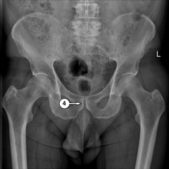
LEFT Iliac crest
5?

LEFT Illium
6?

LEFT Inferior ramus
7?

LEFT Ischial tuberosity
8?

RIGHT Ischial spine
9?

RIGHT Obturator foramen
10?

Symphysis pubis
11?

RIGHT Sacroiliac joint
13?
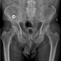
RIGHT Superior ramus
14?

radiopaque objects, clothing removed placed in gown , long hair, oxygen tubing, and pacemaker leads to avoid reduced detail/scatter
during patient preparation, what must be removed?

1. Limit the number of exposures
2. Collimation
3. shielding (led apron)
4. Position correctly
What 4 things help protect you and the patient against radiation?
BEFORE the examination
When must the anatomical marker be placed?
kVp: 80-90
mAs: Depends
Grid: yes
SID: 40in
What technical factors are needed for a good Pelvis (kVp, grid, mA, SID)?
Male: placed over testes (don't cover pubic or ischial areas)
Female: Ovarian contact shield (don't cover coccyx or sacrum)
What are the shielding guidelines for male and female pelvis?
AP
AP Bilateral Frog-Leg (Modified Cleaves Method)
Routine pelvis projections?
What is the evaluation criteria and procedure for an AP Pelvis X-ray?
SID: 40in
kVp: 80-90
Grid: Yes
CR: perpendicular, midway between ASIS and symphysis pubis (2in below ASIS)
Patient position: supine, separate legs and feet and internally rotate 15-20°
Breathing instructions: suspend respiration during exposure
Evaluation criteria: Pelvic girdle, L5, Sacrum & Coccyx, Femoral head, neck & greater trochanters, Lesser trochanters NOT visible, no rotation

What is the evaluation criteria and procedure for an AP Bilateral frog-leg pelvis (Modified Cleaves Method) X-ray?
SID: 40in
kVp: 80-90
Grid: Yes
CR: perpendicular, directed 3 inches below ASIS
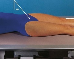
Patient position: supine, Flex both knees about 90 degrees and place plantar surfaces of feet together then let legs fall out laterally 40-45° from vertical
Breathing instructions: suspend respiration during exposure
Evaluation criteria: Femoral heads, neck & trochanteric areas, Acetabulum, no rotation, femoral heads and necks and greater and lesser trochanters should appear symmetric if both thighs were abducted equally

AP Axial Outlet Projection (Taylor method)
AP Axial Inlet Projection
Judet Method (Posterior Oblique Acetabulum)
Special pelvis projections?
What is the evaluation criteria and procedure for a AP Axial Outlet Projection X-ray?
SID: 40in
kVp: 80-90
Grid: Yes
CR: cephalad 20-35° for males and 30-45° for females, midline point 1-2 inches distal to superior border of symphysis pubis or greater trochanter
Patient position: supine, legs extended
Breathing instructions: suspend respiration during exposure
Evaluation criteria: Superior & Inferior rami of pubis, Body & ramus of ischium, no rotation, Demonstration of anterior & inferior pelvic bones with minimal foreshortening, no rotation

What is the evaluation criteria and procedure for an AP Axial Inlet Projection
X-ray?
SID: 40in
kVp: 80-90
Grid: Yes
CR: caudad 40°, midline at level of ASIS
Patient position: supine, legs extended
Breathing instructions: suspend respiration during exposure
Evaluation criteria: Pelvic ring (inlet)(superior aperture) in entirety, Ischial spines fully demonstrated & equal in size & shape, Proper angulation evidenced by superimposed anterior & posterior portions of pelvic ring, no rotation
What is the evaluation criteria and procedure for a Judet Method (Posterior Oblique Acetabulum) X-ray?
SID: 40in
kVp: 80-90
Grid: Yes
CR: BILATERAL AT PELVIC RING, UNILATERAL AT ACETABULUM
Acetabulum:
Affected side down: 2in distal & 2in medial to downside ASIS
Affected side up: 2in directly distal to upside ASIS
Pelvic Ring:
2in inferior to level of ASIS & 2 in medial to upside ASIS
Patient position: Semisupine (affected side up or down), 45-degree oblique (thorax & pelvis) RPO/LPO
Breathing instructions: suspend respiration during exposure

Evaluation criteria for Judet method
Centered to DOWNSIDE acetabulum: Anterior rim of acetabulum, Posterior ilioischial column
Centered to UPSIDE acetabulum: Posterior rim of acetabulum, anterior iliopubic column, Obturator foramen
Pelvic ring: All anatomy demonstrated from both types of centering,
Proper Positioning: Upside obturator foramen open, downside closed, Acetabulum or pelvis centered to IR
1. Anterior rim of acetabulum
2. Posterior ilioischial column
What structures are seen on a Judet Method (Posterior Oblique Acetabulum) X-ray centered to DOWNSIDE acetabulum?

1. Posterior rim of acetabulum
2. Anterior iliopubic column
3. Obturator foramen
What structures are seen on a Judet Method (Posterior Oblique Acetabulum) X-ray centered to UPSIDE acetabulum?

Inlet Pelvis
What kind of projection is this?

AP Pelvis
What kind of projection is this?

Outlet Pelvis
What kind of projection is this?

Judet Pelvis
What kind of projection is this?

Bilateral Frog-leg Pelvis (Modified Cleaves)
What kind of projection is this?

Femur
Largest & Strongest bone in the body, Entire weight is transferred through this bone & associated joints