Radiographic Positioning - Lower Extremity
 Call Kai
 Learn
Learn Practice Test
 Spaced Repetition
 Match
Match Flashcards
 Knowt Play
1/66

There's no tags or description
Looks like no tags are added yet.
Name | Mastery | Learn | Test | Matching | Spaced | Call with Kai | Chat |
|---|
No analytics yet
Send a link to your students to track their progress
67 Terms
Medial Cuniform, right foot
Identify the number (9) in the image below

Navicular, right foot
Identify the number (10) in the image below

Talus, right foot
Identify the number (11) in the image below

Proximal Interphalangeal joint of the fifth digit of the right foot
Identify the number (15) in the image below

Tuberosity of the fifth metatarsal
Identify the number (16) in the image below

Tarsometatarsal joint space TMT
Identify the number (17) in the image below

Lateral Cuniform of the right foot
Identify the number (18) in the image below

Cuboid
Identify the number (19) in the image below

Calcaneus
Identify the number (20) in the image below

Base of the third metatarsal
Where is the central ray placed for a plantodorsal axial projection of the calcaneus?
Dorsoplantar projection
What is another term for the AP projection of the foot?
15 degrees posterior
What CR angle is generally required for the AP standing projection of the foot?
Calcaneus
What is the largest and strongest tarsal bone?
Intermediate cuneiform
The smallest of the cuneiform.
Cuboid
Identify the bone (K) in the image below.

Distal phalanx, first digit, right foot
Identify the bone (A) in the image below.

Interphalangeal joint, first digit, right foot
Identify the bone (B) in the image below.

Proximal phalanx, first digit, right foot
Identify the bone (C) in the image below.

Proximal Interphalangeal joint, first digit, right foot
Identify the bone (D) in the image below.

Medial cuniform
Identify the bone (F) in the image below.

Navicular
Identify the bone (G) in the image below.

Talus
Identify the bone (H) in the image below.

Calcaneus
Identify the bone (I) in the image below.

Lateral Cuniform
Identify the bone (J) in the image below.

Proximal interphalangeal joint
Identify the bone (L) in the image below.

30 degree medial oblique foot
Identify the image below.

Tuberosity of the base of the fifth metatarsal
Identify the area of the foot that has the fracture. The arrow points to it.

Mortise joint
An anterioposterior (AP) oblique ankle projection with the lower extremity rotated medially 15 to 20 degrees demonstrates the:
In 45-degree medial rotation
For the proximal tibiofibular articulation to be opened, the knee must be positioned:
Intercondylar fossa
Examine the image below. What is the primary anatomy of interest?

AP oblique projection of the knee, lateral rotation position
Examine the image below. What projection and position are demonstrated?

Distal Femur
Examine the image below. What anatomy is indicated by the number 1?
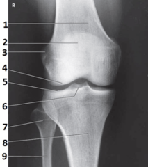
Patella
Examine the image below. What anatomy is indicated by the number 2?

Lateral Epicondyle
Examine the image below. What anatomy is indicated by the number 3?

Lateral Condyle
Examine the image below. What anatomy is indicated by the number 4?

Lateral tibial plateau
Examine the image below. What anatomy is indicated by the number 5?

Intercondylar eminence
Examine the image below. What anatomy is indicated by the number 6?
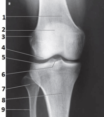
Head of the fibula
Examine the image below. What anatomy is indicated by the number 7?

Proximal Tibia
Examine the image below. What anatomy is indicated by the number 8?

Shaft of the fibula
Examine the image below. What anatomy is indicated by the number 9?

Tangential patella (Settegast method)
Examine the image below. Identify the projection, position, and, if appropriate, method.

Projected over the posterior aspect of the distal tibia.
With a true lateral of the ankle, the lateral malleolus is:
Rotate the lower leg/foot medially 45 degrees
To demonstrate the ankle tibiofibular joint, you would need to:
Condyles
Which femoral structures articulate with the tibia?
0 degrees
The central-ray angle for a lateral projection of the ankle is:
Lateral condyle of the tibia
Identify the anatomy indicated by the number (8).

Medial epicondyle of the femur
Identify the anatomy indicated by the number (1).

Medial condyle of the femur
Identify the anatomy indicated by the number (2).

Medial condyle of the tibia
Identify the anatomy indicated by the number (3).

Tibia
Identify the anatomy indicated by the number (4).

Medial malleolus
Identify the anatomy indicated by the number (5).

Lateral epicondyle of the femur
Identify the anatomy indicated by the number (6).

Lateral condyle of the femur
Identify the anatomy indicated by the number (7).

Head of the fibula
Identify the anatomy indicated by the number (9).
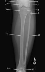
fibula
Identify the anatomy indicated by the number (10).

Lateral malleolus
Identify the anatomy indicated by the number (11).

Medial malleolus
Identify the anatomy indicated by the number (7).

Fibula
Identify the anatomy indicated by the number (1).

Tibia
Identify the anatomy indicated by the number (2).

Tibiofibular joint space
Identify the anatomy indicated by the number (3).

Malleolar fossa
Identify the anatomy indicated by the number (4).

Lateral malleolus
Identify the anatomy indicated by the number (5).

Inferior articular surface
Identify the anatomy indicated by the number (6).

Talus
Identify the anatomy indicated by the number (8).

Osgood-Schlatter disease
Which disease process of the lower extremity is defined as an incomplete avulsion of the tibial tuberosity?
15-to-20 degree medial oblique
For the mortise joint to be demonstrated, the ankle must be positioned in:
40 degrees cephalad
What was the central ray orientation for this projection?
