Exam 2 material Muscles kinesology
 Call Kai
 Learn
Learn Practice Test
 Spaced Repetition
 Match
Match Flashcards
 Knowt Play
1/49

There's no tags or description
Looks like no tags are added yet.
Name | Mastery | Learn | Test | Matching | Spaced | Call with Kai | Chat |
|---|
No analytics yet
Send a link to your students to track their progress
50 Terms
Atlas + bony markings
C1, has a larger articulating surface superiorly to meet with the skull, facet for the dens (C2) to articulate,

C2 Axis + Bony markings
Vertical process on the superior side (DENS) to extend and articulate with C1, lets C1 to spin on top of C2

Joints of CS-atlanto occiptial joint-
atlanto occipital joint- skull and atlas
headflexion and extension (think double chin, head motion, not great range)
Joints of CS- AtlantoAxial
atlas and axis articulate with the dens, cervical rotation, pivot joint

Joints of the CS- Facet joints
between the transverse processes, C2-C7, the rest of the spinal column, functional=diarthrodial, structural=synovial, gliding joints with minimal movement, built for stability and not for mobility
motions: flexion/extension, lateral bending, rotation

Lordosis vs Kyphosis
Lordosis is the convex anterior in middle curvature in C spine, important for force distribution and shock absorption
Kyphosis is what causes the thoracic spine to bend outward, producing a rounded or “hunchback” appearance.

Fibrocartilage discs
joint type- amphidiplrodal, think jelly donut analogy
contains an outer layer=annulus, center jelly layer= nucleus pulposus; layers are important for shock absorption
CS: neuromuscular
The spinal cord goes through foramen in each vertabrae; the nerve root runs through the intervertebral foramen and decreases in size as it branches, sending/receiving sensory and motor information.
CS Muscles: SCM- O, I, action
sternocleidomastoid
originates at the sternum/clavicle
inserts at the mastoid process
actions: head extension/ cervical flexion
Ipsilateral lateral bending
contralateral rotation
elevation of sternum with deep inspiration

CS MUSCLES: Scalene
Anterior: Originates C3-6
Insertion on 1st rib
actions= neck flexion/lateral bending
Middle: Originates C1/2-7
Insertion on 1st rib
Actions = neck lateral bending and first rib elevation
Posterior: Originates at the C4-6/ C5-7
Insertion on the second rib
Actions: 2nd rib elevation

Splenius (Cervicis and capitus combinded)
On the posterior CS/TS
Originates from C7-T6
at the occipital bone, mastoid process and C1-C3
actions Head/cervical extension
ipsilateral lateral bending and rotation

Levator Scapulae
Originates at the C1-C4
Insertion at the medial border of the scapula on the superior angle for scapular elevation

Thoracic spine bones- Vertebrae
12 vertebrae, no transverse foramin, have a costal facet to articulate with the ribs

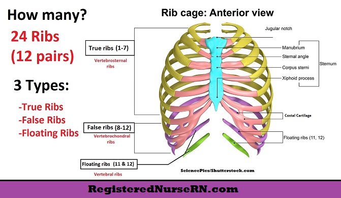
Thoracic spine bones: Ribs
12 pairs for the 12 thoracic vertebrae
True ribs (1- 7)- protect and support the internal organs like the heart and lungs and are directly supported by the sternum
False Ribs (8-10) Protect of organs like the spleen and stomach
Floating ribs (11-12) guard organs like kidneys, are not connected all the way around
Thoracic spine JOINTS- Facet Joints
Joints between the vertebrae, T1-12, these ‘synch’ with C7 and L1, these are Diarthrodial Synovial Gliding joints
Actions: Flexion and extension, lateral bending and rotation

Thoracic spine JOINTS- Costovertebral
joint between the ribs and vertebrae, Diarthrodial synovial gliding joints that can flex and extend

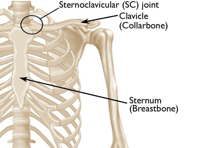
Thoracic spine JOINTS-Sternoclavicular
Joint between the sternumn and clavicle
diarthrodial synovial gliding joint, protraction and retraction @15*, Elevation @45* and depression @15*
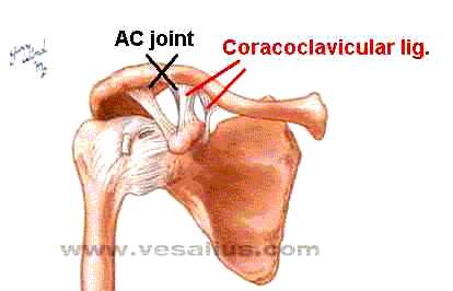
Thoracic spine JOINTS- Acromioclavicular
AKA AC joint, Between the acromion process and the clavicle, diarthrodial synovial gliding, can perform gliding and slight rotation as needed with shoulder movement

Thoracic spine JOINTS- Coracoclavicular
between the coracoid process and the clavicle, a diarthrodial synovial gliding joint, disperses load and stabilizes as needed with shoulder movement
Thoracic spine JOINTS- Scapulothoracic
Between the scapula and the ribcage (thoracic spine)
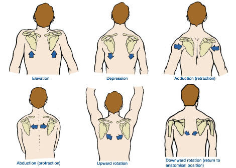
False diarthrodial joints, glides for protraction/retraction, elevation/depression, upward rotation/downward rotation

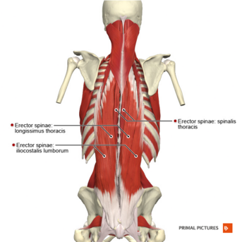
Erector Spinae
Origin= lumbar inferior thoracic aponeurosis (thick band of CT instead of aponeurosis
insertion (3 parts)
Spinalis= superior thoracic
Longissimus = C2-6 Mastoid process
Iliocostalis= Ribs
Actions= spine extension, lateral bending of spine this is specific to the area of pull
Serratus anterior
Orgin-Posterior Lateral Aspect Ribs 1-9
Insertion= Medial Border of Scapula
Actions= scapular stabilozer, protraction of scapula, upward rotation of scapula, (winging scapula applied, why would a weak serratus anterior cause winging scapula?

Internal vs external intercostals
internal intercostals depress the ribs for exhalation, squeezing out excess air
External intercostals elevate the ribs for inhalation they elevate ribs

Pectoralis Major
Origin on clavicle and sternum, plus costal cartilage
Insertion on the lateral humerus
actions
adduction and IR of humerus
flexion, adduction when <90, abduction when >90*

Pectoralis Minor
Origin - Ribs 3-5
Insertion - Coracoid Process
Action - Scapular stabilization, protraction, downward rotation, depression

Trapezius
Origin: C7- T12, skull (occipital)
Insertion: spine of scapula, acromion, clavicle
Actions- lateral bend and extension of the head, elevate the scapula
Upper trap: scapular elevation/extension/rotation of head
middle fibers elevate and retract scapula
lower fibers depress and retract the scapula

Rhomboids
Origin- C7-T5
Insertion- medial border of scapula
Actions- all scapular
Retraction, elevation w/ retraction, downward rotation from upwardly rotated position

Latissimus Dorsi
Origin= T7-L5, Iliac crest, and sacrum
Insertion= Medial humerus
Actions- adduct, extend IR arm, stabilize, and rotate trunk

bones of shoulder girdle
Clavicle (anterior/superior)
Scapula (posterior and superior)
Ribs (medial)
Sternum (rib stability and muscle attachment)

what is the ratio of the scapulohumeral rhythm
1 part scapulothoracic
2 part GH
180 of shoulder abd =120 glenohumeral+60 scapular
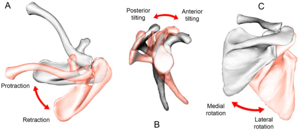
Arthrokinematics of the scapula- Lateral Tilt
Protraction
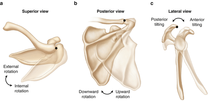
Arthrokinematics of the scapula- Medial Tilt
Retraction
Arthrokinematics of the scapula- extension
anterior tilt

Arthrokinematics of the scapula- flexion
posterior tilt
GH muscles Actions- Latissimus Dorsi
Humeral Adduction, humeral extension, humeral IR, humeral Horizontal abduction

GH muscles Actions- Deltoid
Anterior- GH Abduction, flexion, internal rotation, horizontal adduction
Middle- GH abduction
Posterior- GH Abduction, extension, extenral rotation, horizontal abduction
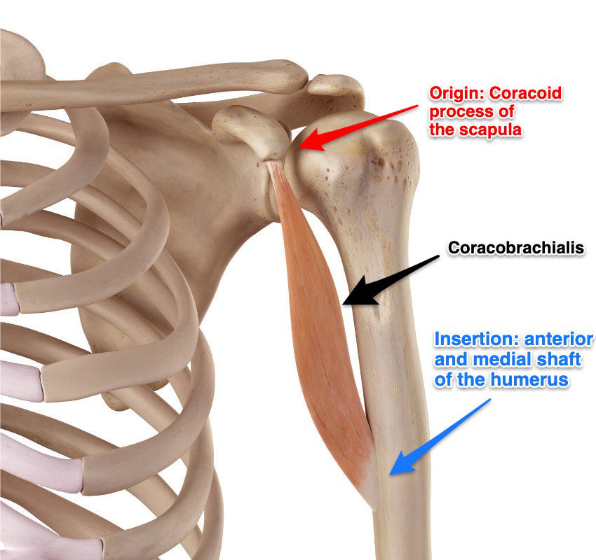
GH muscles - Coracobrachialsis
Origin- Coracoid process of the scapula
Insertion- middle of the border of the humeral shaft
actions - GH Flexion, GH Horizontal Adduction, GH Adduction
GH muscles Actions- Teres Majoir
GH Adduction, GH Extension, GH Internal Rotation

Rotator Cuff Muscles - Supraspinatus
Orgin- Supraspinatus fossa, superior posterior scapula
insertion- greater tubercle of humerus
Actions- GH Abduction, main agonist first 0-15 degrees of abduction, GH stabilization

Rotator Cuff Muscles- Infraspinatus
Origin- posterior medial scapula medial border
Insertion- posterior lateral humerus, greater tubercle
Actions- GH Horizontal abduction, GH ER, GH stabilization

Rotator Cuff Muscles- Teres Minor
Orgin- Posterior Scapula lateral boarder
Insertion- Posterior Lateral Humerus Greater Tubercle
Actions- GH ER, GH Addiction, GH stabilization

Rotator Cuff Muscles- Subscapularis
ORgin- Subscapular fossa anterior scapula, medial boarder
Insertion- Lesser tubercle of the Humerus
Actions- GH IR, GH Adduction, GH Stabilizaiton- Arm raised pulls arm forward and down, scapular neuromuscular control

elbow bones
Humerus, ulna, radius
what are the two elbow joints
both diarthrodial synovial joints
Humeroulnar- medial
radiohumeral- lateral more joint space, less bone on bone more soft tissue provides more mobility
HINGE JOINTS PROVIDIN ELBOW FLEXION AND EXTENTION

Forearm Syndesmosis
interosseous membrane between the ulna and radius, diarthrodial synovial pivot joint, the radius pivots on the ulna for pronation and supination, some stability some moibility

Elbow ligaments- extension
bone on bone locking olecranon process in the fossa, CLOSED PACKED POSITION, less mobile more stable
Elbow flexion
Less stabile more mobile, Ligament support for force transmission/mobility
Ulnar collateral ligament (medial) connects the ulna and humerus in 3 parts, protects from valgus stress (outside in) esp in overhead elbow ext.
Radial Collateral ligament- connects the radius and humerus protect from varus stress- middle out, helps prevent excess lateral rotational instability
Annular Ligament
goes around the head of ligament, hugs radius to the ulna
Elbow muscles- Bicep Brachii
Long- Origin- supra glenoid tubercle- labrum, Insertion- Radial Tuberosity
Short- Origin- Coracoid Process Insertion- Bicep Aponeurosis
Action- Humeroulnar flexion, radioulnar supination, weak GH flexion (elbow flexion)

Elbow Muscles- Brachialis
Origin- Distal Anterior humerus
Insertion- Coracoid Process of Ulna
Actions summarized- Humeroulnar flexion (reverse grip curls)