BISC205 Topic 4
 Call Kai
 Learn
Learn Practice Test
 Spaced Repetition
 Match
Match Flashcards
 Knowt Play
1/44

Earn XP
Description and Tags
we are so cooked
Name | Mastery | Learn | Test | Matching | Spaced | Call with Kai | Chat |
|---|
No analytics yet
Send a link to your students to track their progress
45 Terms
what is a gastrovascular cavity
found in small or thin animals to exchange materials directly with the surrounding medium. it is a central cavity that serves as a two-way digestive tract with a single opening for both food intake and waste removal, while also facilitating the circulation of nutrients and fluids throughout the body.

examples of organisms with a gastrocavity
flatworms, jellyfish, and cnidarians
primary functions of the circulatory system (there are 5)
1) transport oxygen and nutrients to actively metabolizing tissues
2) remove carbon dioxide and other waste products from the tissues
3) transport signalling molecules (including hormones) and immune cells throughout the body
4) protection and healing (ex/ white blood cells, antibodies, platelets)
5) regulation of body temperature: by controlling blood flow to the skin through vasodilation (widening blood vessels) and vasoconstriction (narrowing blood vessels)
open circulatory system vs closed circulatory system: blood
open circulatory system: blood flows freely within body cavities (or sinuses) travelling in any direction to bathe the organs directly. it is pumped in a body cavity called hemocoel, where blood mixes with interstitial fluid to make hemolymph
closed circulatory system: blood is in vessels and is separated from interstitial fluid. it travels in one direction in the blood vessel.
what kind of animals is the open circulatory system best suited for?
animals with a lower metabolic rate.
although the blood pressure is low and oxygen take longer to reach extremities, their need for oxygen is less.
examples: arthropods, bivalves, and snails
what kind of animals is the closed circulatory system best suited for?
animals with a higher metabolic rate.
blood pressure remains high, which means oxygen takes a shorter amount of time to get to body cells in extremities. this is great for animals with a greater need for oxygen.
examples: vertebrates (mammals, fish, birds, reptiles, amphibians), and some invertebrates (earthworms, octopuses, and squids)
open CS vs closed CS: heart shape and function
open circulatory system:
heart is sac-like or tubular
it has Ostia which closes as the heart contracts and opens as the heart relaxes
neurogenic heart
suction pump heart
closed circulatory system:
chambered heart with muscular ventricles
muscular ventricles pump blood in the body with pressure
myogenic heart
pressure pump heart
why does open CS have a suction pump heart and why does closed CS have a pressure pump heart
open CS has a suction pump heart because it acts as a vacuum when blood is sucked into the heart through the ostia
closed CS has a pressure pump heart because the ventricles pump blood in the body with pressure
open CS vs closed CS: neurogenic vs myogenic hearts
open CS has a neurogenic heart because the beating of the heart is set through nerve impulses.
closed CS has a myogenic heart because the rhythm is set in neuromyocardinal cells. the contractions of these hearts are produced spontaneously without requiring external stimulation from nerve cells
Open CS: advantages and disadvantages
advantages
requires less energy for distribution of circulatory fluid
makes animals much less vulnerable to pressure (good for animals that live deep in the sea)
less oxygen is needed since organisms with this system have a lower metabolic rate
disadvantages
less efficient in moving oxygen and nutrients to all body tissues and cells (not good for big organisms)
has low blood pressure, difficult to reach extremities
limited capability to increase or decrease distribution and velocity of blood flow (no vasodilation or vasoconstriction)
better for lower metabolic rates mean slower movements and weaker adaptability to new environments
closed CS: advantages and disadvantages
advantages
more efficient in delivering oxygen and nutrients throughout organism to tissues and cells
more powerful in pressure so blood reaches all extremities of the body
efficient distribution of antibodies, immune responses are stronger, helps body fight off infection effectively
has more capability to increase or decrease distribution and velocity of blood flow
better to sustain higher metabolic rates means faster movements and better adaptability to new environments
disadvantages
requires more energy for distribution of blood (work harder to pump blood faster)
blood is contained in vessels, so a severe injury can resumer in rapid and big blood loss
high pressure system is vulnerable to physical strain and blockages/blood clots in the blood vessels
single circulatory vs double circulatory
single circulatory
1) blood passes through hear once on each loop throughout the body
2) found in bony fishes, rays, and sharks
3) blood leaves heart through arteriosis in bony fish and conus arteriosus in elasmobranches
4) lower blood pressure
double circulatory
1) blood passes heart twice on each loop around the body
2) found in amphibians, reptiles, and mammals
3) deoxygenated and oxygenated blood are pumped separately from each other
4) higher in blood pressure
pulmocutaneous circuit vs pulmonary circuit vs systemic circuit
pulmocutaneous circuit: the circuit through which the oxygen poor blood flows to pick up oxygen through the lungs and skin. found in amphibians
pulmonary circuit: the circuit through which the deoxygenated blood flows to pic up oxygen through the lungs. found in reptiles and mammals
systemic circuit: the circuit through which oxygenated blood delivers oxygen
why do amphibians require a double circulatory system
they need a double circulatory system because they need both a pulmocutaneous circuit and a systemic circuit. the pulmocutaneous circuit will pick up oxygen through the lungs AND skin. This means that when they are underwater, they can redirect blood to the skin for oxygen pickup instead of the lungs.

why do reptiles, mammals, and birds require a double circulatory system
they need a double circulatory system because they need a pulmonary circuit and a systemic circuit because they cannot breathe through their skin, so all gas exchange occurs through the lungs.
additionally, they need higher blood pressure that comes from a circulatory system because they are bigger animals, so their extremities can receive oxygen faster
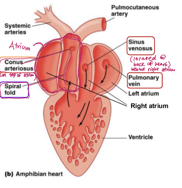
animals with single circulation and the # of atria and ventricles in their hearts
animals: bony fishes and elasmobranchs (sharks, rays, and skates)
number of atria and ventricles: 1 atria and 1 ventricle

fill in the blank: single circuit system
important note:
blood leaves the heart via the bulbs arteriosus in bony fish
blood leaves the heart via the conus arteriosus in sharks, rays, and skates
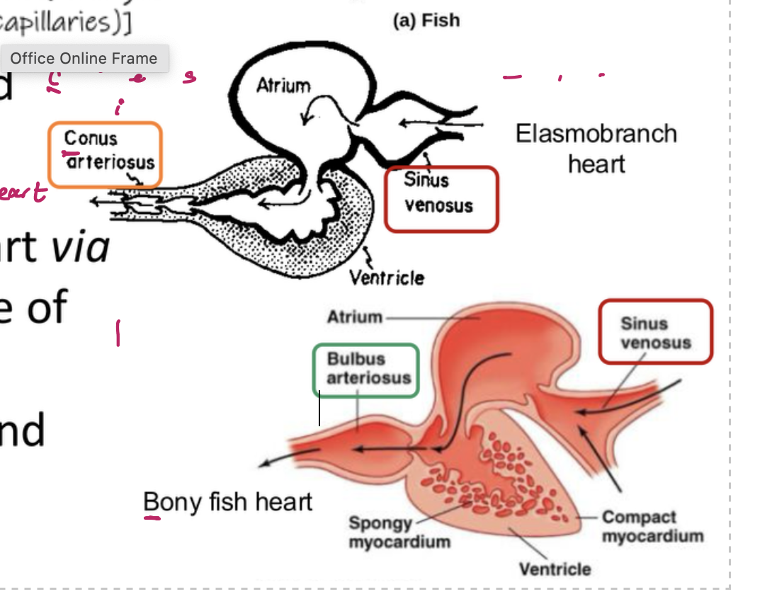
double circuit functions: sinus venous, pulmonary vein, conus arteriosus, and spiral fold
sinus venosus: brings deoxygenated blood from body into right atrium
pulmonary vein: brings oxygenated blood from lungs/skin into the left atrium
conus arteriosus: pumps blood into both pulmoncutaneous and systemic circuits of circulatory system
spiral fold: a hinged, flap-like structure in the conus arteriosus that
1) minimizes mixing of oxygenated and deoxygenated blood
2) helps direct oxygenated blood to systemic circuit and deoxygenated blood to pulmocutaneous circuit
structure of reptilian heart
most reptiles have 3 chambered hearts that include 2 atria and 1 ventricle divided (by partial septa) into three interconnected subchambers
there is mixing of deoxygenated and oxygenated blood
They have the unique structures of
1) CAVUM ARTERIOSUM
2) CAVUM PULMONALE
3) CAVUM VENOSUM
structure of crocodilian heart
crocodilian hearts have 4 chambered hearts that include 2 atria and 2 ventricles (which are separated by a complete septum)
there is mixing of deoxygenated and oxygenated outside of heart
they have a small opening (with a valve-like structure) located at the based of right and left aortas. this is called the FORAMEN OF PANIZZA
structure of amphibian heart
amphibians like frogs, have 2 atria and 1 ventricle (no septum). the two atria receive blood from pulmoncutaneous circuits and systemic circuits.
there is some mixing of the blood in the heart’s ventricle, which reduces efficiency of oxygenation.
this animals has the unique structure of
1) CONUS ARTERIOSUS
2) SPIRAL FOLD
structure of mammal and bird heart
mammals and birds have a four chambered heart, which includes 2 atria and 2 ventricles.
there is no mixing of oxygenated and deoxygenated blood
they have a pulmonary circuit and a systemic circuit

fill in the blanks


fill in the blanks

differences between veins and arteries
veins
under low blood pressure
thin-walled compared to artery of similar size (higher compliance)
blood flows to heart
most veins contain deoxygenated blood, except pulmonary vein
blood moves one direction due to one-way valves, skeletal muscle contraction and smooth muscle contraction
situated near surface of skin
larger lumen compared to artery of the same size
artery
under high blood pressure
thick walled compared to vein of similar size
blood flows away from the heart
most arteries contain oxygenated blood
blood moves in one direction due to high blood pressure and smooth muscle contraction
situated deeper in body
smaller lumen compared to vein of similar size

the three tunica layers
1) tunica intima: inner most layer
2) tunica media: middle layer
3) tunica externa/adventitia: outermost layer
tunica intimate: innermost layer
inner lining is called vascular lining which is supported by internal elastic membrane
provides frictionless pathway for movement of blood

tunica media: inner layer
composed of smooth muscle and elastin
controls vasoconstriction and vasodilation, which regulate body temperature and blood pressure

tunica externa/adventitia
composed of collagen fibres to help support and reinforce shape of blood vessel
has a vasa vasorum and nervi vasorum

differences between arteries and veins: tunica layers
arteries
tunica intima
endothelium is wavy
internal elastic membrane present
tunica media
tunica media is the thickest layer
external elastic membrane is present
veins
tunica intima
no internal elastic membrane because veins are low pressure and no need to stretch like arteries
tunica media
thinner than tunica externa (less smooth muscles = higher compliance of veins)
external elastic membrane is absent in veins (lower elastin = higher compliance of veins)
tunica externa
the thickest in veins bc it prevents walls from collapsing under low blood pressure and it protects from damage
how does blood pressure/hydrostatic pressure and osmotic pressure drive fluid in the capillaries? this involves the arterial end and the venous end
if there is a higher blood pressure than osmotic pressure at the arterial end, this drives fluid from the arterial end into the tissues, and into the venous end of the capillaries
if there is a higher osmotic pressure than blood pressure at the venous end, this drives fluid out from the tissues and into the venous end

law of bulk flow
important notes:
bulk flow occurs when an external force is applied to the fluid
pressure gradient refers to perfusion pressure (Pinlet - Poutlet)

resistance of blood flow

Poiseuille’s equation

assumptions and violations of poiseuille’s equation
assumptions:
tubes are unbranched and rigid
slow is simple and fluid is moving steadily
violations
vessels are actually branched and distensible, changing their diameter as pressure changes
flow us pulsatile (increases and decreases with heartbeat)
fluid is not simple (blood is made of plasma and cells)

perfusion pressure vs transmural pressure
perfusion pressure: pressure gradient refers to perfusion pressure, which is the difference between inlet and outlet pressure
trasmural pressure: the difference between inside and outside pressure

blood velocity

law of Laplace (does not consider thickness of blood vessel wall)

law of laplace (considers thickness of blood vessel wall)

cardiac output equation 1
CO = a measurement of the amount of blood pumped out of each ventricle in one minute (L/min or mL/min)
HR = Heart rate, beats per minute
SV = stroke volume, the volume of blood pumped out of each ventricle per beat

cardiac output equation 2
CO = a measurement of the amount of blood pumped out of each ventricle in one minute (L/min or mL/min)
EDV = the maximum volume of blood in the ventricle at rest. after atrial systole, blood is pumped into the ventricles until they reach EDV
ESV = after ventricular systole, the volume of blood remaining in the ventricle

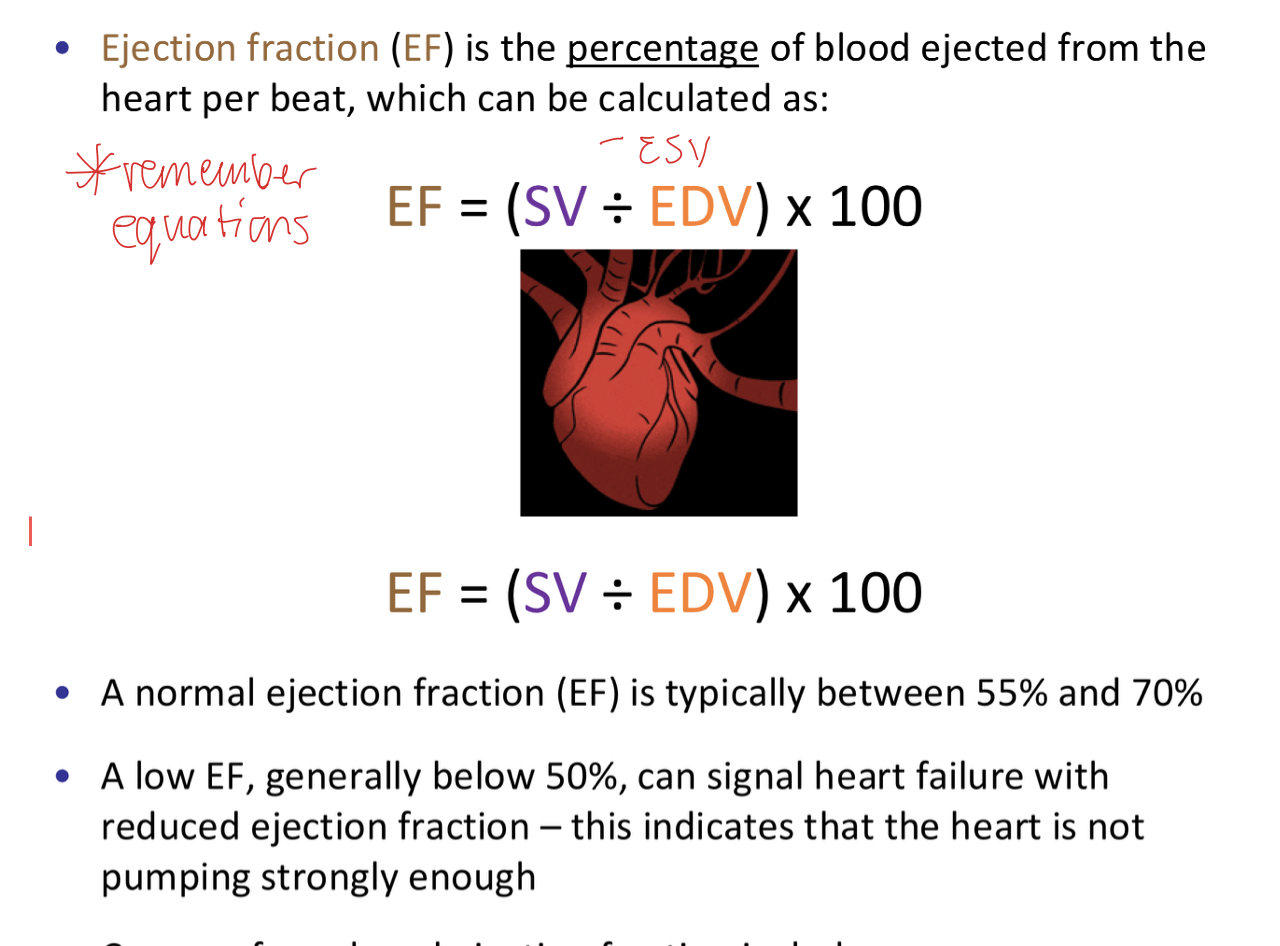
ejection fraction equation
EF = the percentage of blood ejected from the hear per beat
EDV = the maximum volume of blood in the ventricle at rest. after atrial systole, blood is pumped into the ventricles until they reach EDV
ESV = after ventricular systole, the volume of blood remaining in the ventricle
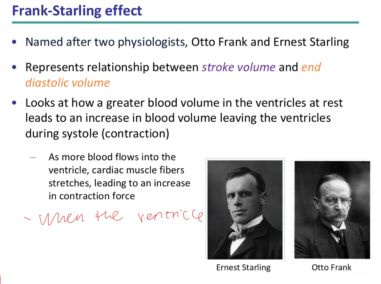
what is the frank starling effect?
the effect in which a greater blood volume in the ventricles at rest leads to an increase in blood volume leaving the ventricles during systole (contraction). this is because as more blood flows into the ventricle, cardiac muscle fibres stretches, leading to an increase in contraction force
why is it not desirable to have an 100% ejection fraction using equation
in a healthy heart, ejection fraction will not reach 100% because ventricles do not completely empty with each heartbeat. the heart’s ventricle would need to remove every drop of blood!

why is it not possible to have an 100% ejection fraction using frank sterling effect
the frank sterling effect has an upper limit. if your cardiac muscle fibres overstretch by an excessive amount of EDV, they would have stretched beyond the optimal length. this decreases contractile force, and thus SV. This makes it impossible to achieve a 100% ejection fraction
