Neuropath and infectious dz
 Call Kai
 Learn
 Practice Test
 Spaced Repetition
 Match
 Flashcards
 Knowt Play
1/138

There's no tags or description
Looks like no tags are added yet.
139 Terms


What is the term for a brain without gyri?
Lissencephaly
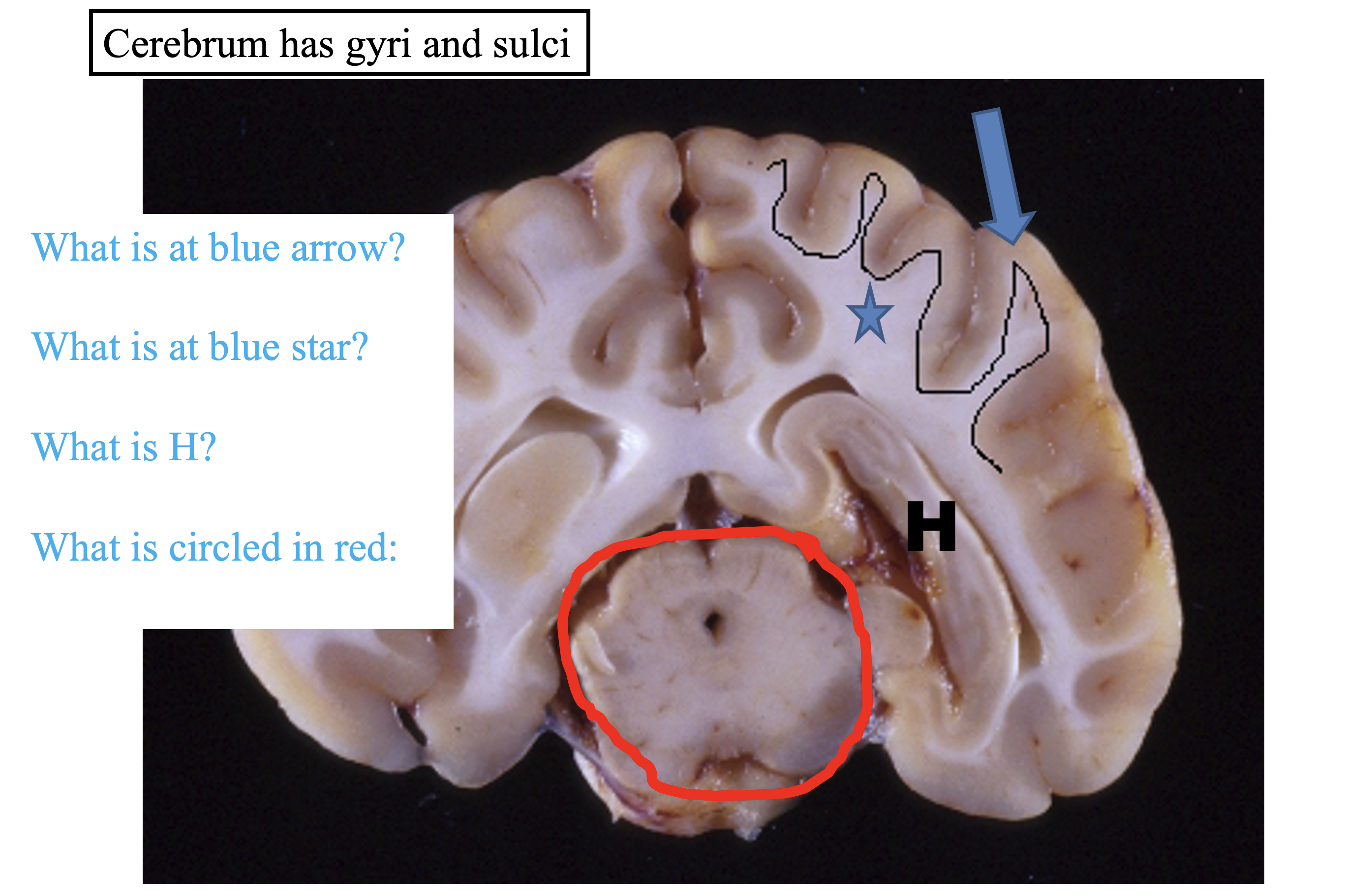

What is in gray matter?
Neurons and neuropil
What is in white matter?
Myelin and axons
Layers of the cerebellar cortex
External granular layer (not present on this image)
Molecular layer - light pink
Purkinje cell layer - large cells on border of pink and purple
Internal granular layer - purple layer
**Inner pink layer is white matter!!!**

Where is the lesion located and is it a polioencephalopathy or a leukoencephalopathy?
Cerebral cortex
Polioencephalopathy
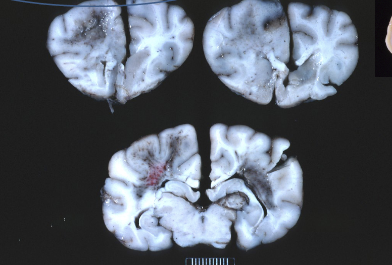
Where is the lesion located and is it a polioencephalopathy or a leukoencephalopathy?
Corona radiata
Leukoencephalopathy
What is malacia?
Softening and liquefactive necrosis of neuroparenchyma

Morphologic diagnosis
Leukoencephalomalacia

Morphologic diagnosis
Poliomyelomalacia
Name the meninges
Dura mater - attached to bone in cranium, not attached to bone in SC
Arachnoid mater - avascular
Pia mater - where vessels are located
What are the leptomeninges
Arachnoid mater and pia mater

What is this
Dura mater

Morphologic diagnosis
Focal symmetrical encephalomalacia
Inflammation of the dura mater
Pachymeningitis

Where is the lesion
Cerebellopontine angle

What is this, what does it do, and what is inflammation here called
Choroid plexus
Produces CSF
Choroid plexitis

Normal CNS neurons

Chromatolysis - cell body swollen with loss of Nissl substance
Degenerative change associated with axonal injury

Ischemic neuronal necrosis
Can be seen with toxins, seizures, degenerative disease
What is the structure separating the lateral ventricles?
Septum pellucidum

What happened here?
Hydrocephalus happened

Neuronophagia
Piecemeal phagocytosis of dead neurons
Can be seen with neuronal infection

What is this inclusion called
Negri body
From a cow with rabies

Spongiform encephalopathy
due to Prion disease
What cells produce myelin in CNS?
Oligodendrocytes
What cells produce myelin in the PNS?
Schwann cells
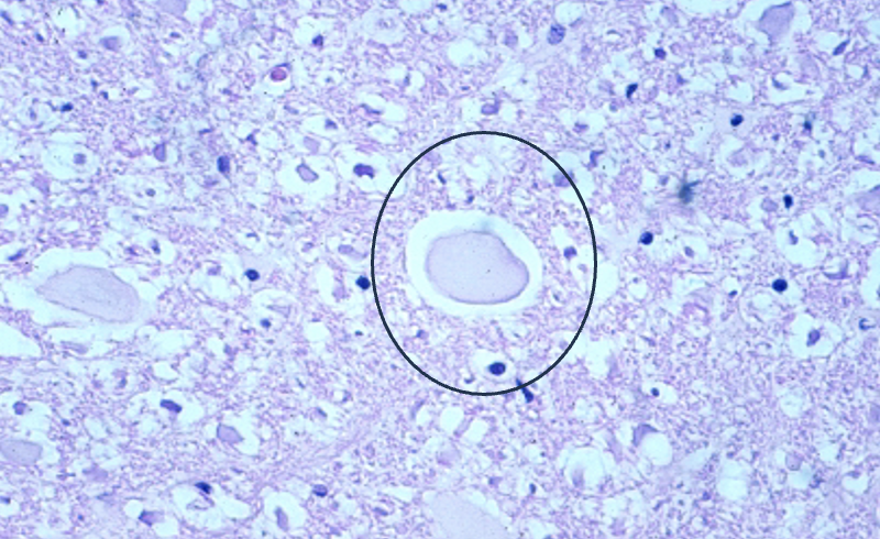
Spheroid (swollen axon)

What is a hydrocephalus lesion in the spinal cord called?
Hydromelia

Wallerian degeneration
Axons die and shrink distal to damage
Remnants of myelin sheathes form vacuoles and “digestion chambers”
Functions of astrocytes
“Brain glue”
Structural support - fibrous tissue
Repair
Blood-brain barrier
Increase in number of astrocytes
Astrocytosis

Increase in astrocytic cell processes
Astrogliosis
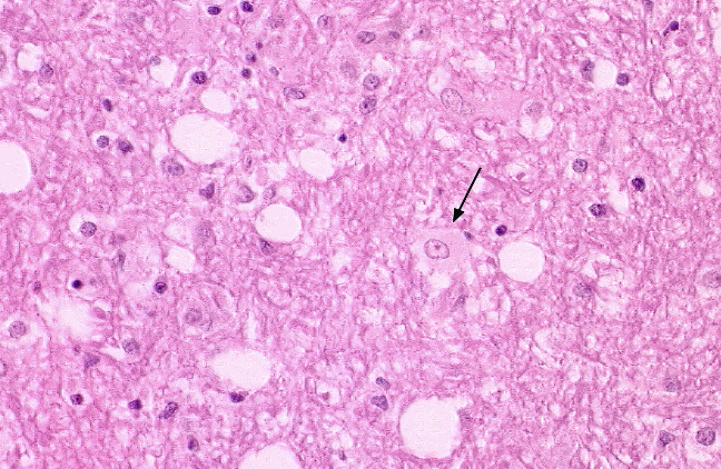
Gemistocyte - reactive astrocyte with visible eosinophiolic cytoplasm

Alzheimer type II cell
Astrocyte with clear, swollen nuclei
Seen in hepatic and renal encephalopathies
Role of oligodendrocytes
Myelin formation in CNS

Demyelination
Function of microglia
Macrophages in brain
Phagocytic
Immune defense

What is the single layer of epithelium lining the ventricles and central canal?
Ependyma

Ependymitis - inflammation and necrosis of ependyma
Causes obstructive hydrocephalus
Arrow is pointing to irregular surface in cerebral aqueduct

Microgliosis - proliferation of microglia
Focal proliferation = glial nodule → viral encephalitis
What are gitter cells?
Foamy, lipid-laden macrophages in brain
Have ingested a lot of stuff
Sources of gitter cells
Circulating macrophages
Resident microglia

Where are these hematomas
Epidural

Where are these hematomas
Subdural

Where is this hematoma
Subarachnoid
Term for hemorrhage in the spinal cord
Hematomyelia
What type of lesion is associated with an in-utero BVD infection in a calf?
Hypomyelinogenesis
What is hypomyelinogenesis
Deficient myelin formation
What is leukodystrophy
Abnormal myelin formation or maintenance
Ex) Globoid cell leukodystrophy (Krabbe’s disease)
What is demyelination
Removal of normal myelin
Primary: removal of myelin only, no axon damage - often inflammatory
Secondary: removal of myelin following axonal damage (Wallerian degeneration)
Examples of primary demyelinating diseases?
Coonhound paralysis
Distemper
CAE
Causes of purulent/neutrophilic inflammation
Bacterial infection
Bad viral infection (EEE, WNV)
Causes of lymphoplasmacytic inflammation
Viral
Immune mediated
Causes of granulomatous inflammation
Protozoa
Mycobacteria
Fungi
Causes of eosinophilic inflammation
Parasites
Causes of pyogranulomatous inflammation
Fungi
FIP
Routes of bacterial infection in the brain
Hematogenous spread
Spread from nearby structure (bone, middle ear)
Spread via peripheral nerves (Listeria encephalitis)
Penetrating wound (deer fights)
How does Listeria encephalitis invade the brain?
Enters peripheral nerve fibers via abrasion orally or nasally
V (trigeminal), VII (facial)
Travels to brain stem
General and specific features of viral infections
General features: mononuclear cells, perivacular cuffing, glia, mild meningeal inflammation
Specific features: neuronal degeneration (neuronophagia), viral inclusion bodies, demyelination

What is this and what might it indicate?
Negri body in neuron
Rabies
Characteristic features of canine distemper
Lymphoplasmacytic meningoencephalitis
Intranuclear and intracytoplasmic inclusions
Demyelination

What is happening here
Hydrocephalus due to cerebral aqueduct obstruction from FIP
High protein exudate clots the CSF
What type of hypersensitivity is FIP
Type III
What kind of virus is CAE and how is it transmitted
Lentivirus, through colostrum and milk
Causes asymmetrical lesions in brain stem/spinal cord with demyelination
Difference between phaeohyphomycosis and aspergillosis
Phaeo: usually starts in the cerebellum, large areas of discolored necrosis
Asper: likes vessels, often secondary to gastroenteritis
Difference between blastomycosis and cryptococcosis
Blasto: broad-based budding
Crypto: narrow-base budding, snotty capsule
What is parelaphostrongylosis
Parasitic disease in small ruminants and camelids
Causes spinal cord necrosis and hemorrhage
What is cuterebriasis
Parasitic larvae of botfly that enters through nose in cats
Often in cats
Eosinophilic meningoencephalitis
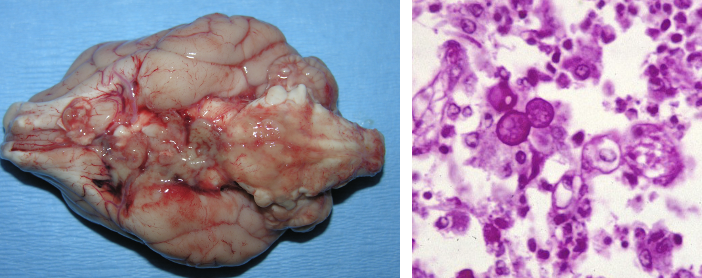
Wah eh
Blastomycosis

Wat
Phaeohyphomycosis

wah eh
Cryptococcosis

What
Aspergillosis
Potential agents of protozoal encephalitis
Toxoplasma
Neospora
Sarcocystis
Common bacterial causes of otitis media/interna
Staphylococcus
Streptococcus
E. coli
Pseudomonas
Proteus
Mycoplasma bovis (calves)
What is discospondylitis and how is it caused
Inflammation of vertebral disc and adjacent vertebral bodies
Caused by hematogenous spread
Common bacteria involved in discospondylitis
Staphylococcus
Streptococcus
E. coli
Brucella canis
Common bacteria involved in ulcerative keratitis (corneal ulcer)
Staphylococcus
Streptococcus
Pseudomonas
E. coli
Gram staining characteristics of Listeria monocytogenes and where it is found?
Gram-positive rod
Everywhere in nature, often in spoiled food
Pathogenic features of Listeria monocytogenes
Facultatively intracellular; can invade many cells
Hemolysin enzyme
Can spread to adjacent cells without exiting them
Polymerizes cell actin to form pseudopods
Transmission of Listeria
Primary route = ingestion (spoiled silage)
Disease is rare, more common in immunocompromised hosts and in winter
Inhalation also possible but less common
Pathogenesis of CNS Listeria infection
Ingesion/inhalation → entry through damaged mucosa → access to peripheral nerve endings → ascend nerve sheaths → CNS disease
Often infects trigeminal n. (CN V)
Primarily seen in ruminants
Clinical presentation of Listeriosis CNS disease
“Circling disease”/meningoencephalitis
Fever, CN deficit, circling, head tilt, nystagmus, head pressing
Fatal if left untreated
ZOONOTIC
Diagnosis of Listeriosis
Histopathology showing multifocal microabcesses in brain
Immunohistochemistry → Ab present for listeria
Culture/PCR from blood, infected brain, or abortive tissues
Treatment for Listeriosis
Antibiotics
No vaccine; avoid spoiled silage, lunch meats, dairy
Characteristics of Clostridium spp.
Gram-positive anaerobic rods
Produce spores and neurotoxins
Where is Clostridium botulinum found
Soil, decaying organic matter & surrounding plant material
Transmission of C. botulinum
Ingestion of pre-formed neurotoxin
Wound contamination also possible
Pathogenesis of Botulism
Toxin absorbed from GI tract → enters bloodstream → reaches peripheral nerves → taken up by presynaptic nerves at synapse → inhibits ACh release → flaccid paralysis
Clinical signs of Botulism in horses/ruminants
Dysphagia, slow to eat
Muscle weakness, altered gait, recumbency
Mydriasis (dilated pupils)
Decreased gut motility
Bladder distension
“Shaker Foal syndrome” in foals (similar presentation + muscle tremor)
Diagnosis of Botulism
Normal CBC/chem/UA
Toxin needs ID by laboratory (i.e. ELISA)
Treatment of Botulism
Antitoxin → restricts circulating toxin but does not help already ruined nerves
Recovery via rest & new nerve growth
5 types of edema in CNS
Vasogenic edema
Cytotoxic edema
Intramyelinic edema
Interstitial edema
Osmotic edema
What type of edema is associated with trauma, inflammatory lesions, and hemorrhagic lesions?
Vasogenic edema
Disruption of the BBB, buildup of extracellular fluid
What type of edema is associated with hypoxia, metabolic disturbance, and some toxins?
Cytotoxic edema
No BBB disruption, intracellular fluid buildup
What kind of edema is associated with bromethalin toxicity and hepatic/renal encephalopathy?
Intramyelinic edema (type of cytotoxic edema)
What type of edema is associated with hydrocephalus?
Interstitial edema (increased interstitial fluid)
Increased ventricular pressure → disruption of ependyma → CSF enters white matter interstitium → myelin is dissolved + atrophy