Bio Lecture Exam 2
 Call Kai
 Learn
Learn Practice Test
 Spaced Repetition
 Match
Match Flashcards
 Knowt Play
 Podcast
1/136

There's no tags or description
Looks like no tags are added yet.
Name | Mastery | Learn | Test | Matching | Spaced | Call with Kai | Chat |
|---|
No analytics yet
Send a link to your students to track their progress
137 Terms
What is histology defined as?
The study of tissues
What are the four primary tissue types in the human body?
Epithelial, Connective, Muscle, and Nervous (Memory: ECMN).
What are the structural characteristics of epithelial tissue?
Cells are tightly packed, little extracellular matrix, has an apical and basal surface, usually avascular, often regenerates quickly, and is supported by a basement membrane.
In epithelial tissue naming, what do the first and second parts of the name signify?
The first part indicates the number of cell layers (simple = one layer, stratified = multiple layers), and the second part indicates the shape of cells at the apical surface (squamous = flat, cuboidal = cube-shaped, columnar = tall).
What are the primary functions of simple squamous epithelium?
Diffusion and filtration.
What are the primary locations of simple squamous epithelium?
Alveoli of lungs and lining of blood vessels (endothelium).
Locations of simple cuboidal epithelium?
Kidney tubules and small gland ducts.
What is the function of simple cuboidal epithelium?
Secretion and absorption
What is the function of simple columnar epithelium
Secretion and absorption
Location of simple columnar epithelium
Much of the digestive tract.
What specialized structures may be present in simple columnar epithelium, and what are their roles?
Microvilli (increase surface area) and goblet cells (secrete mucus).
Why is pseudostratified columnar epithelium classified as a single layer?
It appears multilayered but is actually one layer because all cells contact the basement membrane.
Astrocytes
maintain chemical environment in neuron
Locations of pseudostratified columnar epithelium
Respiratory tract
What specialized structures may be present in pseudostratified columnar epithelium, and what are their roles?
Goblet cells (produce mucus) and Cilia (help move mucus)
What are the functions of stratified squamous epithelium
Protection
Location of keratinized stratified squamous epithelium
In the epidermis
Location of nonkeratinized stratified squamous epithelium
mouth, esophagus, and vagina.
What is transitional epithelium specialized for
Specialized to stretch
where is transitional epithelium found
urinary system- bladder
What are the functions of Connective Tissue
Support, Binding, Protection, Transport
What three elements are usually contained in connective tissue?
Cells, protein fibers, and ground substance.
What are the three types of connective tissue fibers and their functions?
Collagen fibers (strength), elastic fibers (stretch and recoil), and reticular fibers (supportive network).
What is areolar connective tissue
A loose connective tissue that wraps and cushions structures
Where is areolar connective tissue found
Beneath many epithelial tissues
What are the functions of adipose tissue?
Energy storage, insulation, and protection/cushioning.
Dense regular connective tissue
tendons and ligaments- run in the same direction
Dense irregular connective tissue
Found in the dermis of the skin, running in multiple directions
Why is blood classified as a connective tissue and what does it do?
It has a liquid extracellular matrix called plasma and transports substances throughout the body.
What are the characteristics of hyaline cartilage?
Most common type
Where is hyaline cartilage found
found at the ends of many bones, the nose, and the trachea.
What are the characteristics of fibrocartilage?
Strong and resistant to compression
Locations of fibrocartilage
found in intervertebral discs and the pubic symphysis.
What are the characteristics of elastic cartilage?
Flexible
Where is elastic cartilage found
found in the external ear
Connective tissue function
protection
framework
binding structure
storage
immune protection
transport
Elastic Fibers
Thin, branching protein fibers that stretch and recoil readily, allowing structures like blood vessels to expand and relax.
Collagen Fibers
Long, unbranched, flexible protein fibers resistant to stretching that make up 25% of all protein in the human body.
Reticular Fibers
Thin, branching protein fibers that form a delicate woven framework (stroma) within organs possessing abundant space.
Cartilage
Semi-rigid connective tissue; flexible and resilient
Lacunae
Small spaces within the bone matrix that house osteocytes.
Chondroblasts
produce matrix
Chondrocytes
surround by matrix
Interstitial growth (cartilage)
Growth of cartilage from within, where chondrocytes divide and secrete new matrix, allowing the tissue to expand.
Appositional growth (cartilage)
Cartilage increases in thickness by adding new matrix layers to the surface of existing cartilage.
Function of Bone
support and protection
movement
hemopoiesis
storage of mineral and energy reserves
Hematopoiesis
The process of blood cell formation occuring in the bone marrow,
Osteoprogenitor Cells
Stem cells in the bone marrow that differentiate into osteoblasts, responsible for bone formation.
Osteoblasts
Bone-building cells that synthesize and secrete the bone matrix ("blasts build").
Osteoclasts
Large cells responsible for bone resorption and breakdown ("clasts crush").
Osteocytes
Mature bone cells situated within lacunae that maintain existing bone tissue.
Long Bones
Longer than they are wide
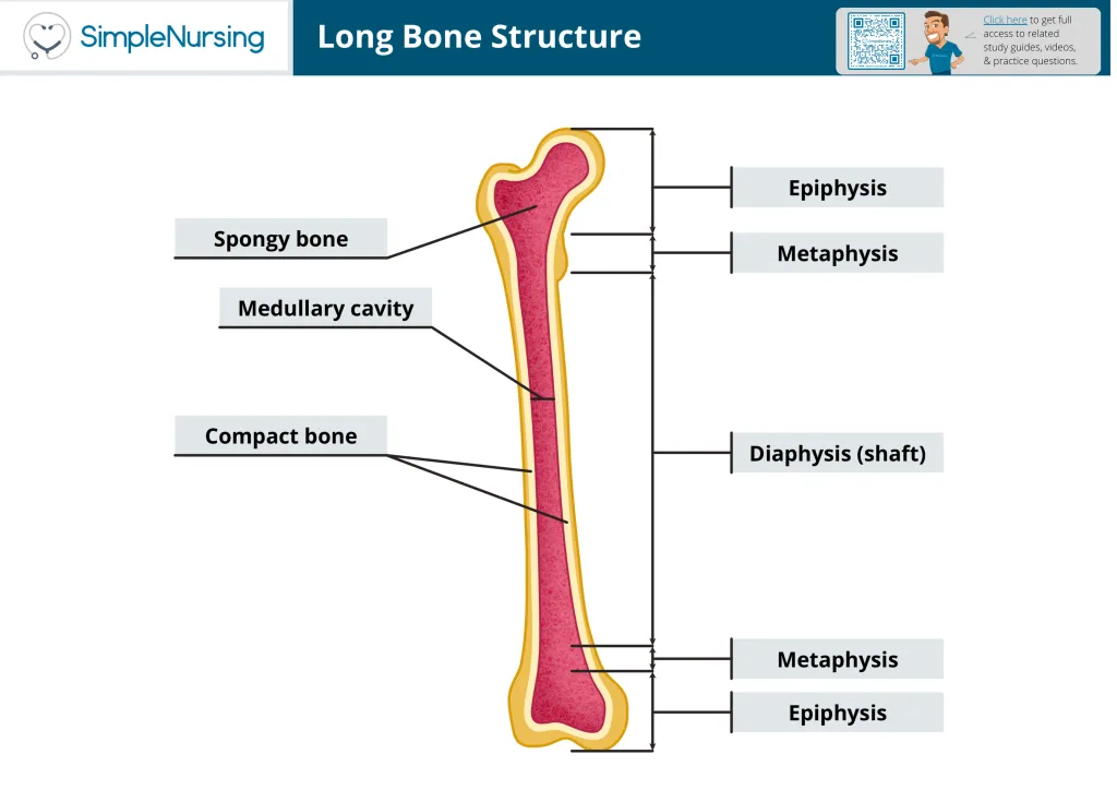
Epiphysis
The end of a long bone
Diaphysis
The shaft of a long bone- surrounds the hollow medullary cavity.
Metaphysis
The transitional region between the diaphysis and epiphysis that contains the growth plate in growing bones.
Periosteum
A tough outer connective tissue covering of bone containing blood vessels, nerves, and osteogenic cells. critical for bone growth, repair, and tendon/ligament attachment, but absent over articular cartilage.
Endosteum
A thin membrane that lines the internal bone surfaces. Purely metabolic
Compact Bone
solid and dense- on the external surfaces of long and flat bones
Spongy Bone
open lattice of narrow plates called trabeculae- internal surface of bones
Osteon
fundamental structural unit of compact bone
Central Canal - part of the compact bone
At the center of the osteon; carries blood vessels and nerves
Concentric Lamellae - part of compact bone
rings of bone around central canal
osteocytes- part of compact bone
housed in lacunea between concentric
Canaliculi- part of the compact bone
tiny interconnecting channels within bone that extend between lacunea- allows osteocytes to connect and communicate
perforating canals- part of compact bone
run perpendicular to and help connect multiple central canals, passageways for blood vessels and nerves.
Circumferential lamellae- part of the compact bone
surrounds the outer surface and contributes to the bone's strength and structure.
interstitial lamellae- part of the compact bone
leftover parts of osteons that have been partially reabsorbed
Ossification
formation and developement of bone
Intramembranous Ossification
Bone develops directly from mesenchymal tissue; it forms many flat bones, certain skull bones, and part of the clavicle.
Endochondral Ossification
Begins with hyaline cartilage model and produces of majority in the body
Epiphyseal Plate
large layer of hyaline cartilage at the boundary of the epiphysis and diaphysis; site for interstitial growth (5 distinct zones)
Resting Zone
Small chondrocytes in hyaline cartilage- farthest from medullar cavity and nearest epiphysis
Proliferation Zone
The growth plate zone where chondrocytes undergo rapid mitotic division on the epiphyseal side.
Hypertrophic Zone
The growth plate zone where older chondrocytes enlarge and mature.
Calcification Zone
The growth plate zone where the surrounding cartilage matrix calcifies and chondrocytes die.
Ossification Zone
The growth plate zone where osteoblasts replace the calcified cartilage framework with new bone on the diaphyseal side.
Interstitial Growth of bone
bone grows in length- occurs in the epiphyseal plate
Appositional Growth
Bone growth in width and diameter occurring in periosteum
Organic Component of Bone Matrix
The matrix fraction composed mostly of collagen fibers that provides bone with flexibility and tensile strength, preventing it from becoming brittle.
Inorganic Component of Bone Matrix
The mineral fraction composed mainly of calcium phosphate crystals that provides hardness and compression resistance, preventing excessive flexibility.
Hematoma Formation
The initial step of bone fracture repair where ruptured blood vessels produce a localized blood clot around the break site.
Soft Callus
A fibrocartilaginous bridge that forms during fracture repair to temporarily stabilize the fractured bone ends.
Hard Callus
A rigid bony collar formed during fracture repair when osteoblasts replace the fibrocartilaginous soft callus with trabecular bone.
Bone Remodeling
A continuous coupled process where osteoclasts remove older or damaged bone and osteoblasts form new bone to adapt to stress, repair microdamage, and maintain calcium balance.
Articular Cartilage function
Reduces friction and absorbs shock in moveable joints
Medullary cavity
Cylindrical space inside diaphysis- contains yellow bone marrow
Red Bone Marrow
Bone marrow responsible for producing blood cells; adult sites include the skull, vertebrae, ribs, sternum, pelvis, and proximal ends of certain long bones.
Yellow Bone Marrow
Bone marrow consisting mostly of adipose tissue that stores triglycerides and fat for energy.
Greenstick Fracture
an incomplete bone break where the bone bends and cracks on one side only without snapping into separate pieces
Part of nervous system
neuron
glial cells
Neuron
Excitable cells that initiate and transmit nerve impulses
glial cells
non excitable cells that support and protect neurons
astrocytes
maintain chemical environment inside neuron
Microgillia
immune system cells
Schwann cells and oligodendrocytes
helps transmit more quickly
Ependymal cells
produce spinal fluid
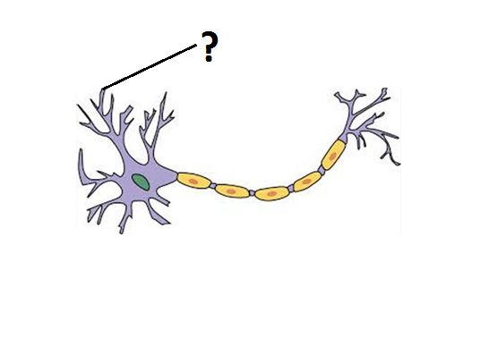
Dendrite
receives sensory input
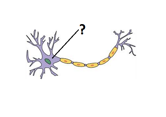
Soma
the cell body of a neuron, containing the nucleus and organelles.
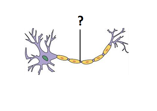
Axon
carries impulses away from cell body
Axon hillock
trigger zone for action potentials
