Module 2
 Call Kai
 Learn
Learn Practice Test
 Spaced Repetition
 Match
Match Flashcards
 Knowt Play
1/31

There's no tags or description
Looks like no tags are added yet.
Name | Mastery | Learn | Test | Matching | Spaced | Call with Kai | Chat |
|---|
No analytics yet
Send a link to your students to track their progress
32 Terms
What are the 2 division of the skeleton and what do they consist of?
Axial Skeleton: skull, vertebral column, ribs, and sternum
Appendicular Skeleton: the rest of the bones (upper limbs and lower limbs)
What are the main functions of bones?
Support: Skeleton provides framework that anchor all soft organs
Movement: where they meet we have joints that allow movements also muscles use the bones as levers
Protection: strong (ex: ribs and skull protect lungs, heart, brain etc)
Storage of minerals and fat
Hematopoiesis: Production of blood cells, occurs within the marrow cavities of the bone
What are the types of bones?
Flat: muscle attachments and protection of the organs
Long: longer than they are wide most commonly found in appendicular skeleton
Short: stability and some movement
Sesamoid: embedded in tendon ex: patella
Irregular: bone shape doesn’t fit in the other categories
What is the basic composition of bone?
2 layers:
Outer cortical layer: compact bone, hard and strong
Inner cancellous layer: spongy bone, porous and sponge-like
Blood cells are produced by the bone marrow in the medullary cavity.
Medullary cavity = hollow central space inside the shaft of a long bone that contains bone marrow. It is not a layer of cancellous bone.

What is the structure of bones?
Epiphysis: Knobby, enlarged regions at the end, they form joints and serve as attachment sites for tendons and ligaments
Metaphysis: region between the diaphysis and epiphysis
Diaphysis: elongated cylindrical shaft
Periosteum: protects the bone and contains blood vessels and nerves that supply the bone. Also has cells forming new bone tissue.

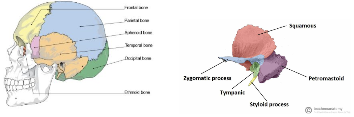
What are the types of cranial bones?
22 bones total in the skull
Frontal (1bone)
Parietal(2bones)
Temporal(2 bones) - Zygomatic process, external auditory (acoustic) meatus (ear hole) and mastoid process
Occipital(1bone)
Sphenoid(1bone)
What are the different sutures? And where are they?
Coronal = frontal and parietal bones
Sagittal= Parietal bones
Lambdoid= occipital and parietal bones
squamous= temporal and parietal bones

What are parts of the cranium?
Cranial Vault:
Encases the brain and is formed by the frontal, parietal, and occipital bones (flat bones)
Cranial base:
Fossa is defined as a depression in a bone- a big dip or hole
Contains 3 fossa:
Anterior cranial fossa
Middle cranial fossa
Posterior cranial fossa
What are the facial bone of skull?
Maxillary bone (maxillae)- paired, upper jaw bones (2 bones)
Nasal bones - paired, articulate with the frontal bone, form bridge of nose (2 bones)
Zygomatic bones- paired, form the cheekbones, include the Zygomatic process (2 bones)
Mandible ( single, forms the lower jaw
Body
Ramus
Angle

What is the division of the vertebral column?
Made of 26 bones in total.
Cervical (7 vertebrae)
Thoracic (12 vertebrae)
Lumbar (5 vertebrae)
Sacrum ( 5 fused vertebrae)
Coccyx (4 fused vertebrae)

What is the vertebrae structure?
Body (anterior)
Vertebral arch (posterior): extending into the spinous and transverse processes:
Spinous processes extending posteriorly
Transverse process: paired, extending laterally
Vertebral foramen (canal): houses the spinal cord.

Where can you find the Atypical vertebraes? How can they affect your movement?
Atypical vertebrae are C1 (atlas) and C2 (axis) which are the first and second cervical vertebrae.
Movement:
The skull rests on top of the lateral masses of the C1 with the occipital condyles. This joint allows the “yes” movement of the head. Under the C1, the dens of the C2 attach with the anterior arch of C1. This allows for the “no” movement of the head.
What is the structure of the C1 vertebrae?
Anterior arch: provides surface for articulation with the dens (a feature of C2)
Lateral masses: provide surfaces for articulation with the occipital condyles of the occipital bone.

What is the structure of the C2 vertebrae?
The main feature is the dens, which rest within the anterior arch of the C1.

What is vertebral articulation? what are the structures?
Vertebrates articulate to stack on top of one another.
Structure:
Intervertebral Disc:
between 2 vertebrates and acts as a shock absorber.
Intervertebral Foramina:
Lateral openings between adjacent vertebrae that allow for the passage of spinal nerves

What are the parts of your ribs?
you have 12 ribs total
True ribs ( pairs 1-7)
False ribs (pairs 8-10)
Floating ribs(11-12)

What is the structure of your ribs?
A typical rib is long, flat, and twisted, consisting of head, neck, tubercle, shaft (body) and angle (where the shaft is bent sharply anteriorly).
Costal groove- is the sharp inferior border which is grooved.

What are the parts of the sternum?
Makes up the anterior portion of the thoracic cage.
3 parts:
Manubrium: Articulation with rib 1
Body: Articulates with ribs 2-7
The xiphoid process

What is the thoracic cage?
ribs
thoracic vertebrae posteriorly
sternum
What is the pectoral girdle? What does it consist of?
Connects upper limb to the axial skeleton and is formed by the clavicle and scapula
Clavicle: is an S shaped bone. Joins with the manubrium of the sternum medially (proximally) and the scapula laterally (distally)
Scapula
Anterior surface: is the corracoid process. The acromion and coracoid process help stabilize the shoulder joint
Posterior surface: Acromion and spine
Lateral surface: the glenoid fossa, articulates with the head of the humerus (arm bone).

What is the humerus? And what is the structure?
Articulates with the glenoid fossa to form a shoulder joint and with the radius and ulna to form the elbow joint.

Where can I find the radius? What are it’s features?
In the forearm, Laterally placed (thumb side)
Features:
Head: articulates with capitulum and with ulna
Shaft
Neck
Distal end: wide and flat articulates with the carpal(wrist) bones form wrist joints. Styloid process

Where can I find the Ulna? What are it’s features?
Found in forearm, Medially placed (little finger side)
Features:
Articulates with distal end of the humerus
Olecranon: fits into the olecranon fossa of the humerus
Trochlear notch: C-shaped depression which interlocks with the trochlea of the humerus
Shaft
Neck
Head (distal end)
Styloid process

What is the structure on the bones in your wrist and hand?
Wrist 8 carpal bones (short bones)
Palm/hand: 5 metacarpal bones (long bones)
Digits/finger: 14 phalanges (long bones)
What is the pelvic girdle? What does it consist of?
Attaching the lower limb to the axial skeleton is formed by the hip (innominate) bones.
Hip bone consists of the ilium, ischium,and pubis

What are the features of the pelvis?
Greater sciatic notch: allows the passage of major nerves and vessels from the pelvic cavity into the posterior region of the lower limb
Lesser sciatic notch: allows the passage of structures from the pelvic cavity to the genital region
Acetabulum: deep curved depression for articulation with the head of the femur
Obturator foramen: passage of nerves and blood vessels

What are the femurs features?
Features:
Head
Neck
Shaft
The proximal end:
The head articulated with the pelvic griddle forming hip joints
The femur has bumps on it that serves as surfaces for muscle attachment
The distal end:
Articulates with the tibia and patella forming knee joints.
Features:
Medial and lateral condyles ( they form the knee joints)
Patellar- groove for the patella

Where can i find the Tibia? what’s it’s structure?
Found in the leg
Is the only weight-bearing bone of the leg
Proximal end:
Medial and lateral condyles: articulate with the medial and lateral condyles of the femur
Tibial tuberosity
Distal end:
Medial malleolus

Where can I find the fibula? What are its features?
Does not bear or transfer weight
Features:
Proximal end:
Medial head which articulates with tibia
Neck
Shaft
Distal end:
Lateral malleolus: provides lateral stability to ankle joint
What is the structure of the ankle and foot bones?
Ankle
7 tarsal bones
(short bones)
Foot
5 metatarsal bones
(long bones)
Digits/toes
14 phalanges
(long bones)
What is a synovial joint?
Synovial joint: A freely movable joint with a fluid-filled space between the bones that allows smooth movement
What are the movments made at Synovial joints
Gliding: the motion of bones sliding on one another
Angular: a motion that changes a joint angle
Flexion- decreasing the angle between articulating bones (bending)
Extension- increasing the angle between articulating bones (straightening)
Abduction-movement away from the midline of the body
Adduction- movement toward the middling of the body
Rotation: pivots around its own axis
Medial rotation- towards the midline
Lateral rotation-away from the midline
Special movements:
Supination and pronation: rotation of the radius over the ulna. The supination forearm rotates so the palm faces anteriorly. Pronation forearm rotates and faces posteriorly
Inversion and eversion: Twisting motion of the ankle. Inversion twisted so the soles of the feet turn medially. Eversion when the soles turn laterally.
Circumduction: Movement that combines flexion, abduction, extension and adduction in succession