Chapter 6: Membrane Potentional & Synaptic transmission
 Call Kai
 Learn
 Practice Test
 Spaced Repetition
 Match
 Flashcards
 Knowt Play
1/56

There's no tags or description
Looks like no tags are added yet.
Name | Mastery | Learn | Test | Matching | Spaced | Call with Kai | Chat |
|---|
No analytics yet
Send a link to your students to track their progress
57 Terms
What are the two major divisions of the nervous system?
Central nervous system (CNS)
Peripheral nervous system (PNS)
What structures make up the central nervous system (CNS)?
Brain
Spinal Cord
What is the peripheral nervous system (PNS)?
The portion of the nervous system outside the brain and spinal cord that connects the CNS with the rest of the body.
What are the two major divisions of the peripheral nervous system shown in the figure?
Somatic Nervous System
Autonomic Nervous System
What does the somatic motor nervous system control?
Voluntary control of skeletal muscles
What does the autonomic motor nervous system control?
Involuntary control of smooth muscle
Cardiac Muscle
Glands
What are the two divisions of the autonomic nervous system shown in the figure?
Sympathetic
Parasympathetic
How do the somatic motor and autonomic motor nervous systems differ?
Somatic motor controls voluntary skeletal muscle, while autonomic motor controls involuntary smooth muscle, cardiac muscle, and glands.
How is the nervous system organized from its major divisions to their subdivisions?
Nervous system → CNS and PNS
CNS → brain and spinal cord
PNS → somatic and autonomic
somatic → sensory and motor
autonomic → sympathetic and parasympathetic
What is the general direction of information flow through a neuron?
Dendrites → cell body (soma) → axon → synapse


What is the function of dendrites in a neuron?
They receive incoming signals and carry information toward the cell body.

What is another name for the cell body of a neuron?
The soma
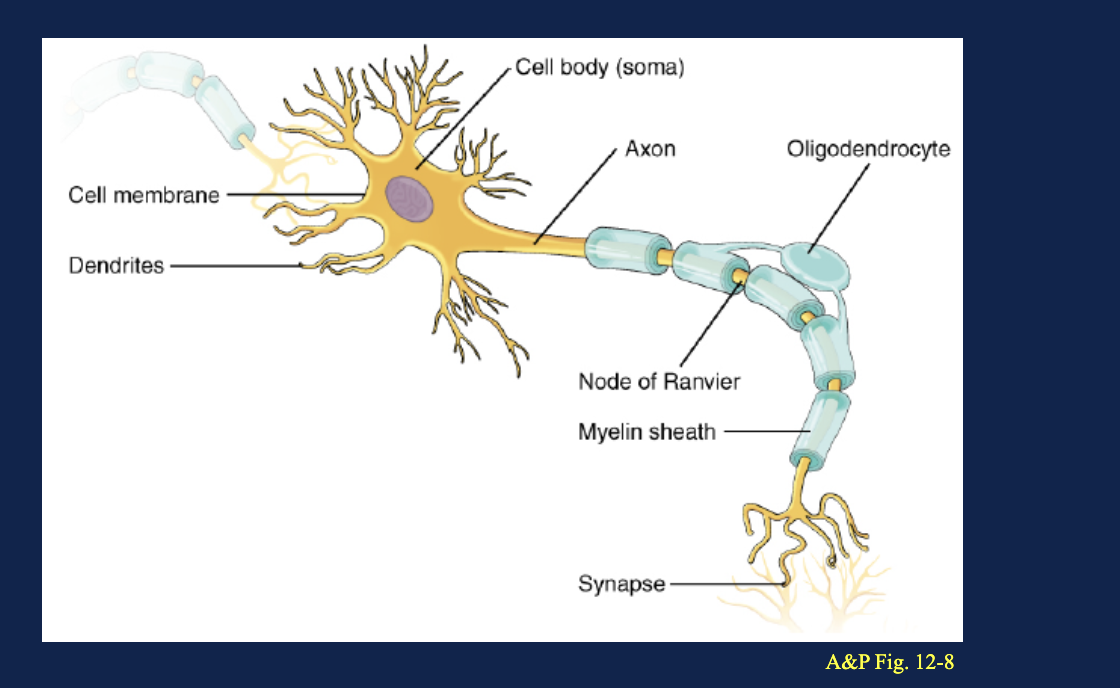
What is the function of a neuron's axon?
It carries signals away from the cell body
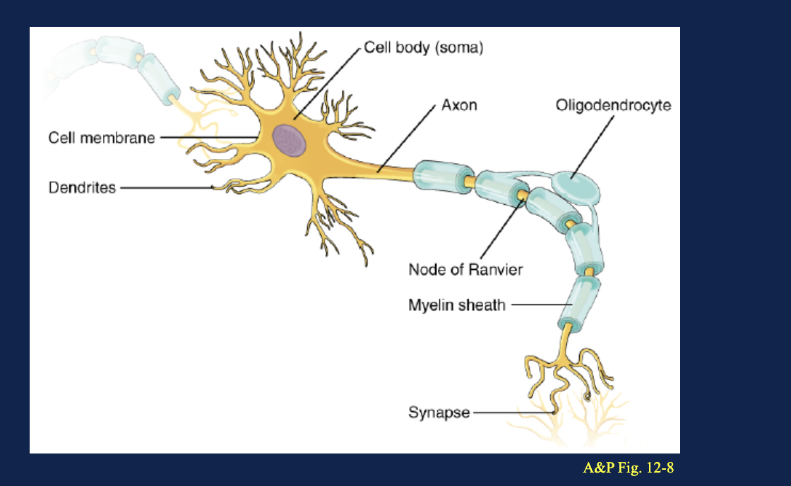
What is the function of the myelin sheath around an axon?
It insulates the axon and increases the speed of signal conduction

What are nodes of Ranvier?
Gaps between adjacent sections of myelin along an axon

What type of signal conduction allows a signal to travel rapidly from node to node along a myelinated axon?
Saltatory conduction

What cell forms myelin around axons in the central nervous system?
An oligodendrocyte

What is a synapse?
The site where a neuron communicates with another cell

On an unlabeled neuron diagram, how can dendrites and the axon be distinguished?
Dendrites are branched structures that receive signals toward the soma, while the axon is the long extension that carries signals away from the soma
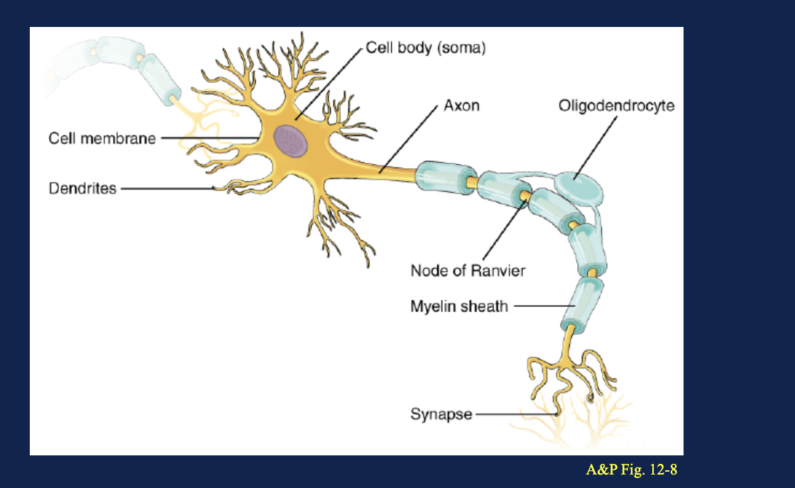
On an unlabeled myelinated neuron, what are the small gaps between sections of myelin?
Nodes of Ranvier
ow are oligodendrocytes, myelin, and the CNS related?
Oligodendrocytes are glial cells that form myelin around axons in the CNS
What is the difference between a presynaptic and postsynaptic neuron?
The presynaptic neuron sends the signal, while the postsynaptic neuron receives it.
What direction does an afferent neuron carry information?
Toward the central nervous system
What direction does an efferent neuron carry information?
Away from the central nervous system to a target.
What is the main function of interneurons?
They process and relay information between neurons within the CNS.
What is the basic pathway of neural communication from a sensory receptor to a target?
Sensory receptor → afferent neuron → CNS → efferent neuron → target
What can be a target of an efferent neuron?
A muscle, gland, or another neuron
What memory trick distinguishes afferent from efferent neurons?
Afferent ARRIVES at the CNS; efferent EXITS the CNS

In a neural pathway diagram, how can an afferent neuron be identified?
Its signal travels from a sensory receptor toward the CNS.

In a neural pathway diagram, how can an efferent neuron be identified?
Its signal travels away from the CNS toward a target
Can the same neuron be both postsynaptic and presynaptic?
Yes. It can receive a signal at one synapse and send a signal at another synapse.
What is membrane potential?
The difference in electrical potential across the cell membrane.
What does a membrane potential of −70 mV mean?
The inside of the cell is 70 mV more negative than the outside
Where is the reference electrode placed when measuring membrane potential?
In the extracellular fluid outside the cell
Where is the recording microelectrode placed when measuring membrane potential?
In the cytosol inside the cell
What does a negative membrane potential indicate about the inside of a cell?
The inside is electrically negative relative to the outside
A reference electrode is outside a neuron and a recording electrode is inside it. The voltmeter reads −70 mV. How should this result be interpreted?
The inside of the neuron is 70 mV more negative than the outside
How are ion gradients related to membrane potential?
Unequal ion distributions and selective ion movement across the membrane create an electrical difference between the inside and outside of the cell
What is the approximate resting membrane potential of a neuron?
About −70 mV
Where are Na+ and K+ primarily concentrated in a resting neuron?
Na+ is concentrated outside the neuron, while K+ is concentrated inside
What does the Na+/K+ ATPase transport during each cycle?
3 Na+ out of the cell and 2 K+ into the cell using ATP
What concentration gradient does K+ have in a resting neuron?
K+ is high inside and low outside, so its concentration gradient favors movement out of the neuron.
What concentration gradient does Na+ have in a resting neuron?
Na+ is high outside and low inside, so its concentration gradient favors movement into the neuron.
What is the approximate ratio of K+ leak channels to Na+ leak channels at rest?
Approximately 25:1
Why does K+ have a greater influence than Na+ on the resting membrane potential?
The resting membrane has many more K+ leak channels and is therefore much more permeable to K+
How does K+ leakage contribute to the negative resting membrane potential?
K+ leaks out of the neuron, causing positive charge to leave and making the inside relatively negative.
What two forces make up an electrochemical gradient?
The concentration gradient and the electrical gradient

A membrane diagram shows high Na+ outside, high K+ inside, many K+ leak channels, and a Na+/K+ ATPase. How do these features produce the resting membrane potential?
The pump maintains Na+ and K+ gradients, while abundant K+ leak channels allow K+ to leave, making the inside relatively negative.
What is the equilibrium potential for an ion?
The membrane potential at which the ion's chemical and electrical forces balance, producing no net movement of that ion
In the equilibrium potential example, which ion can cross the membrane?
K+ can cross, while Na+ and Cl− cannot
Why does K+ initially move from compartment 2 to compartment 1?
K+ moves down its concentration gradient from high K+ concentration to low K+ concentration.
What happens to charge as K+ moves from compartment 2 to compartment 1?
Compartment 1 becomes relatively positive and compartment 2 becomes relatively negative.
What two forces act in opposite directions on K+ as equilibrium potential develops?
The chemical concentration gradient and the electrical gradient.
What does "no net movement of K+" mean at equilibrium?
K+ still moves in both directions, but equal amounts move each way
Does equilibrium potential require equal ion concentrations on both sides of the membrane?
No. Ion concentrations can remain different because the chemical and electrical forces are balanced.
For K+ in a resting neuron, which directions do the chemical and electrical forces favor?
The chemical gradient favors K+ movement out, while the negative intracellular electrical potential attracts K+ back in.

In an equilibrium potential diagram, K+ initially diffuses down its concentration gradient until charge separation creates an opposing electrical force. What happens when the two forces become equal?
K+ reaches its equilibrium potential and there is no net K+ movement.
