Final practicum (Kriska)
 Call Kai
 Learn
 Practice Test
 Spaced Repetition
 Match
 Flashcards
 Knowt Play
1/117

There's no tags or description
Looks like no tags are added yet.
118 Terms
Hard Palate
bony part of the roof of the mouth
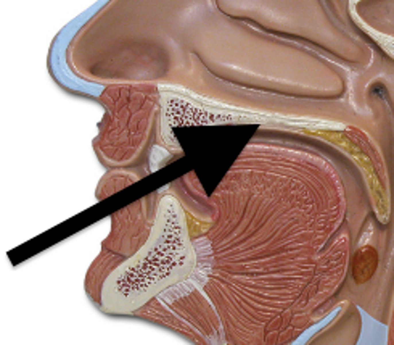
Soft Palate
soft part of the roof of the mouth
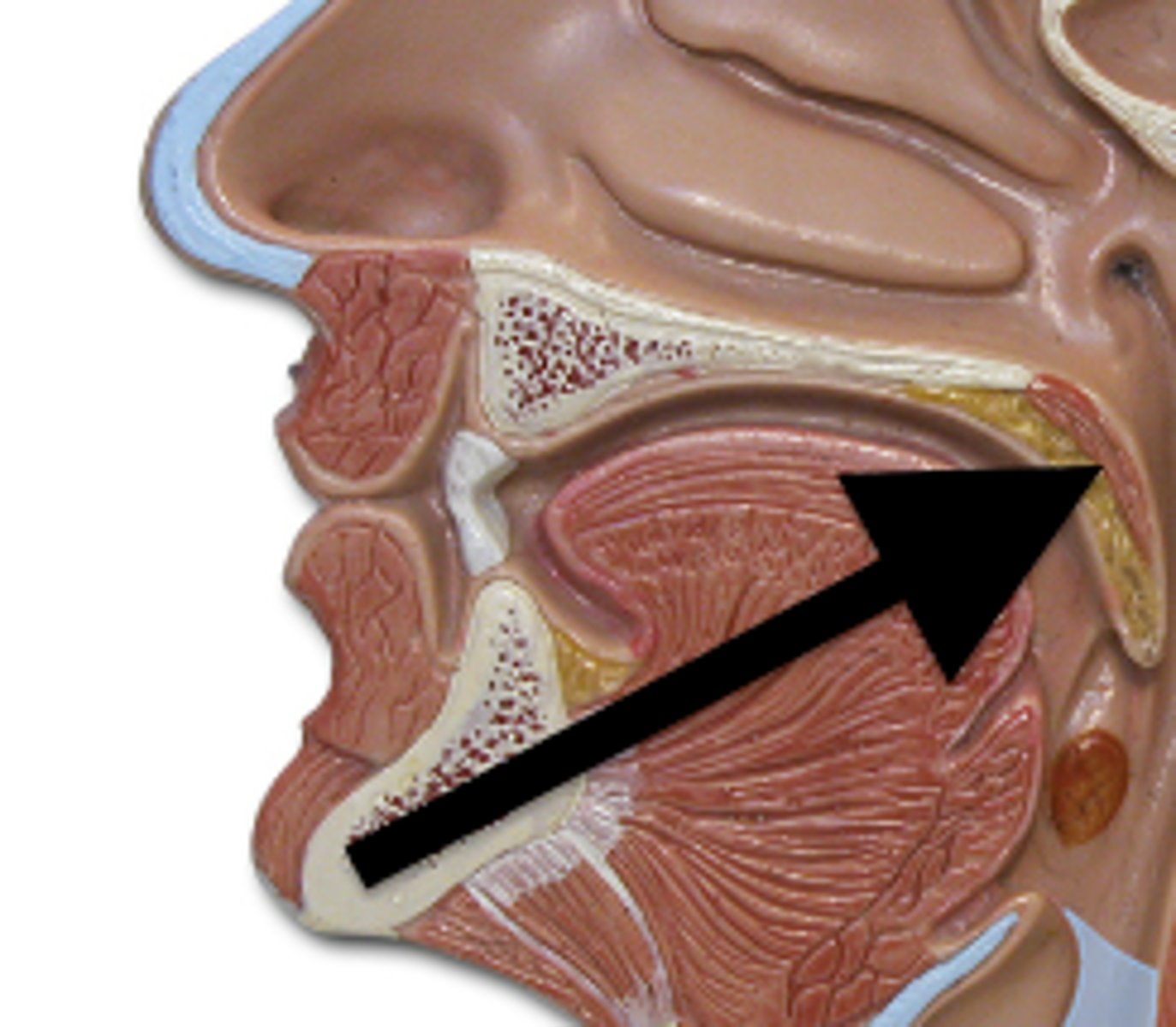
Pharynx
where respiratory & digestive tracts meet
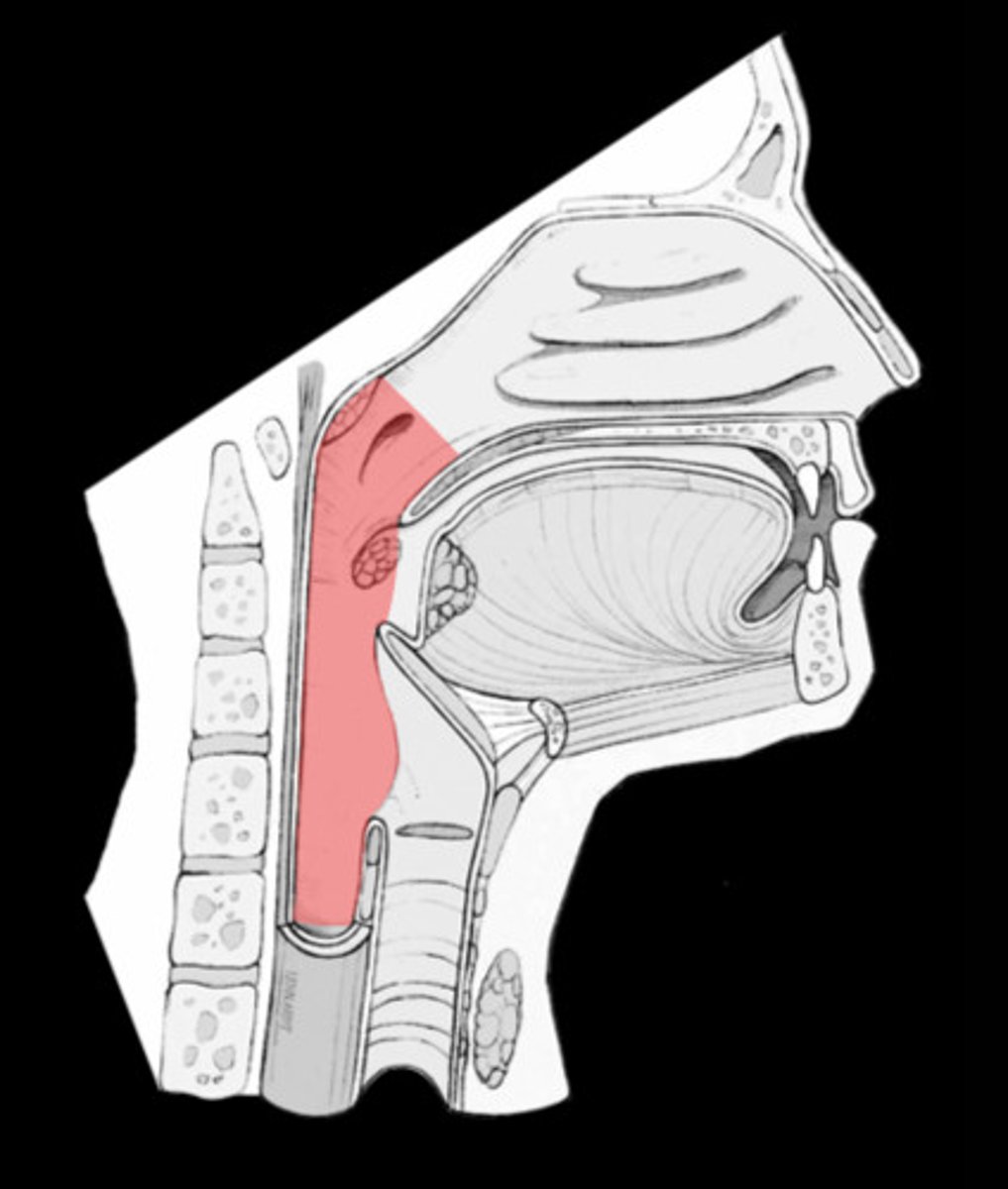
Nasopharynx
upper portion behind nasal cavity
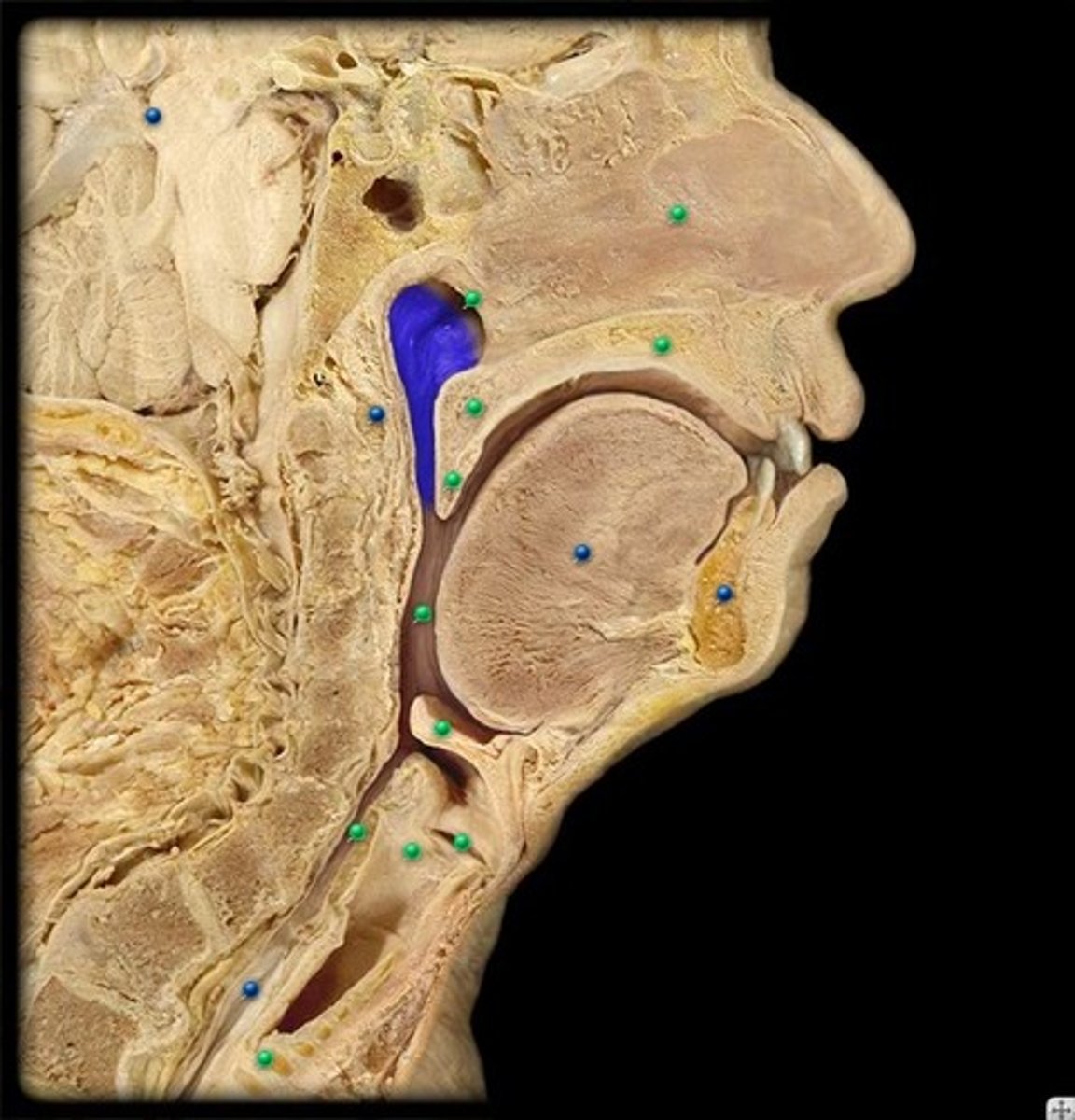
Oropharynx
middle portion behind oral cavity
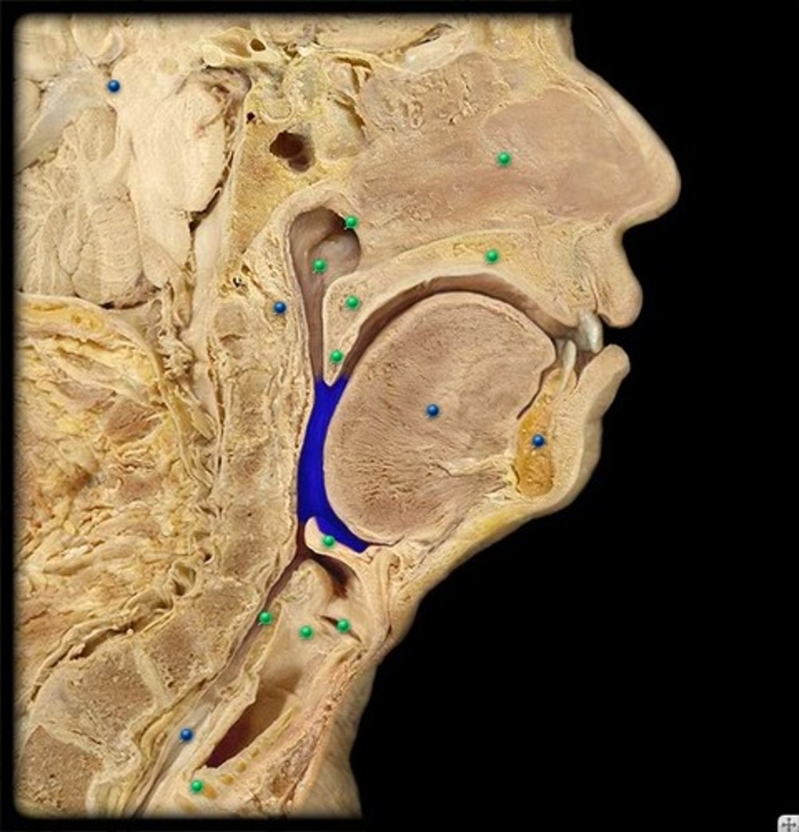
Laryngopharynx
bottom portion
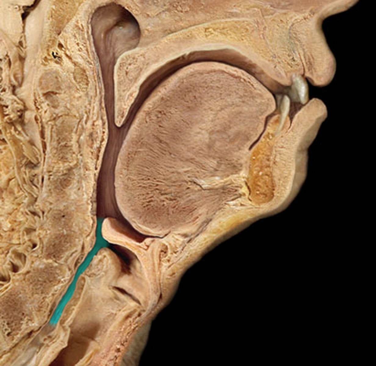
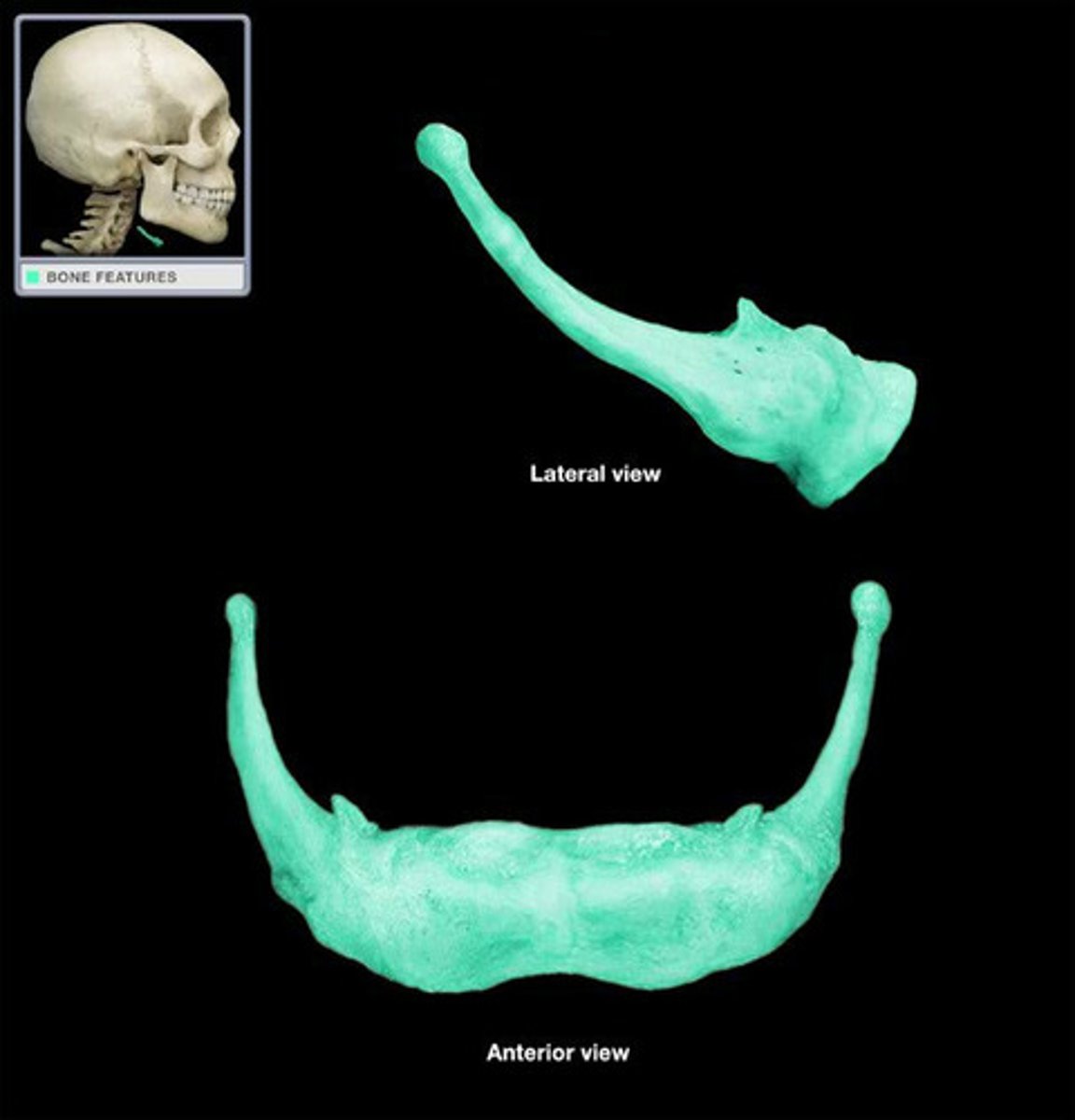
Hyoid Bone
Aids in tongue movement & swallowing
Larynx
contains:
- thyroid cartilage
- epiglottis
- vocal cords (folds)
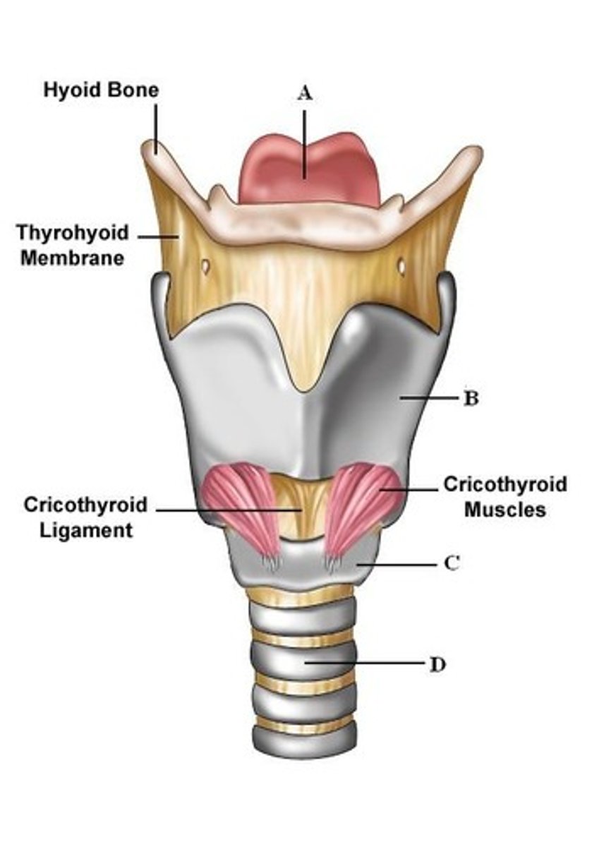
Thyroid Cartilage
large piece of cartilage of the larynx
-angle of changes@ puberty
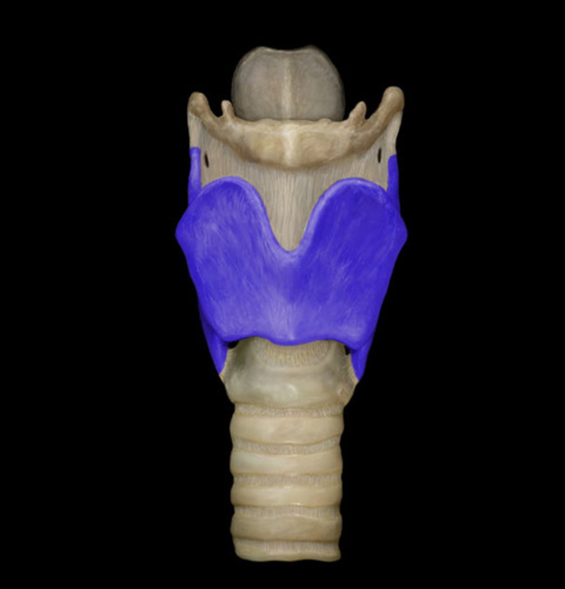
Epiglottis
cartilage that covers to the entrance of the larynx
-folds DOWN when swallowing -prevents choking
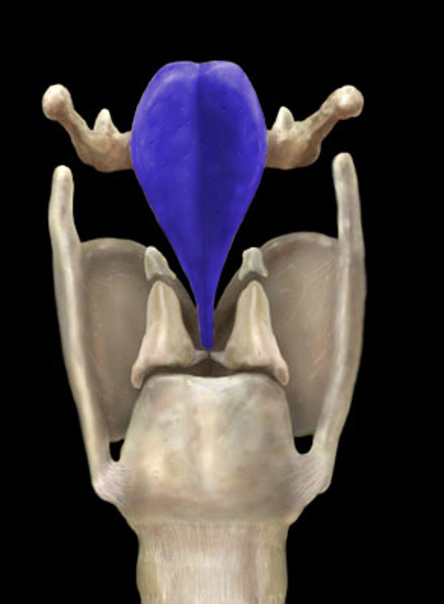
Trachea
- C-shaped pieces of cartilage
-protects!
- keep trachea open
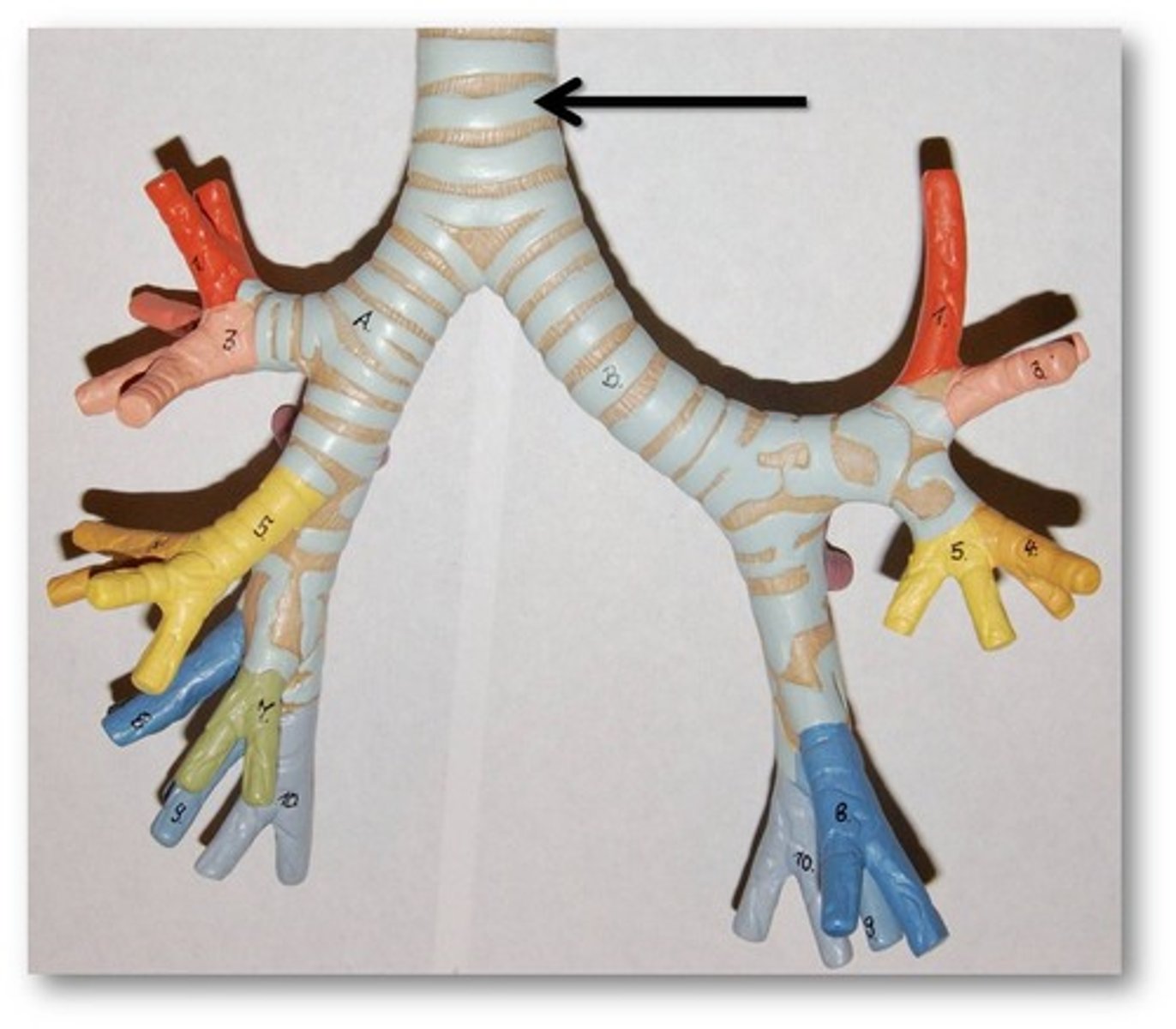
Primary Bronchus
-right/left primary bronchi
lead air into the lungs
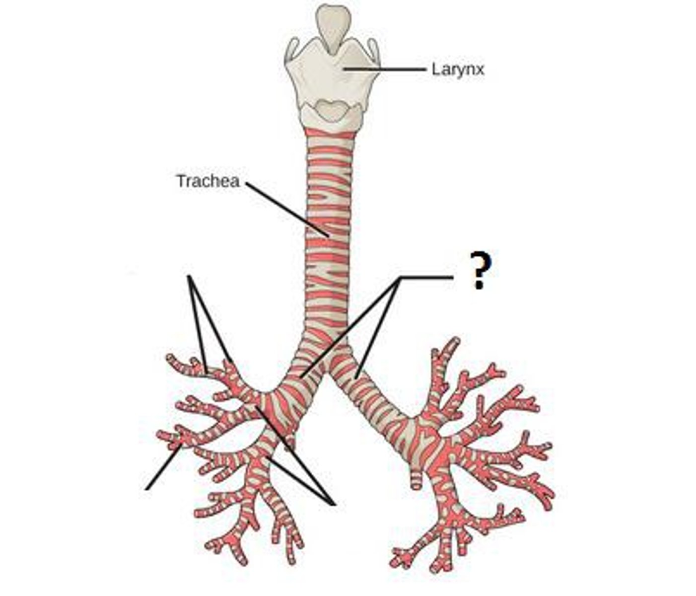
Bronchioles
- narrowest air tubes!
-Attached to alveoli
-Made up of smooth muscle, no cartilage
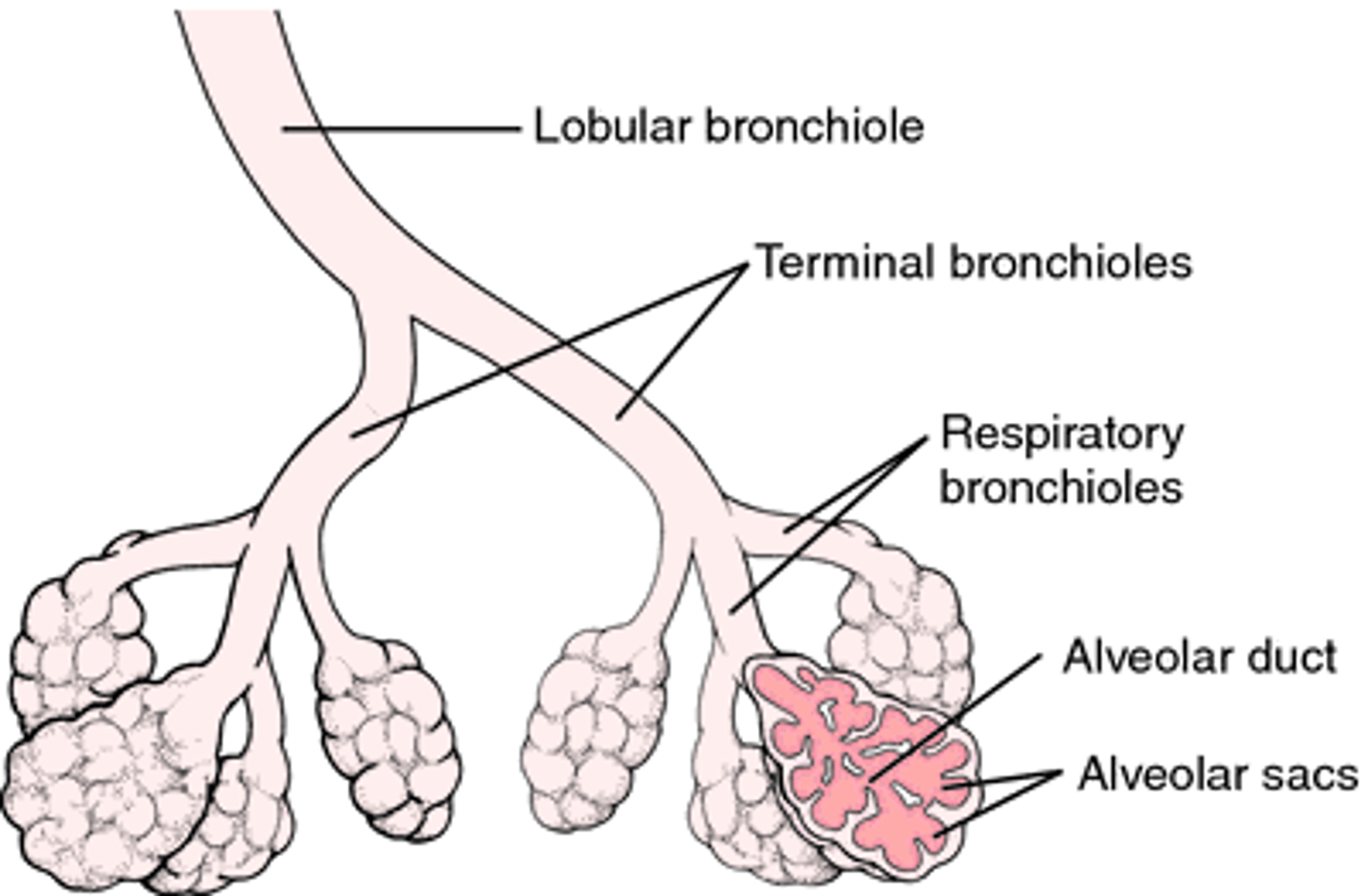
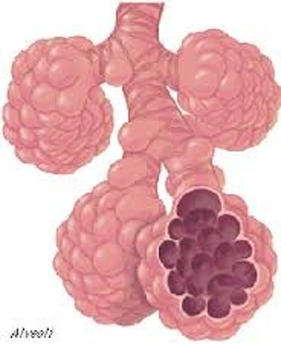
Alveoli
- air sacs
-Surrounded by capillaries
- Site of gas exchange
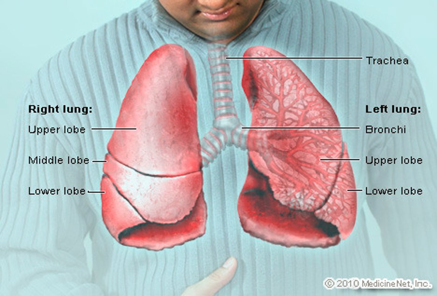
Lung
-Supportive tissue of respiration
-Right lung: 3 lobes
-Left lung: 2 lobes
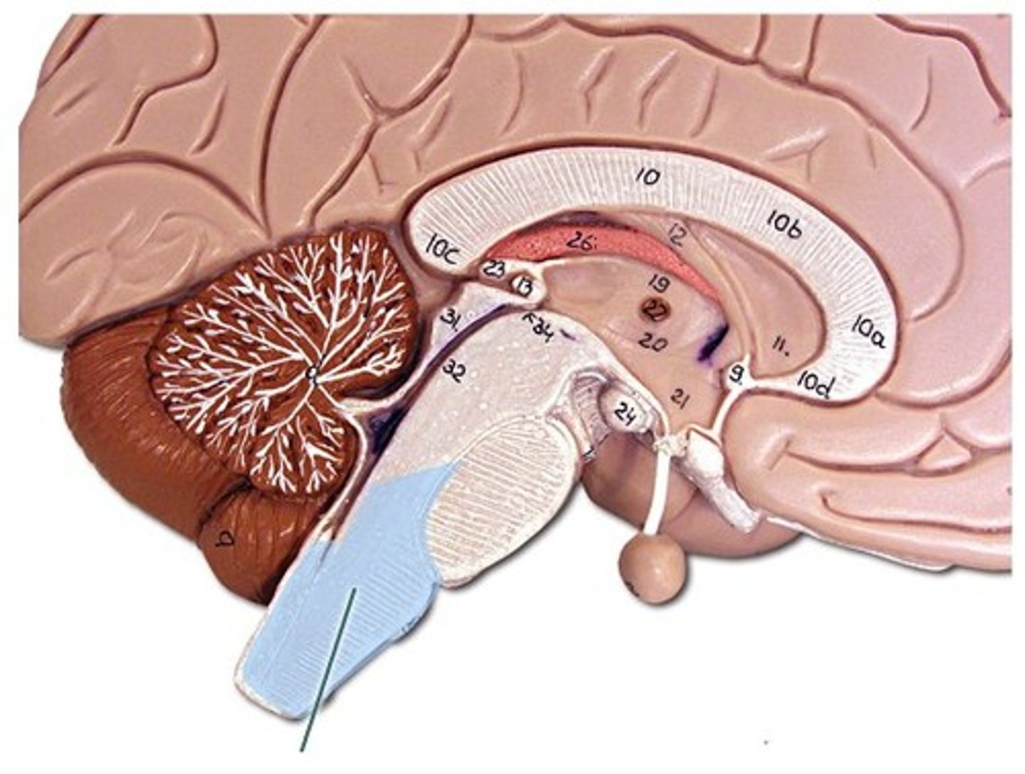
Medulla oblongata
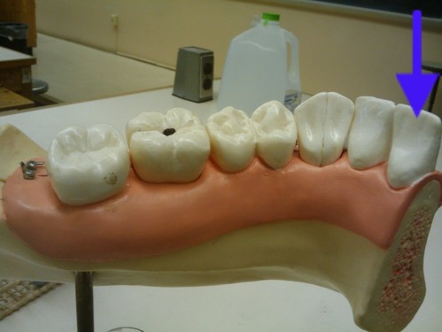
Incisor
front teeth
-tears food
-1 root
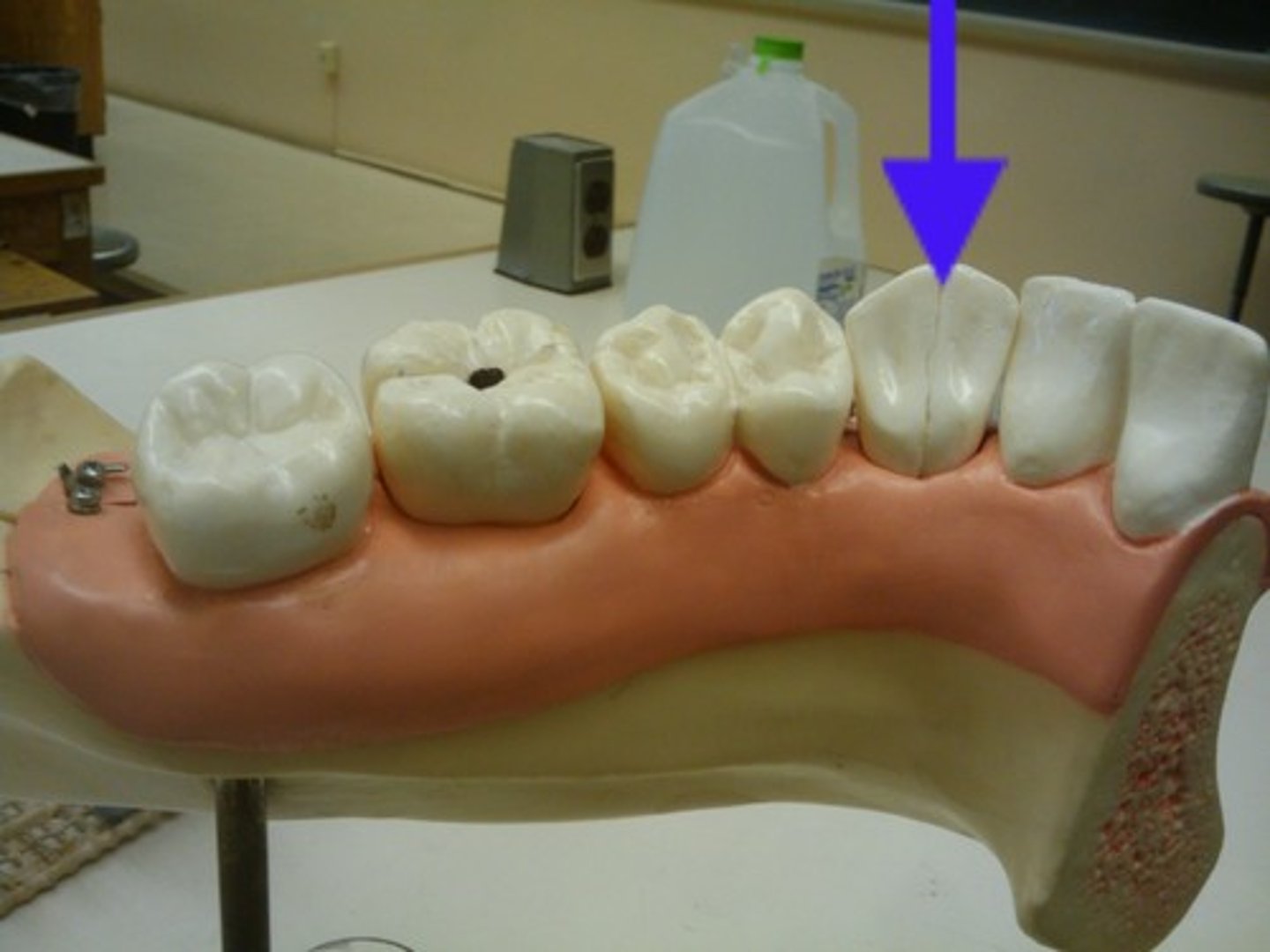
Canine
sharp and pointed teeth
-holds food
-1 root
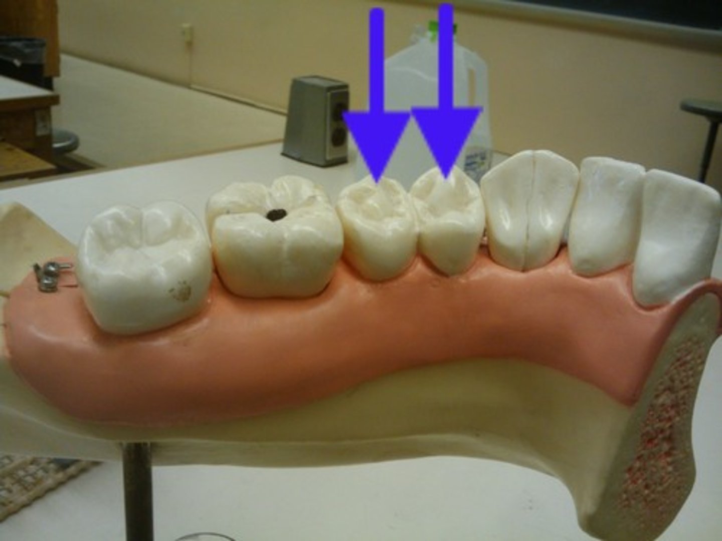
Premolar
(bicuspid)
-two cusps
-1-2 roots
Molar
has3-4 cusps
grinding & crushing food
2-4 roots
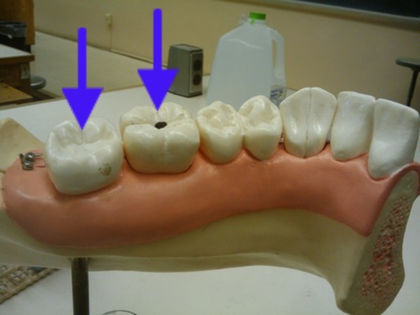
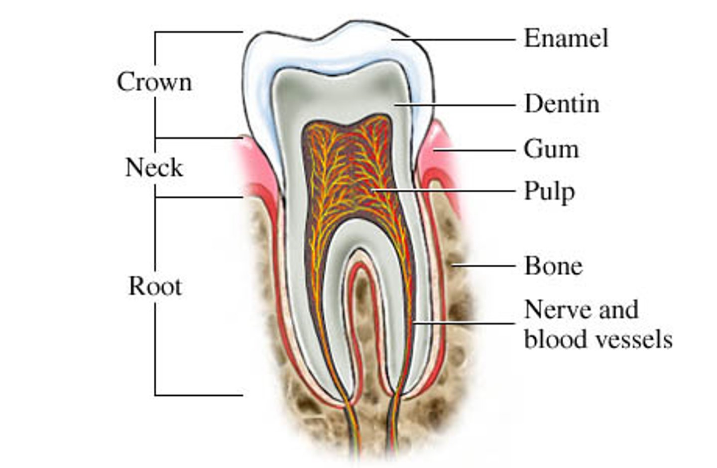
Crown
visible part of the tooth
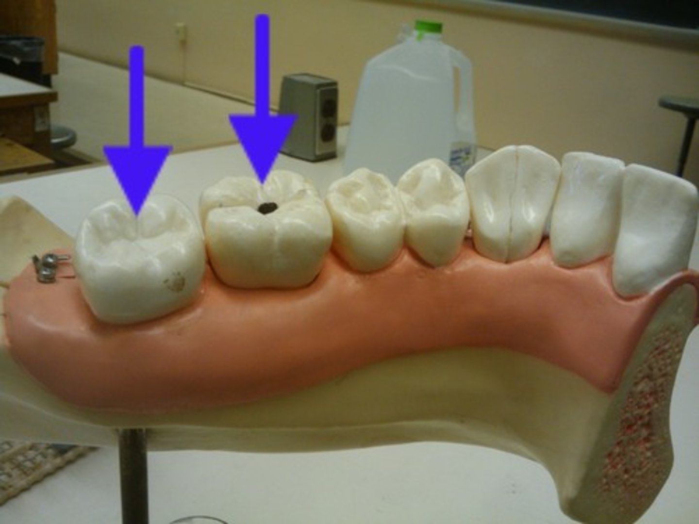
Root
with in jaw bones
-covered by the gums
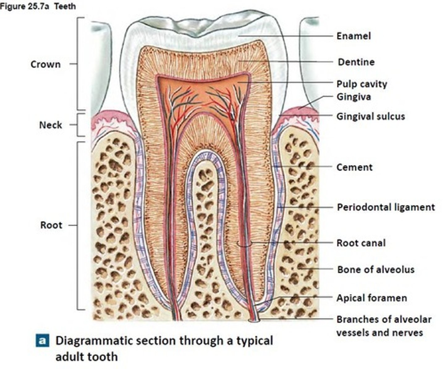
Gingiva
(gums)
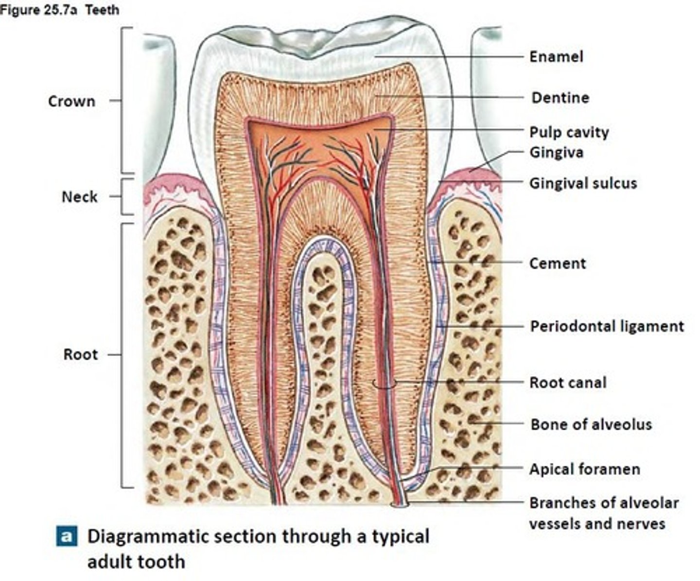
Enamel
hardest part of the tooth!
-white outer part = calcium phosphate
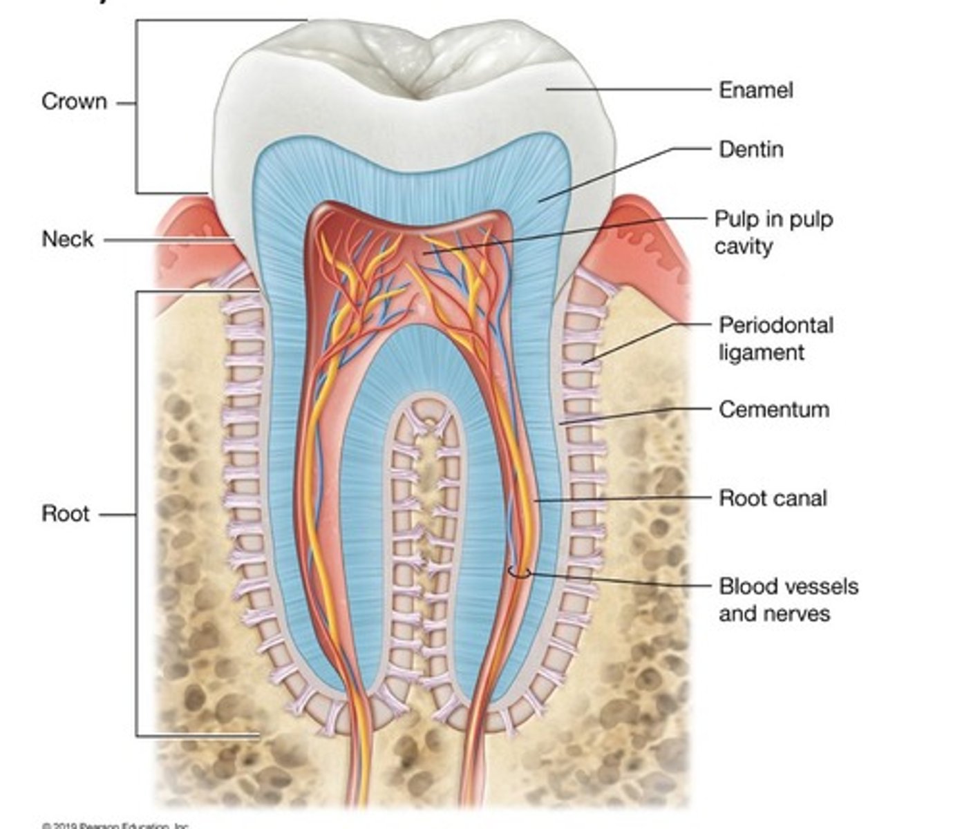
Dentin
deep to enamel/
- hardish part
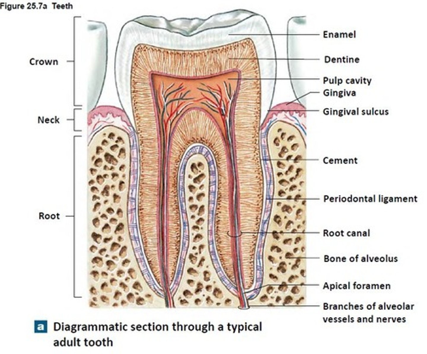
Cementum
helps "cement" teeth to jaw bones
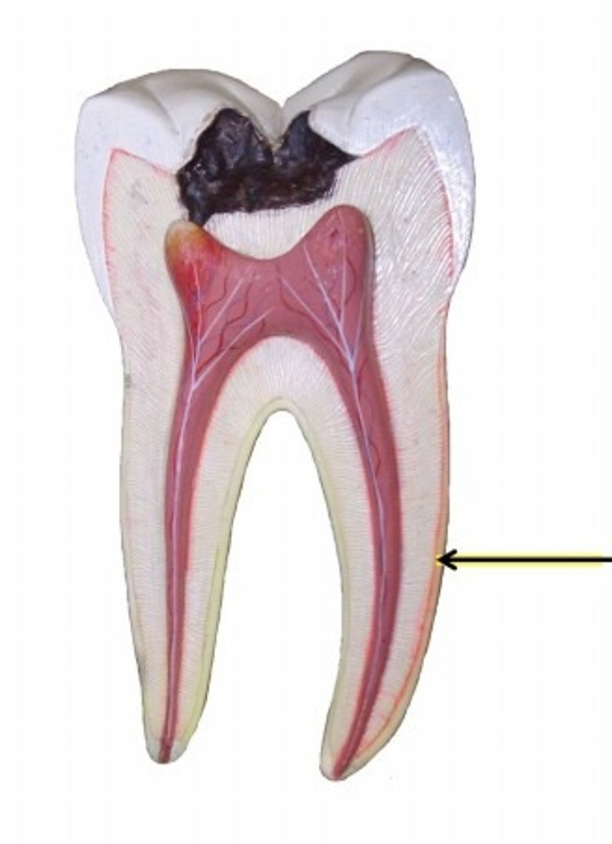
Pulp Cavity
softer inner part
blood vessels & nerves
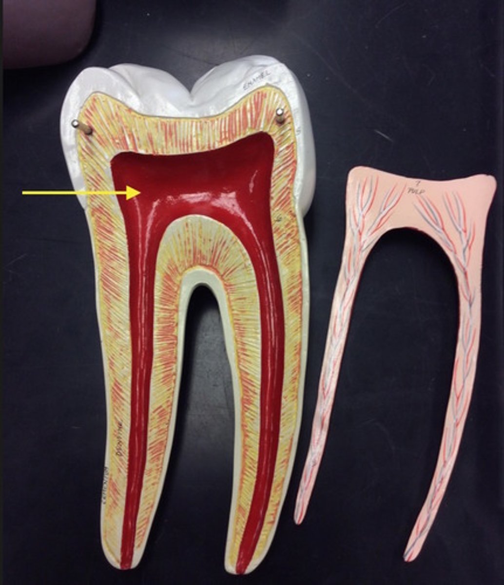
Root Canal
the space within the root of the tooth
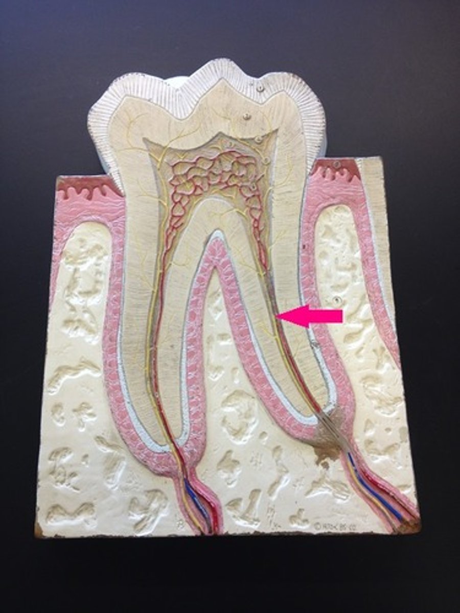
Sensory Nerve
innervates the pulp cavity
- exits via root canal
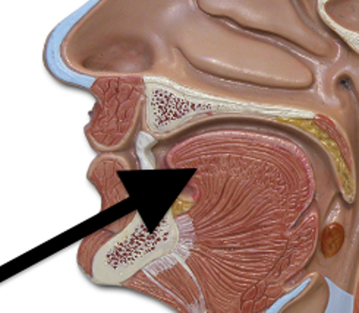
Tongue
skeletal muscle!!!
-food manipulation
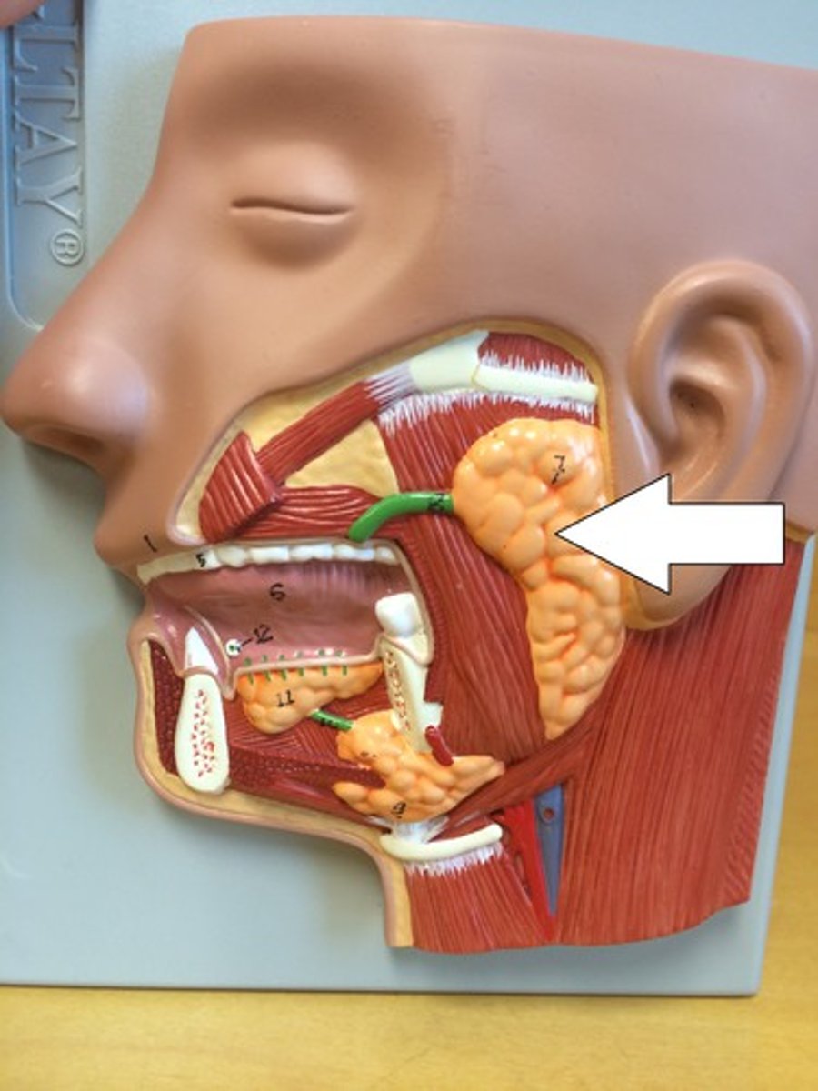
parotid gland
in front of the ear
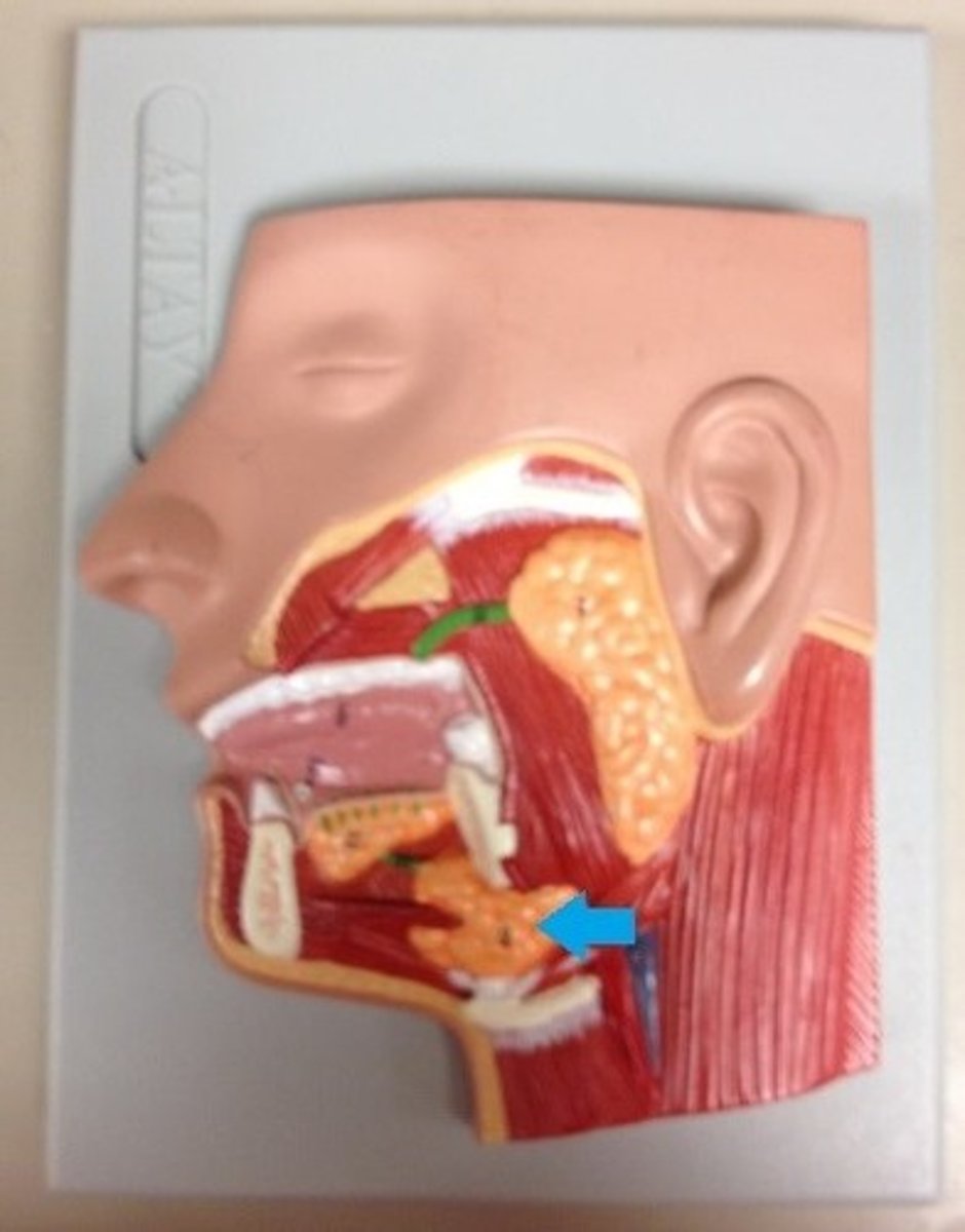
Submandibular gland
under the mandible
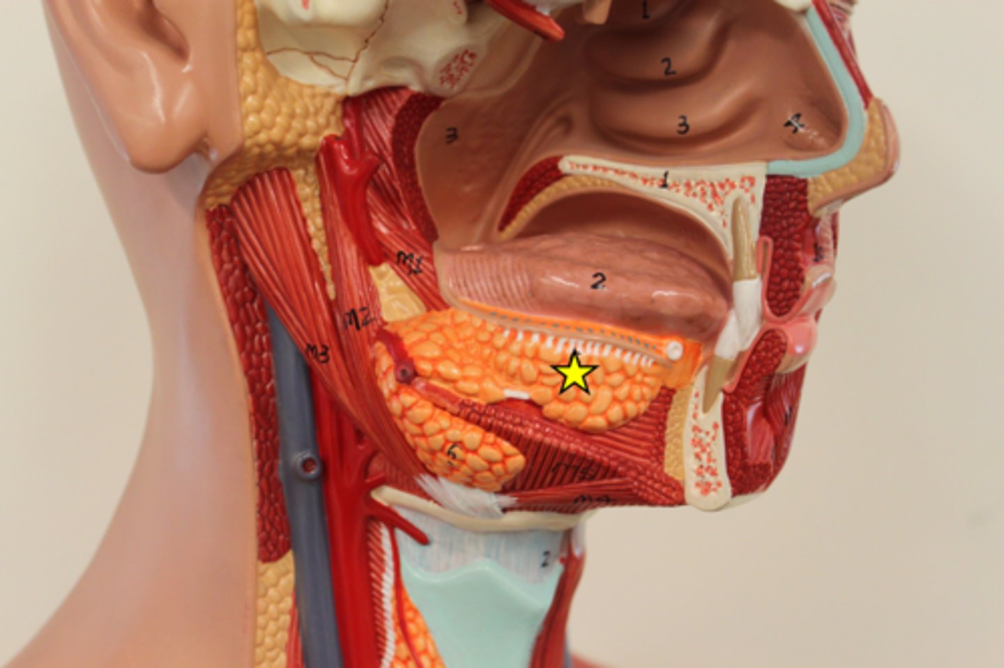
Sublingual gland
under the tongue
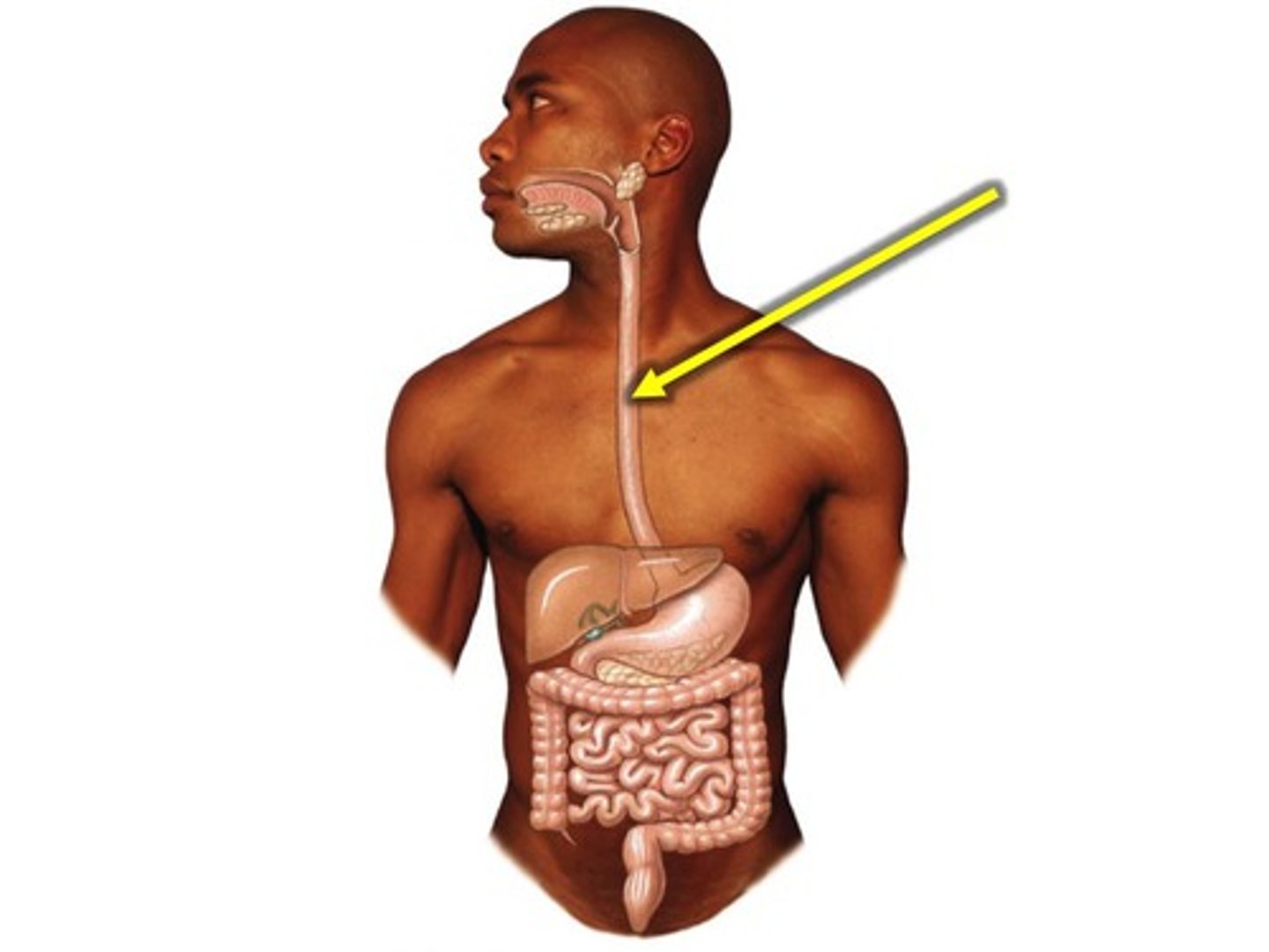
Esophagus
- tube that connects the throat to the stomach
- peristalsis
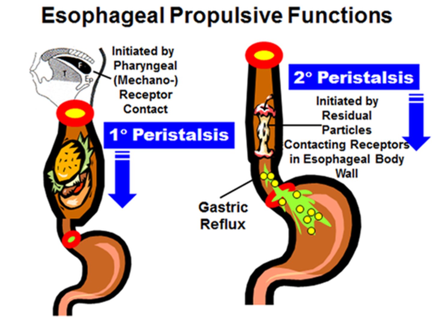
Peristalsis
contraction & relaxation of a muscular tube
move food in 1 direction
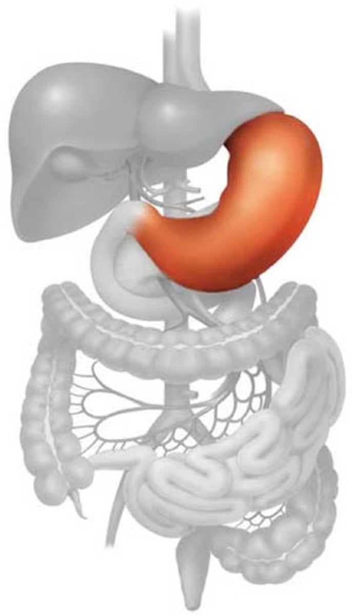
Stomach
- sac which holds, churns, & crushes food
- HCl & enzymes
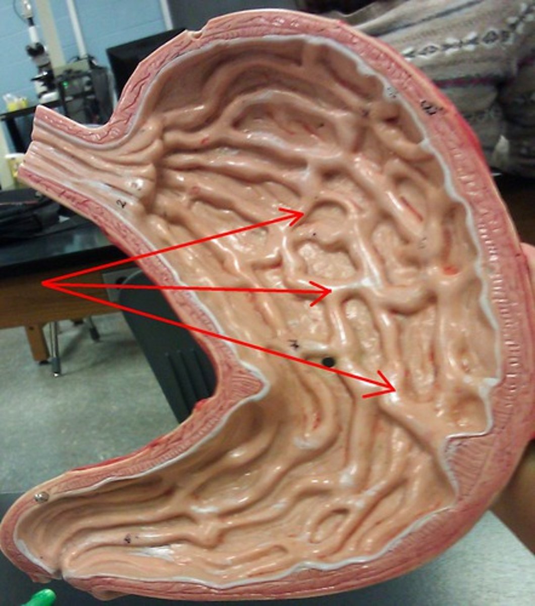
Rugae
folds of the mucosa (in the stomach)
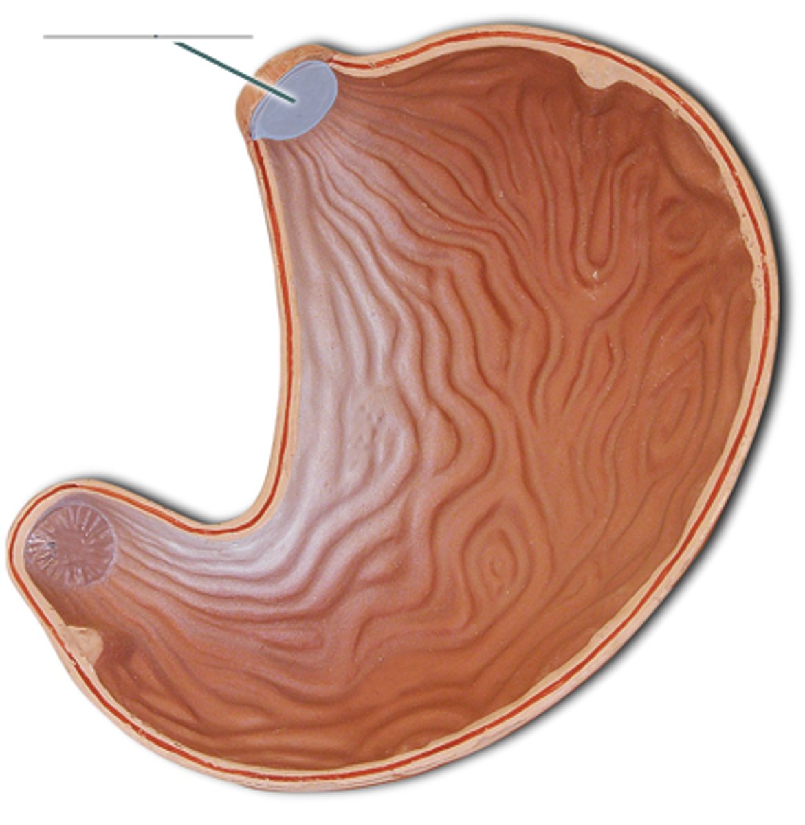
Gastroesophageal Sphincter
circular muscle!!!
controls speed of bolus
keeps acidic inside!
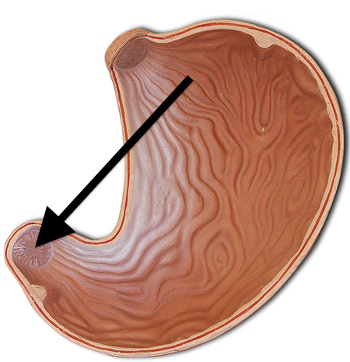
Pyloric Sphincter
controls the speed of chyme
Small intestine
-Primary site of nutrients absorption & chemical digestion
-22 feet long
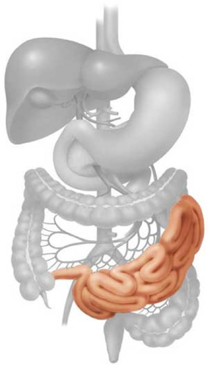
Duodenum
first & shortest part
- "C" shaped
-Receives liver & pancreatic juices
-TONS of digestion happens here
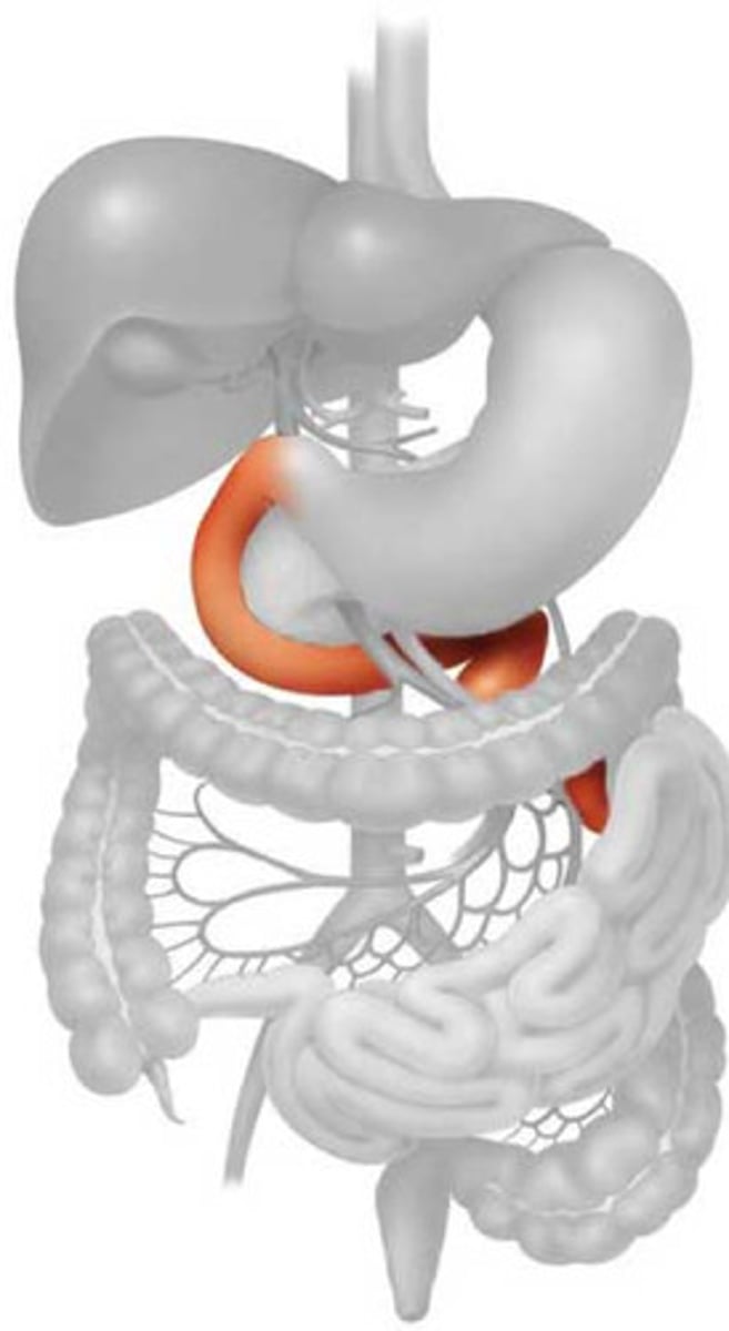
Jejunum
where most absorption happens
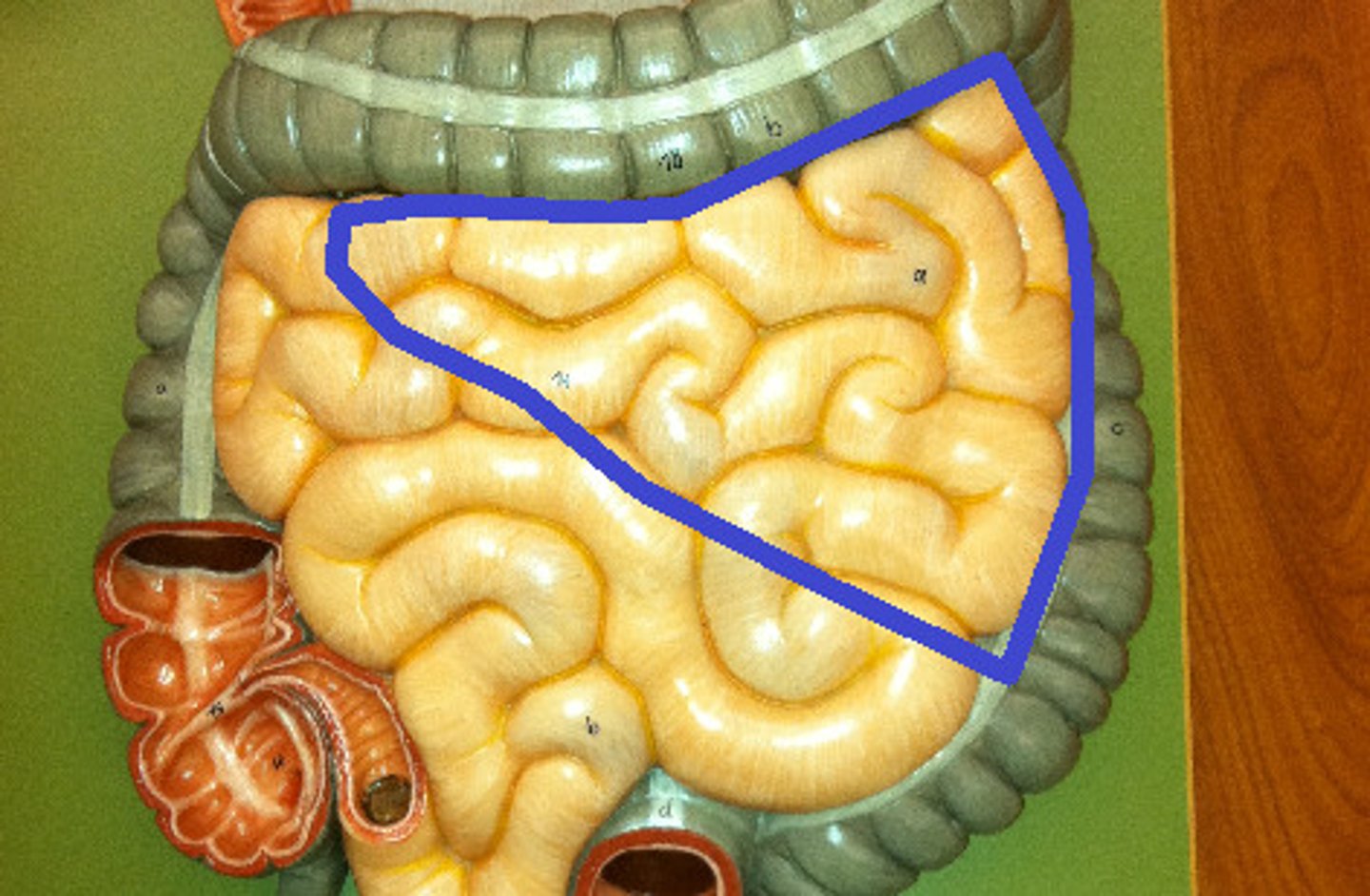
Ileum
last part!!
- connect w/the large intestine
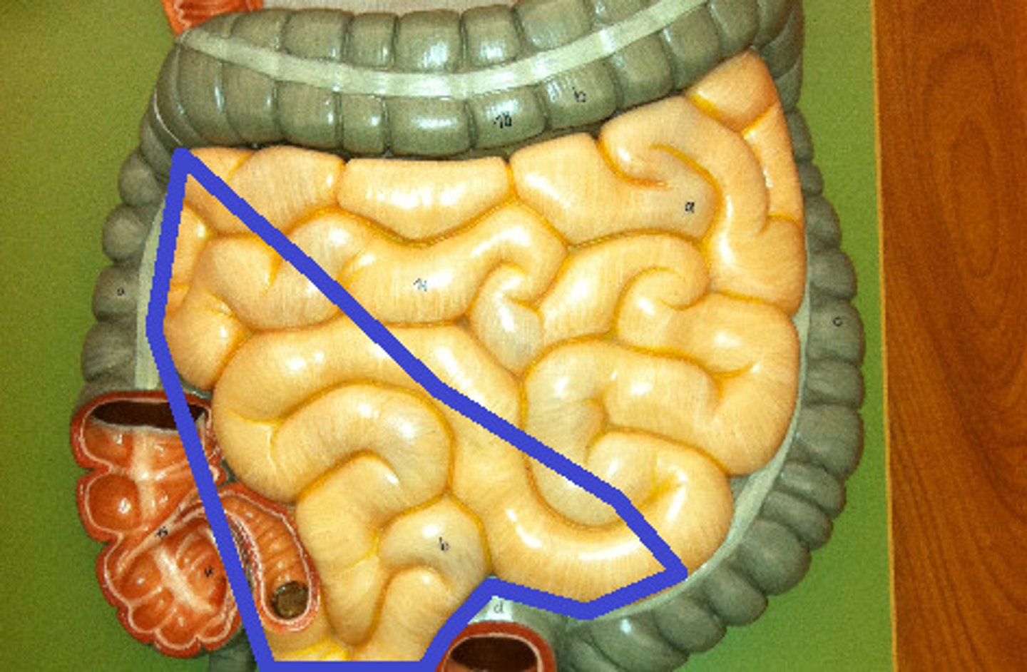
Ileocecal Valve
sphincter that controls the flow into the cecum
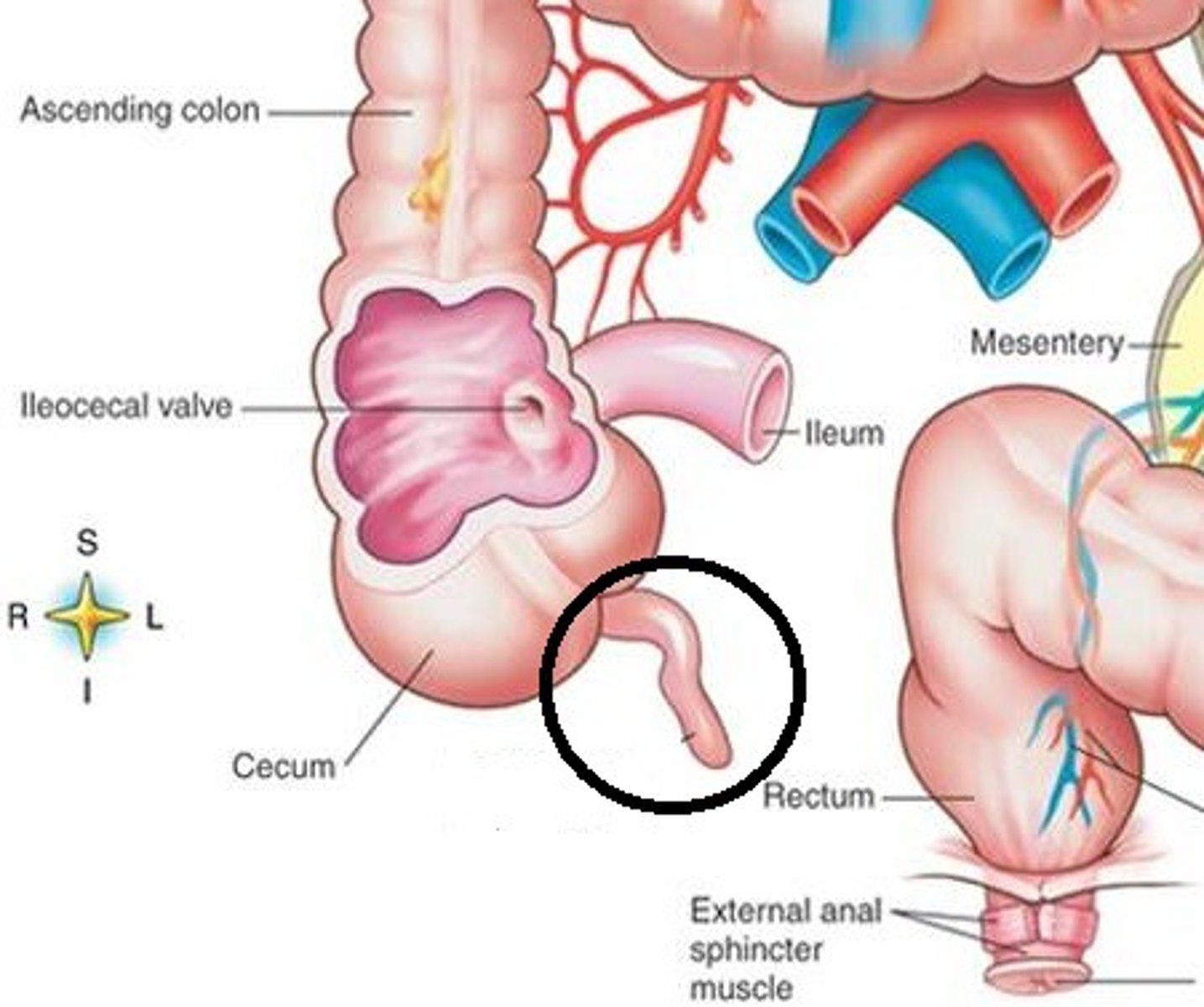
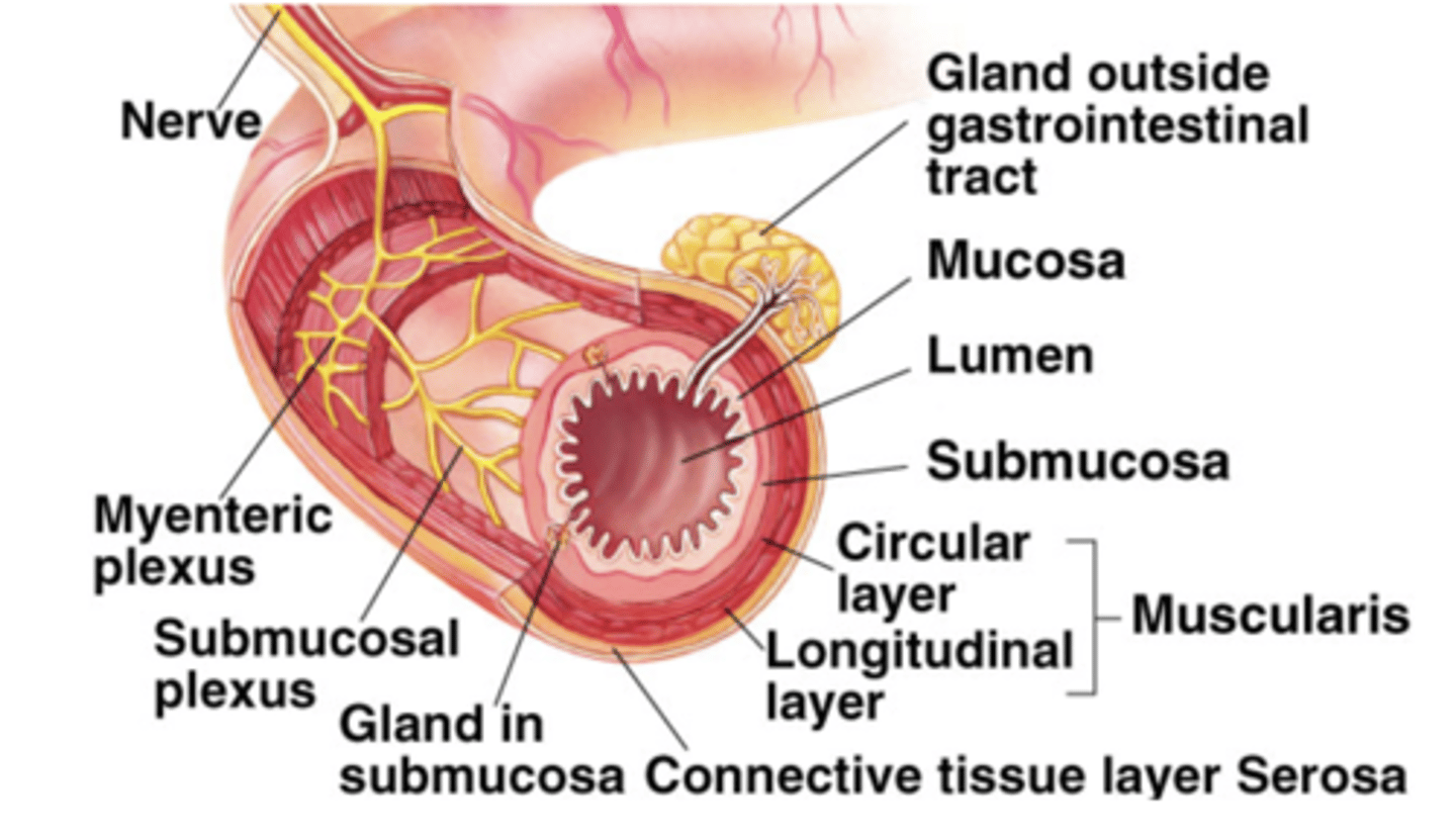
Mucosa
innermost layer covered in cells specialized for absorption
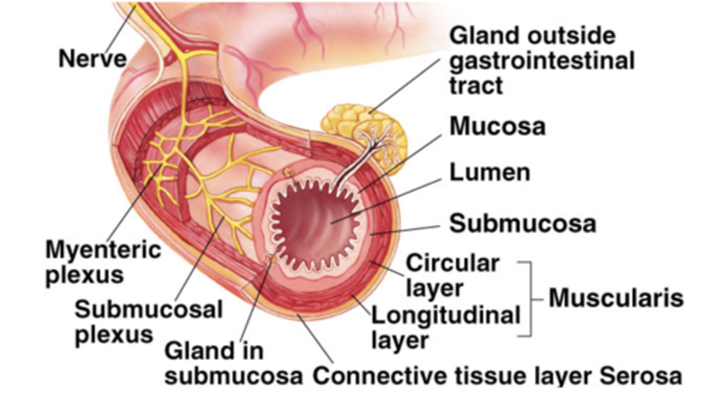
Submucosa
thin supportive mucosa containing nerves & blood vessels
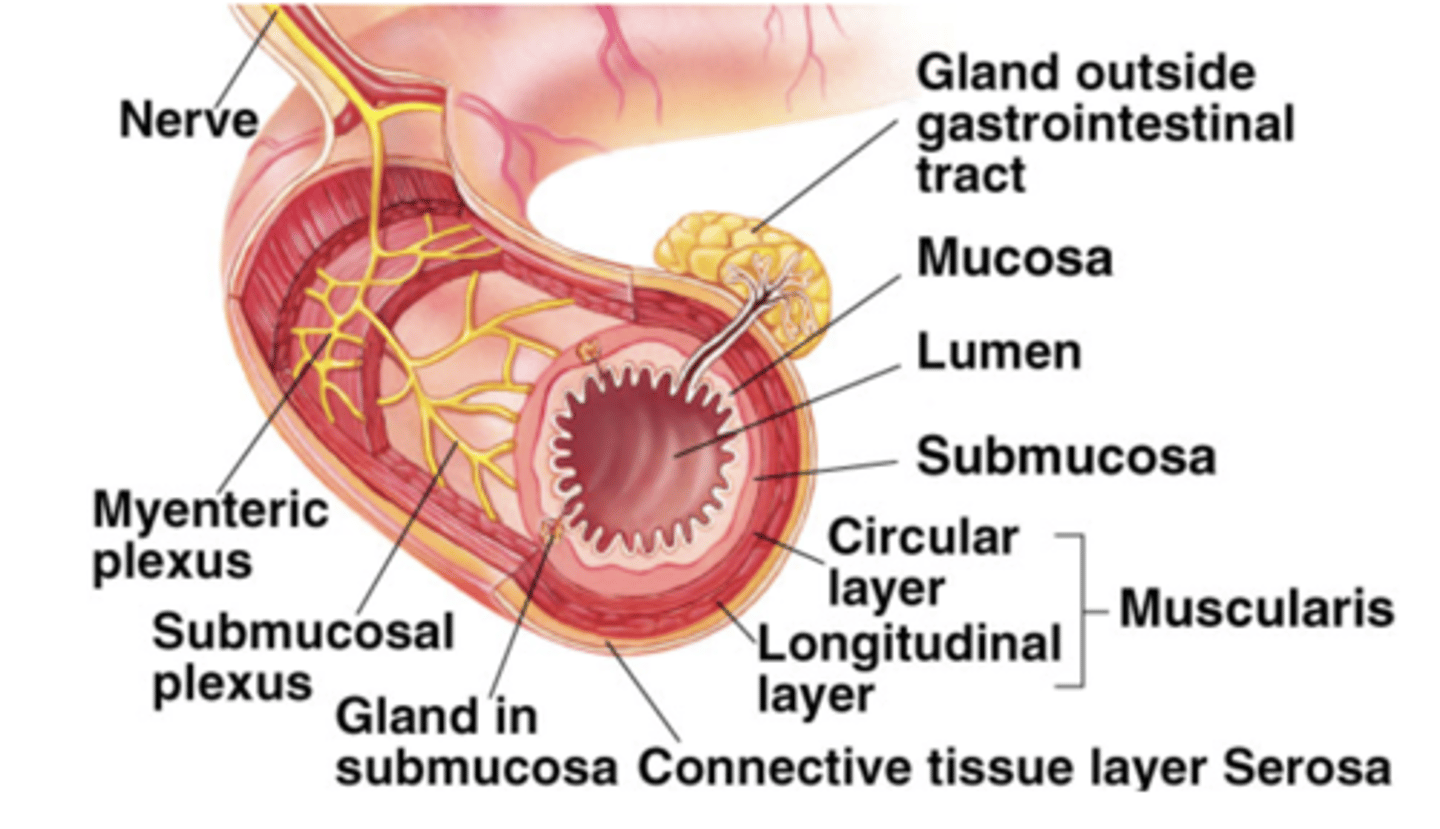
Muscularis
smooth muscle layer that causes peristalsis
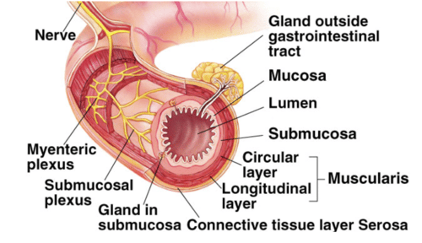
Serosa
outermost layer, thin connective tissue
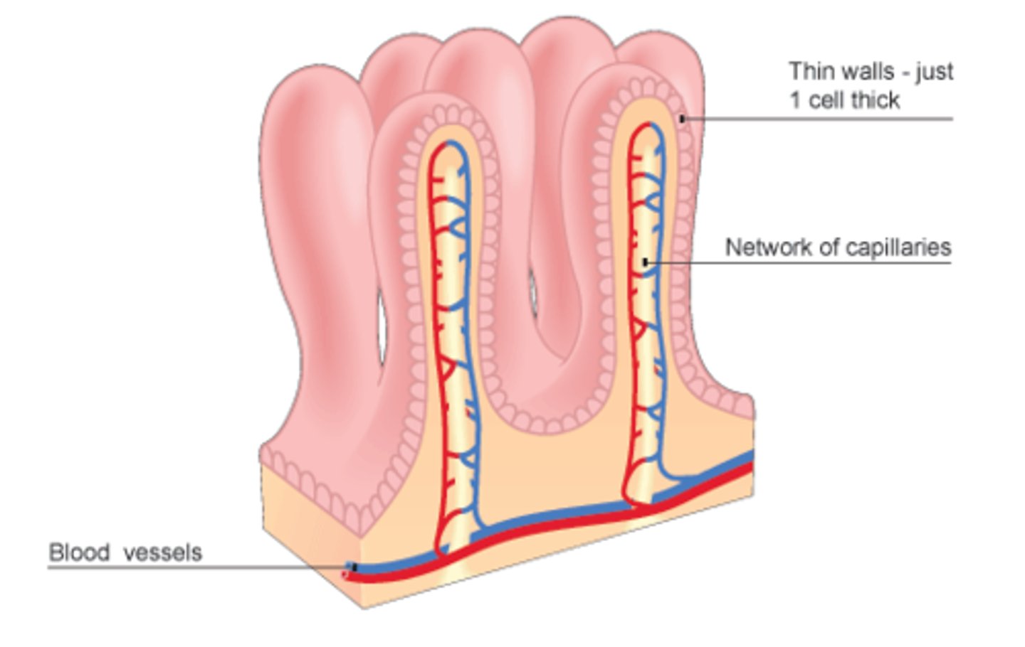
Villus
finger-like projections covered by microvilli
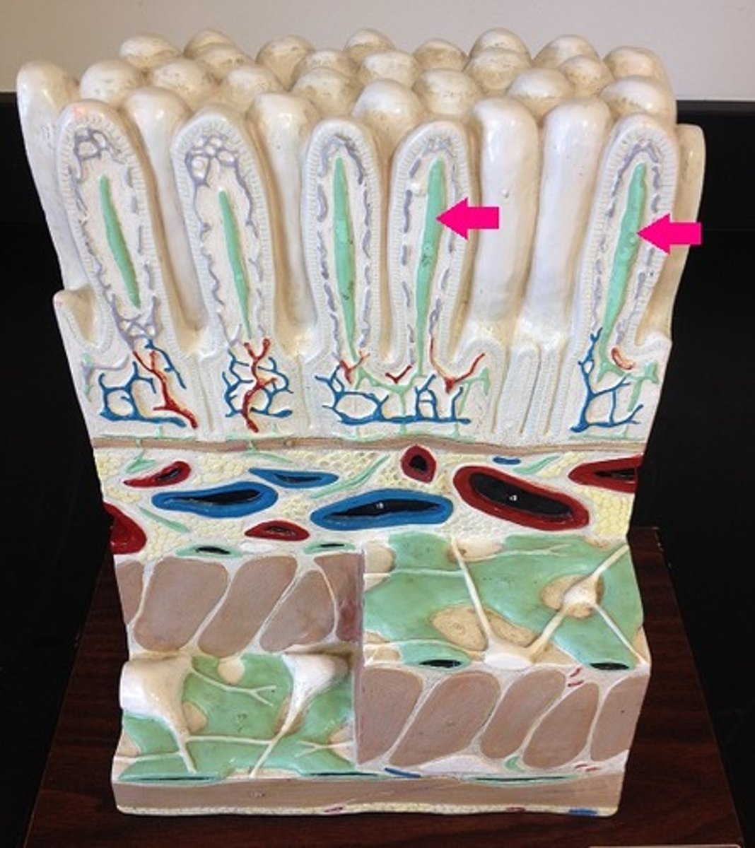
Lacteal
lymphatic vessel
-helps with in fatty absorption
Large intestine
water absorption!
- bacteria make vitamins !
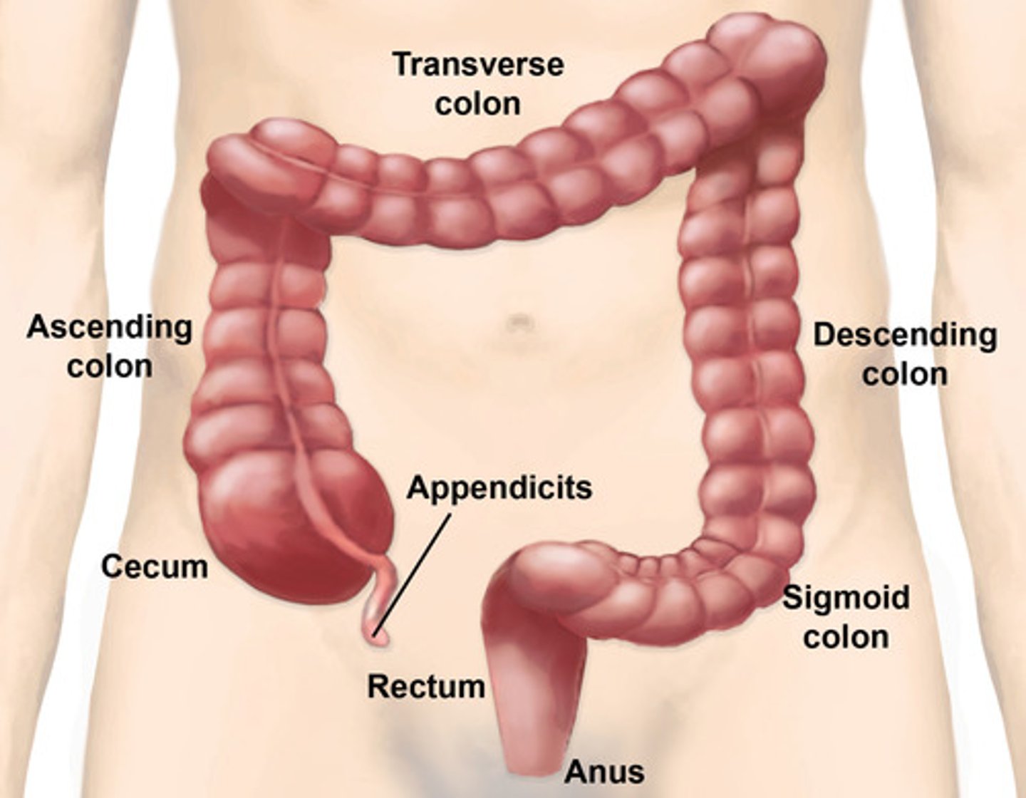
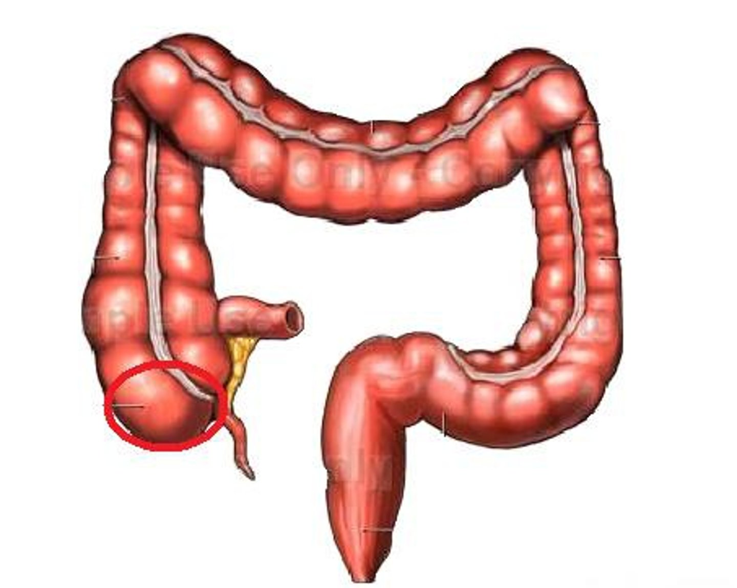
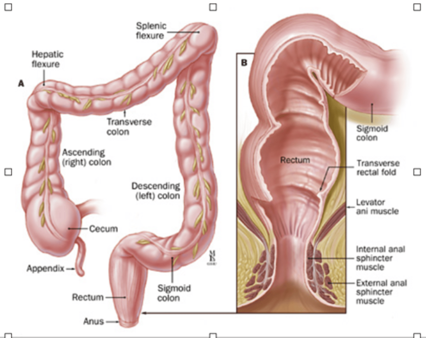
Cecum
transitionary pouch
- receives fecal matter!
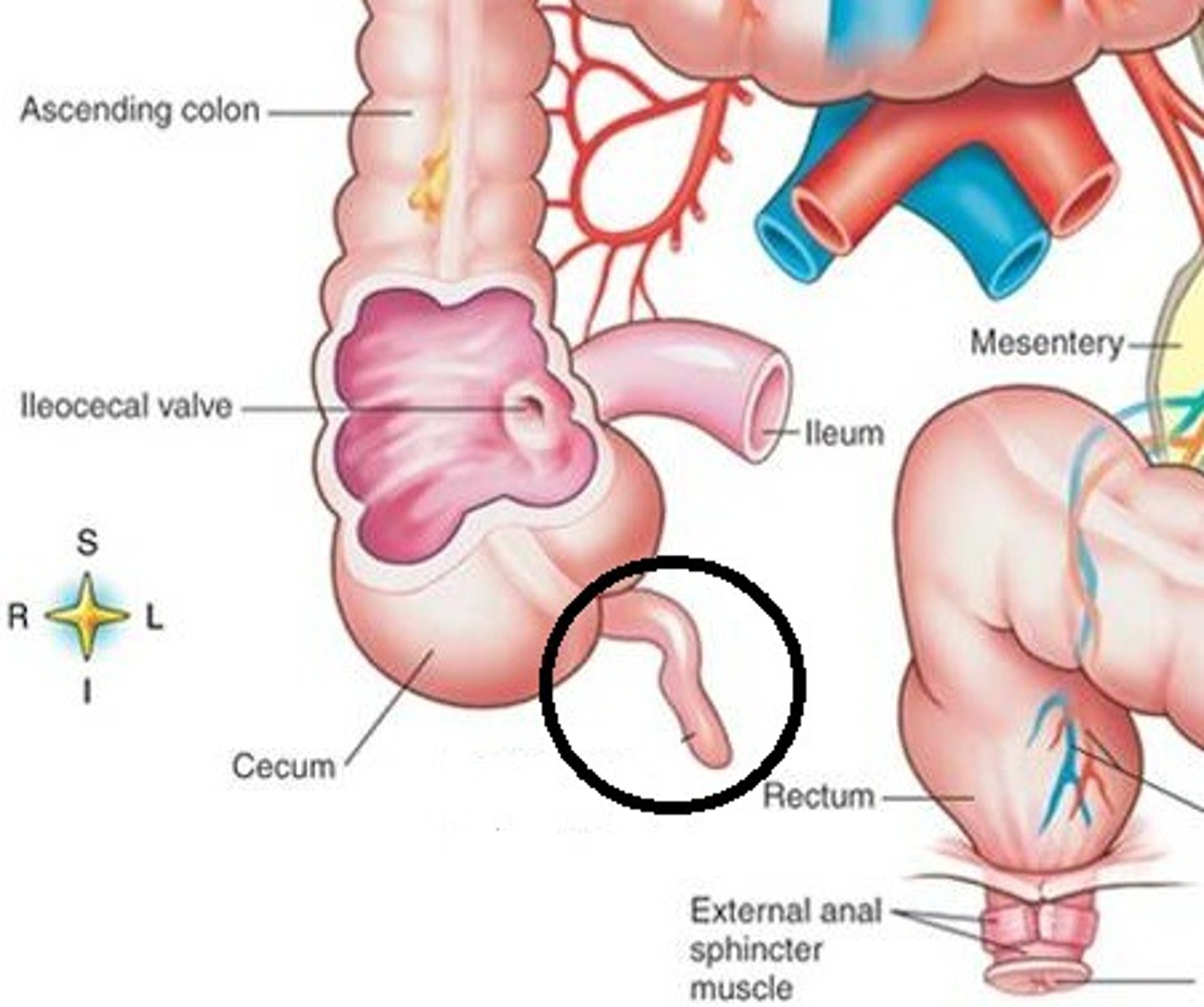
Vermiform Appendix
vestigial structure
-May hold helpful bacteria
-appendicitis!
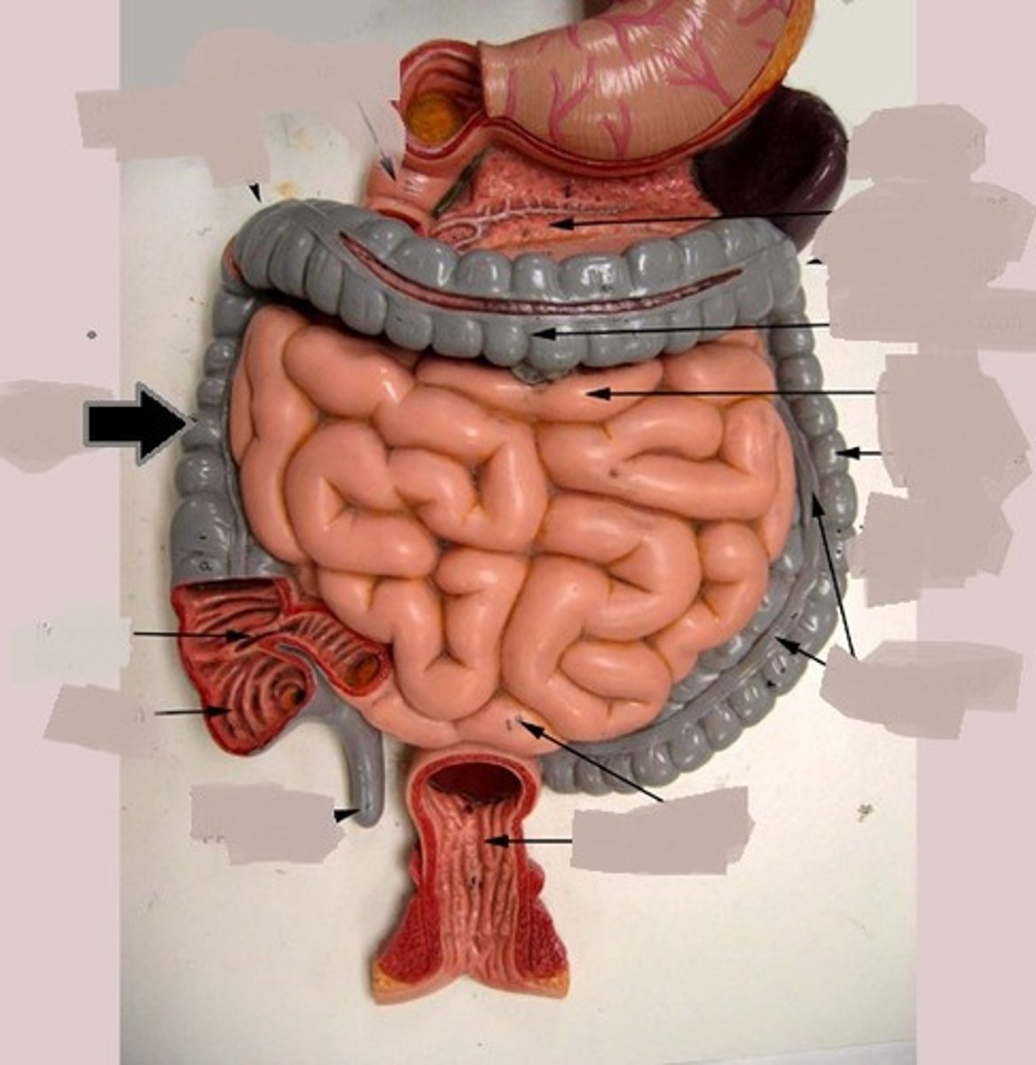
Ascending Colon
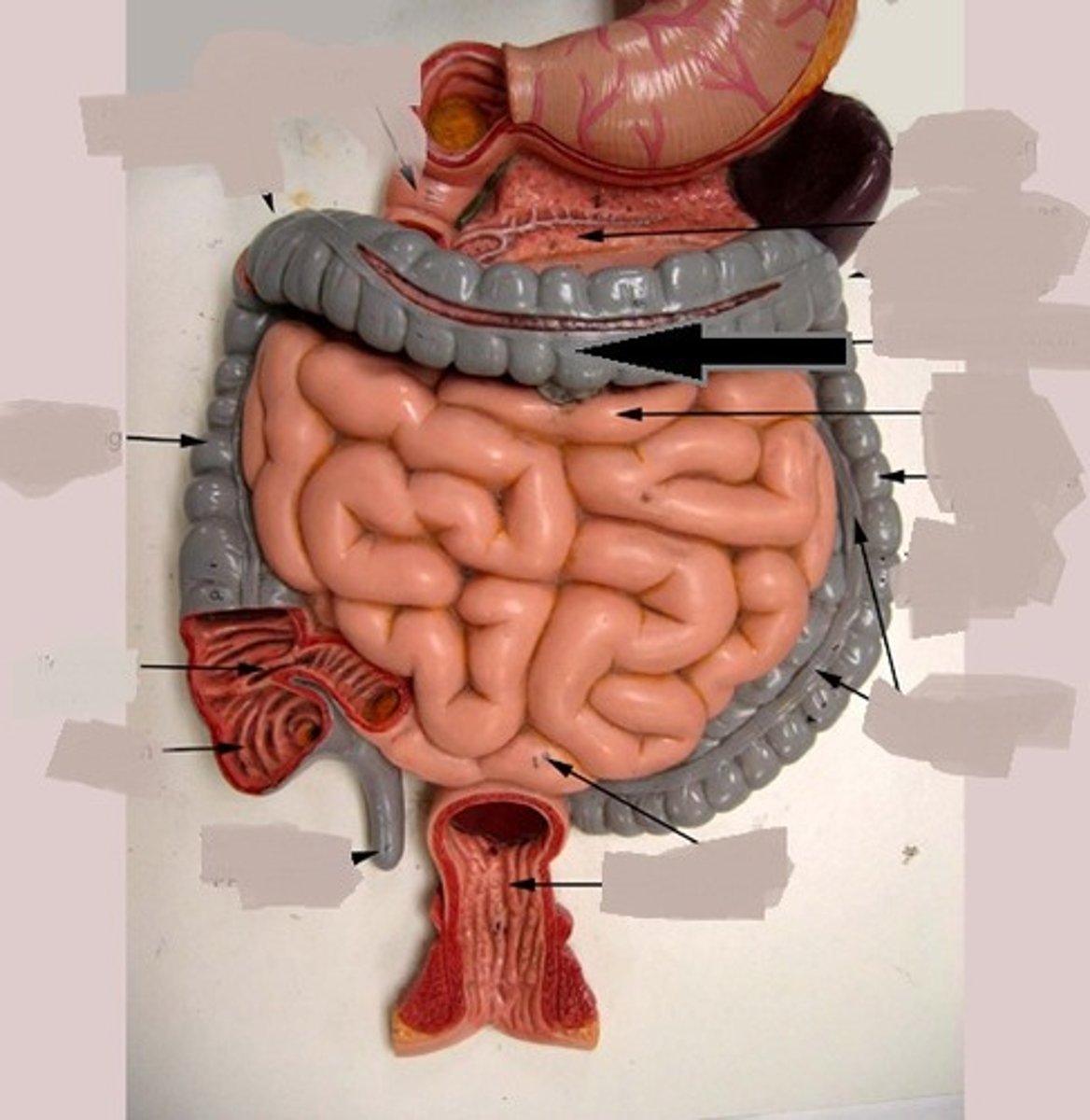
Transverse colon
descending colon
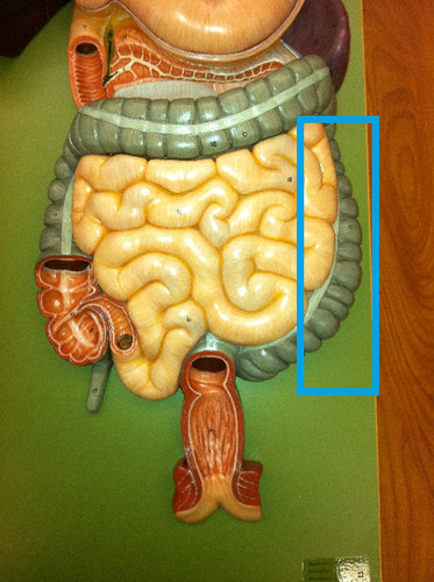
Sigmoid Colon
"S" shaped section of the large intestine
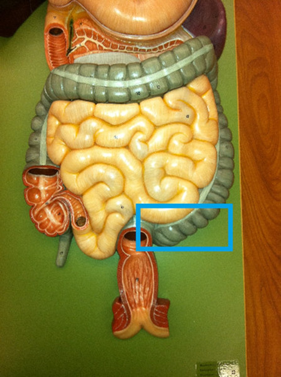
Rectum
straight portion of the large intestine
-holds feces Temporary
-(stretch receptors~!!!)
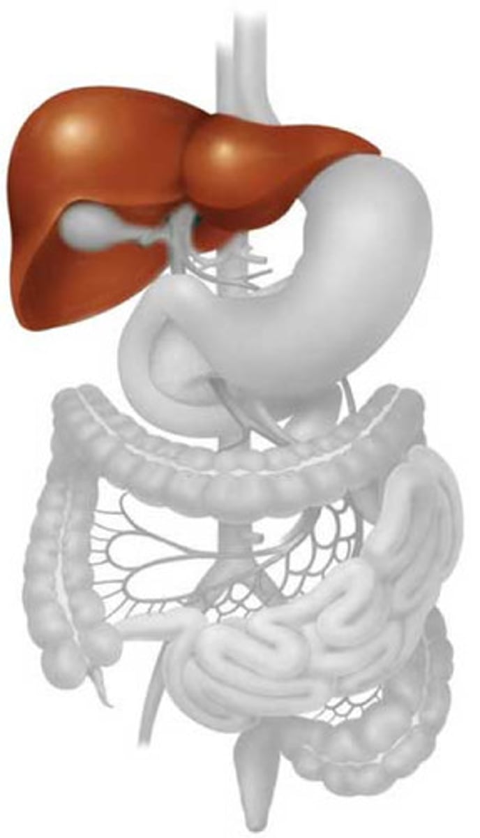
Liver
makes bile
- aid in fat absorption
-detoxification : EX: beer!
Gallbladder
stores bile
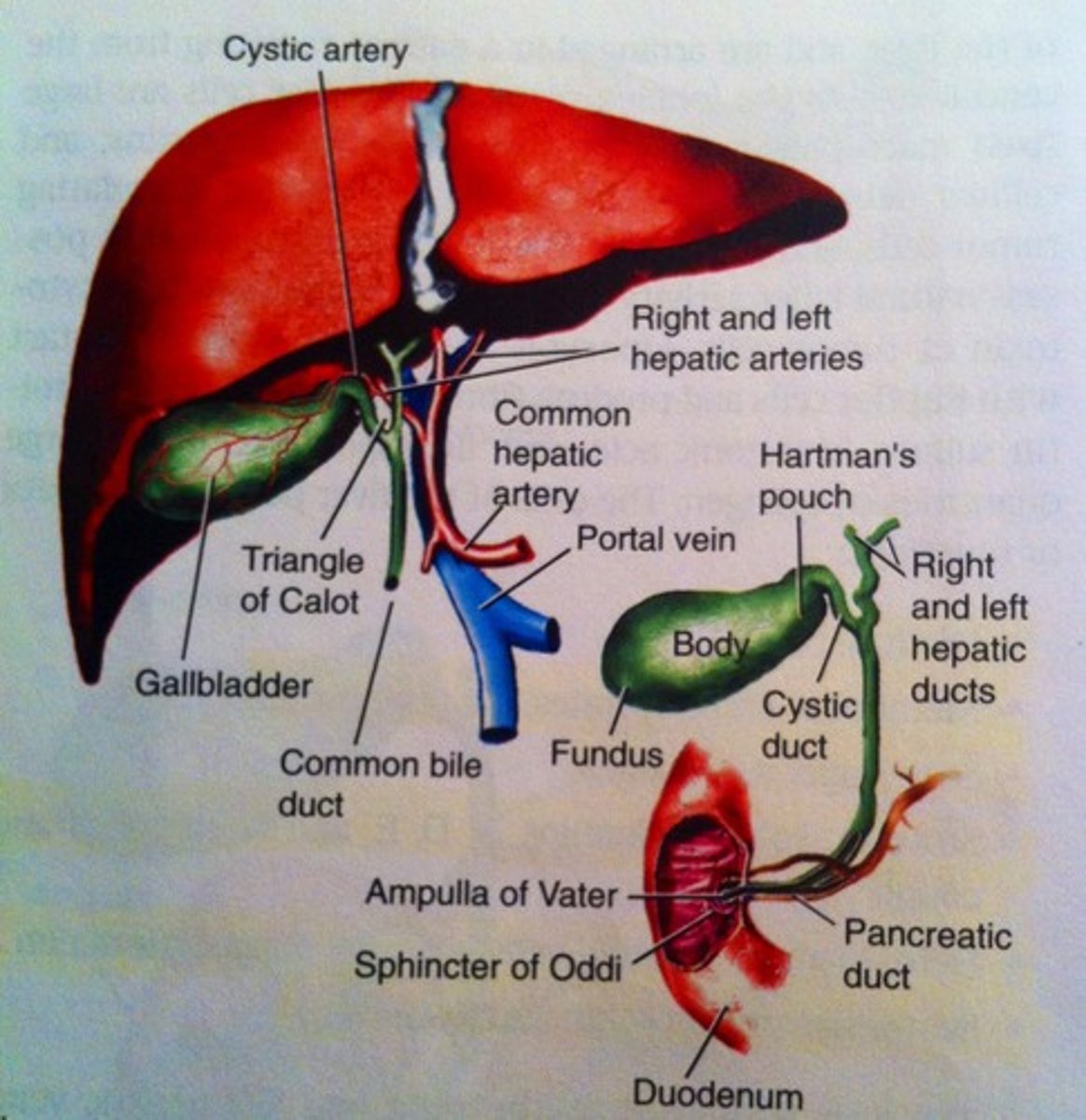
pancreas
-MAkes digestive enzymes that break down all macro molucues
-glucose homeostasis
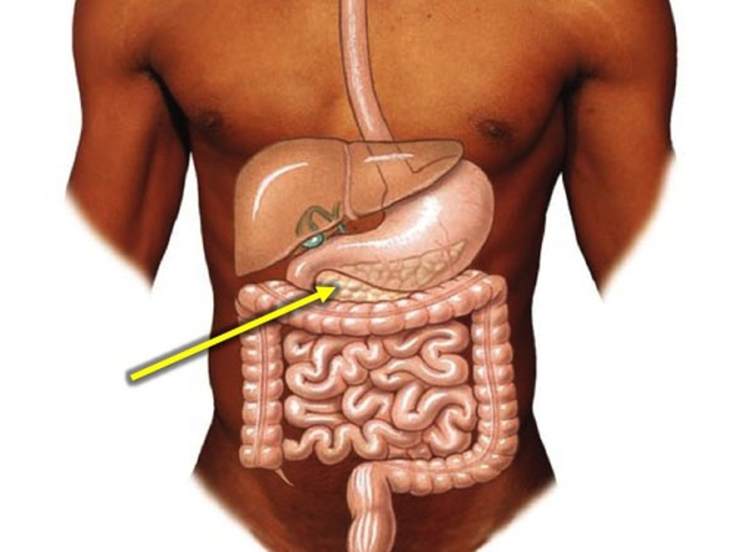
Common Bile Duct
connects the hepatic and cystic duct
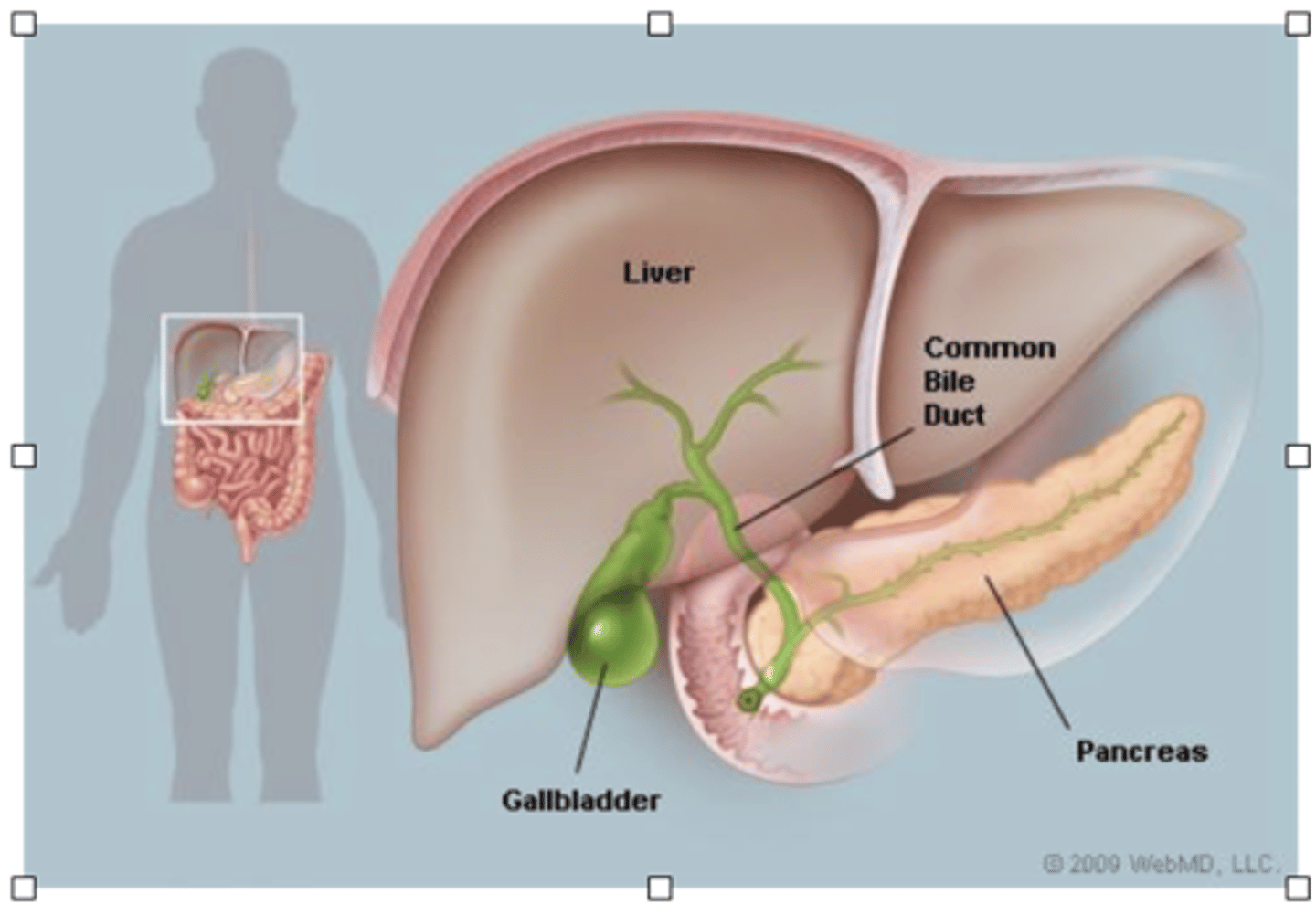
Pancreatic Duct
connects pancreas to duodenum
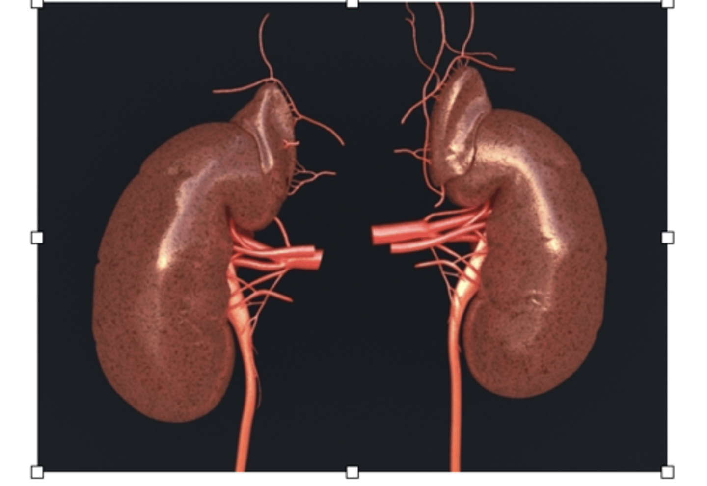
kidney
2 layers cortex (outside) medulla (inside)
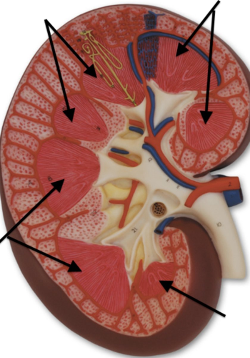
Renal Pyramid
Pyramid shaped structure within the kidney
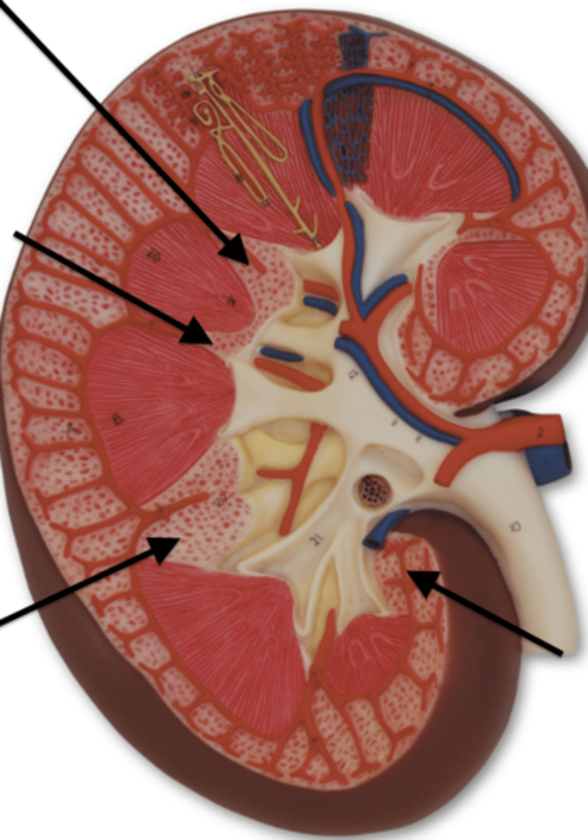
Renal Column
cortex tissue that is in between the pyramids
Calyx
funnel that catches urine
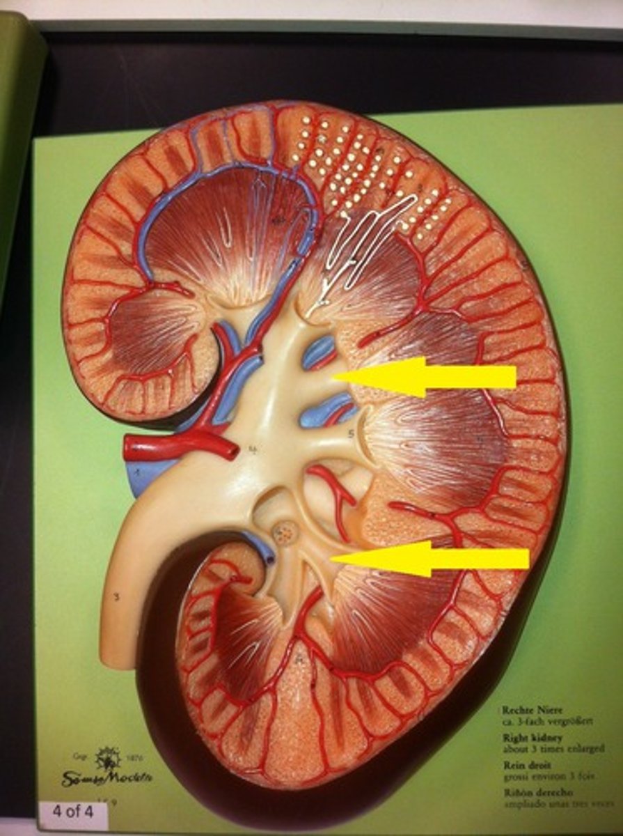
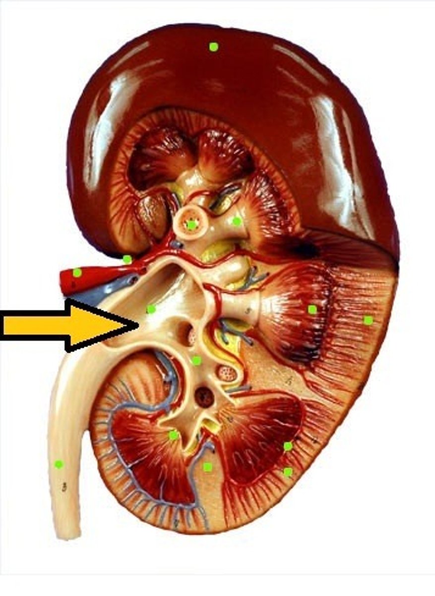
Renal Pelvis
collects urine
- connects to ureters
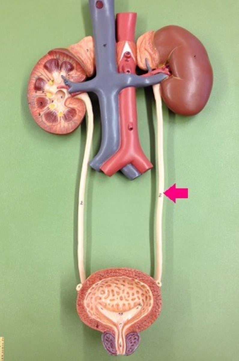
Ureter
extends kidney to bladder
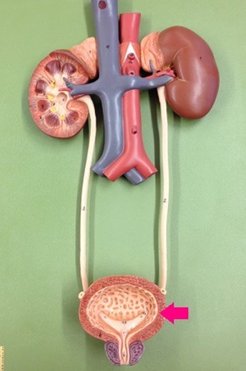
Urinary Bladder
urine storage
- rugae for expansion!!!
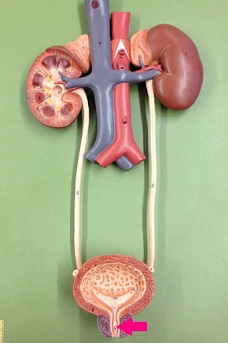
Urethra
carries urine outside of the body
-size difference!
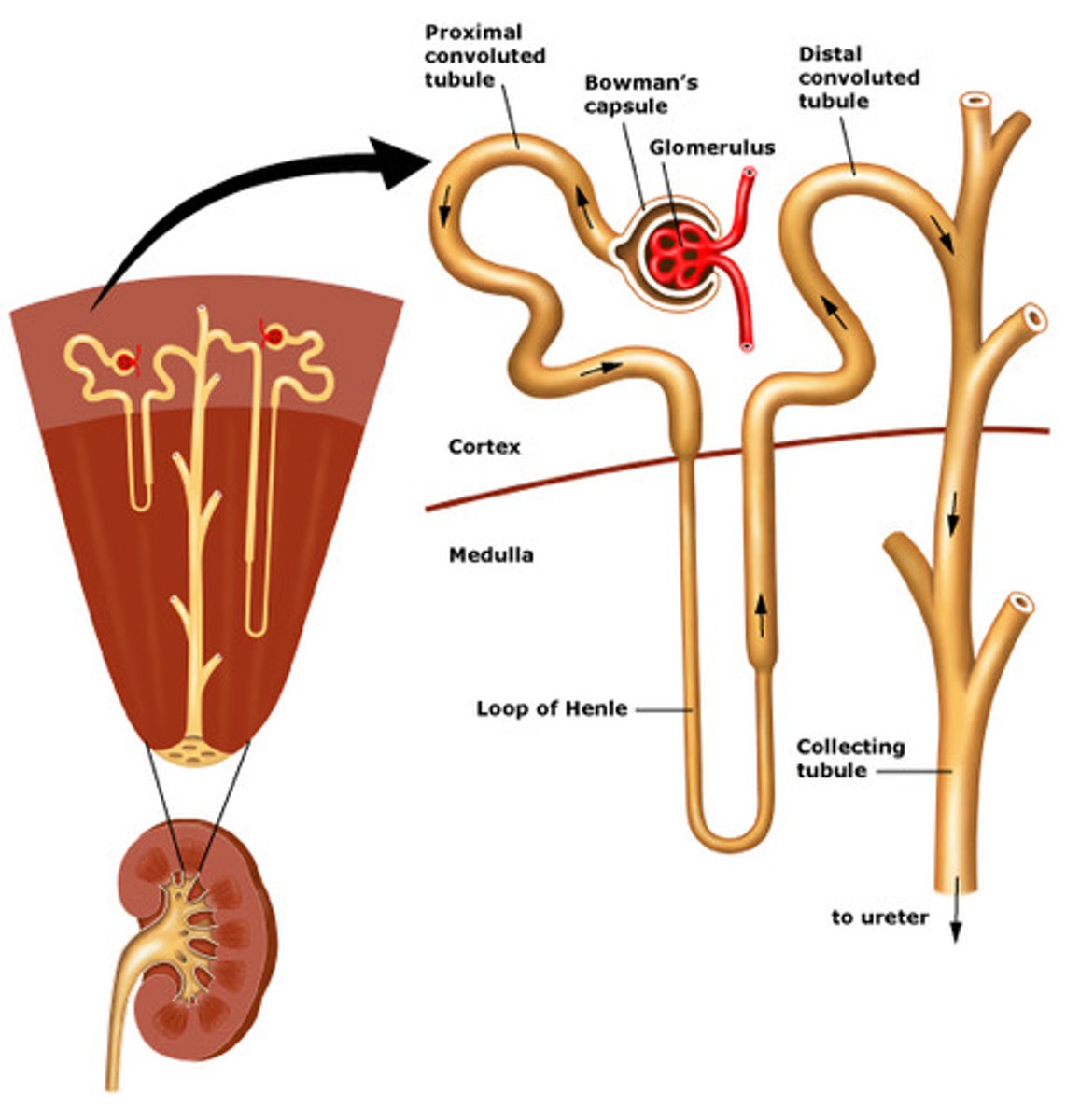
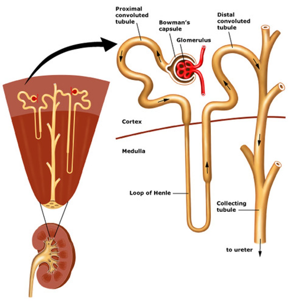
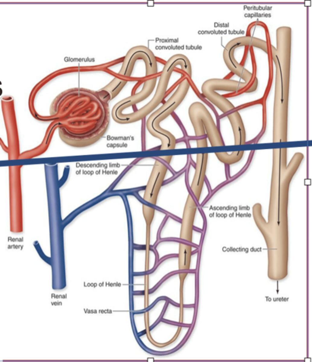
Nephron
the functional unit of the kidney
-filters blood
-makes urine
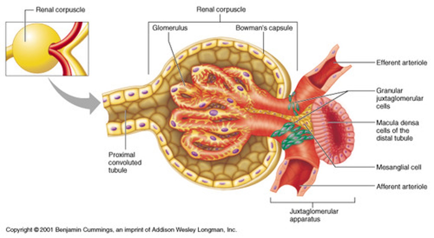
Glomerulus
ball of blood vessels
Glomerular Space
area between glomerulus & capsule
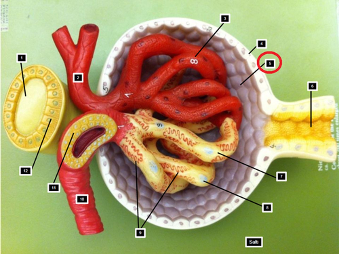
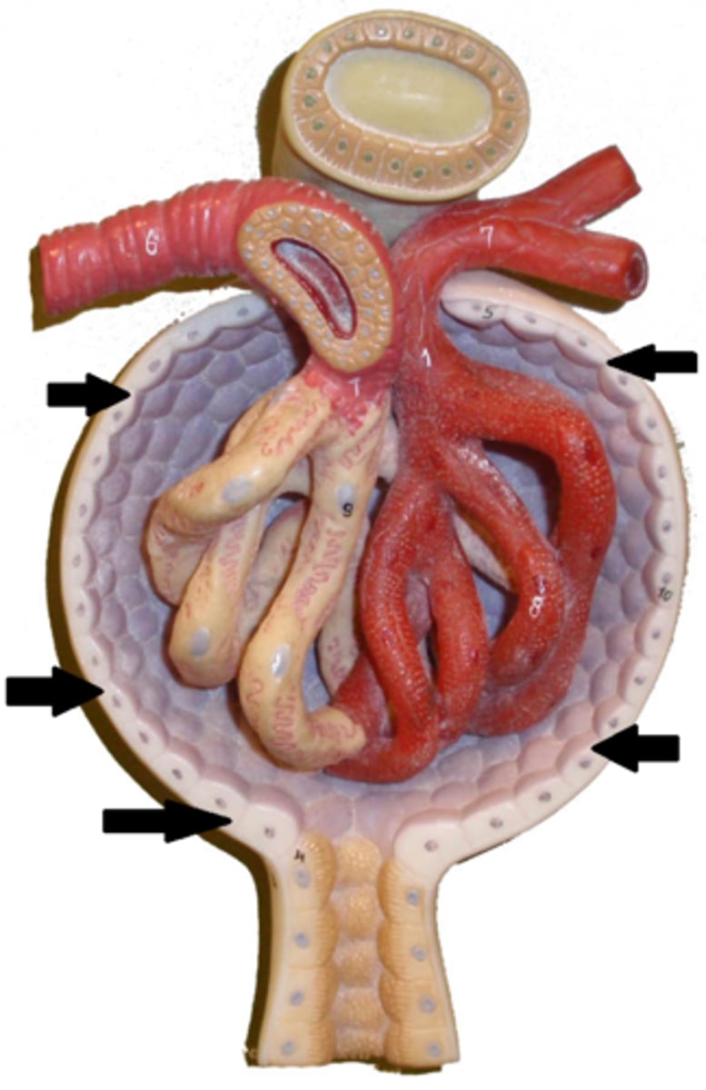
Glomerular Capsule
outside layer, covers the glomerulus
Proximal Convoluted Tubule
first tubule
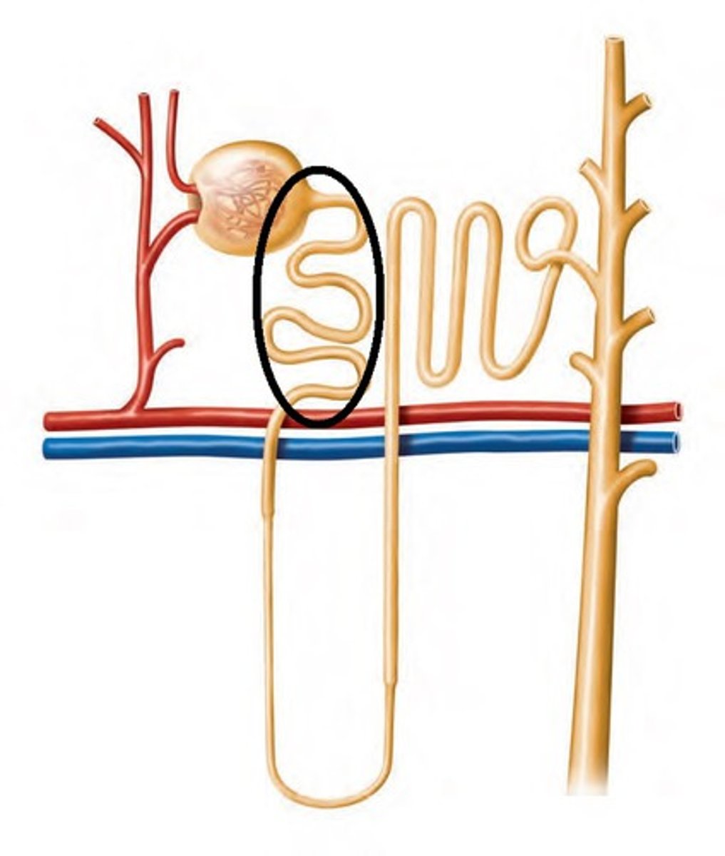
Descending Limb of Loop of Henle
part that dips the renal medulla
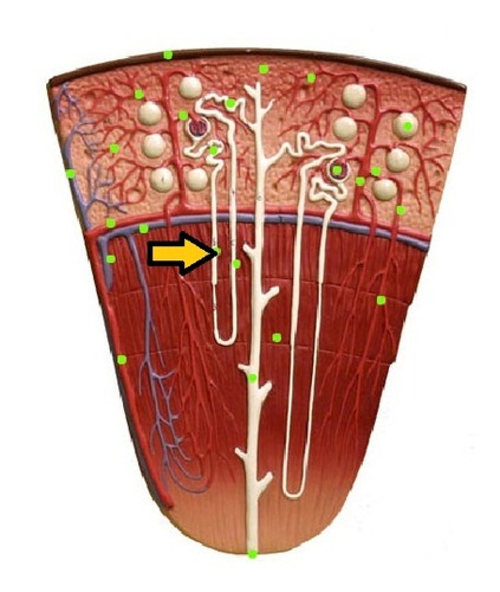
Ascending Limb of Loop of Henle
part of the loop of that goes to go up
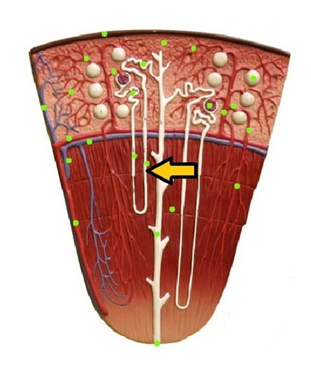
Distal Convoluted Tubule
reabsorption of Na+ & H2O
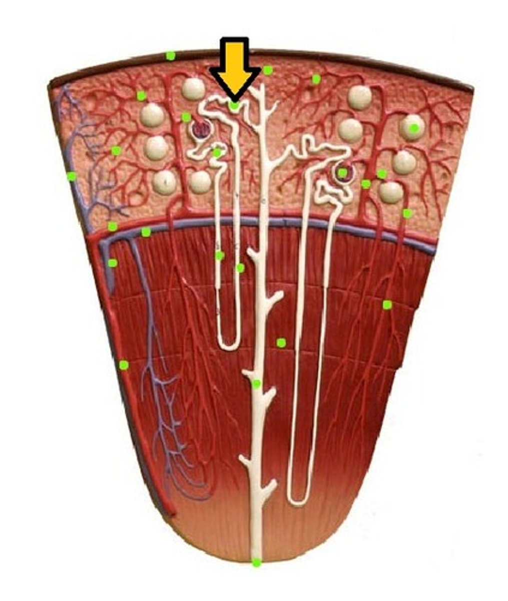
Peritubular Capillaries
surround the glomerulus & proximal /distal tubules
-in the cortex
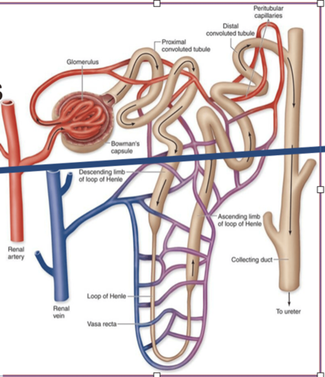
Vasa Recta
straight capillaries
-in the medulla
Afferent Arteriole
"approaches" the glomerulus (thick)
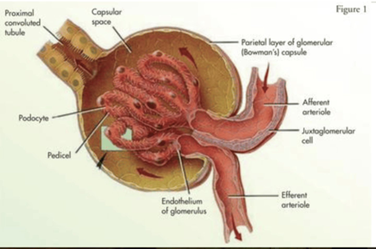
Efferent Arteriole
"exits"the glomerulus (thin)
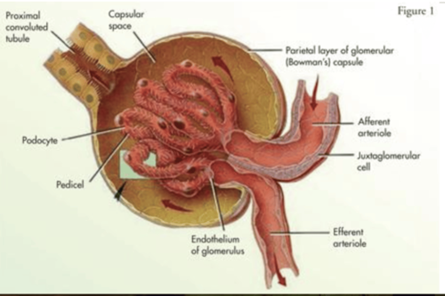
Juxtaglomerular Apparatus
-Includes:
juxtaglomerular cells (JG cells)
macula densa cells
-helps blood pressure homeostasis
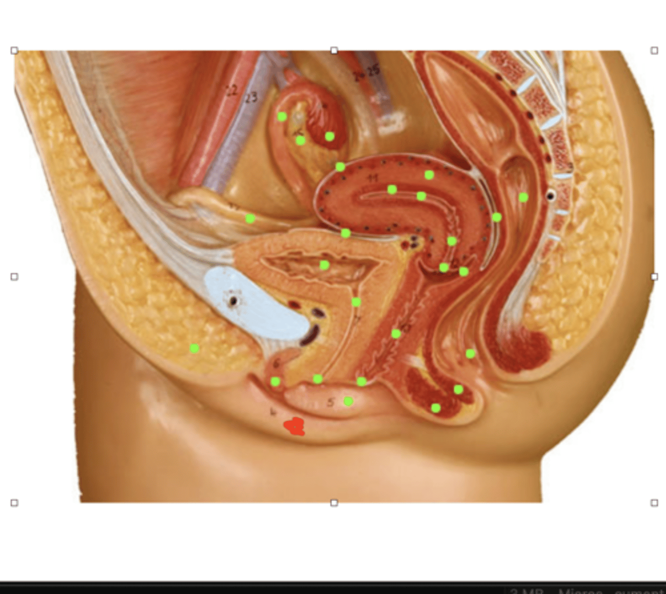
Labia Majora
two folds of hair covered skin extending from the mons pubis ---> perineum
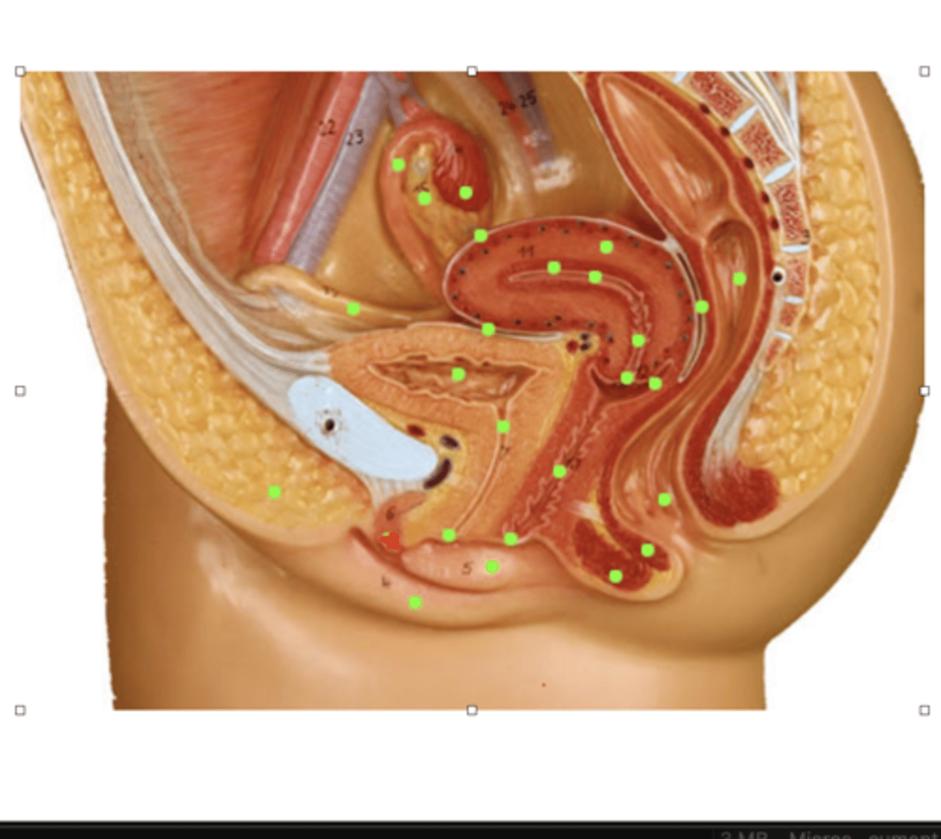
Labia Minora
pair of hairless skin folds
covers the opening of vagina & urethra
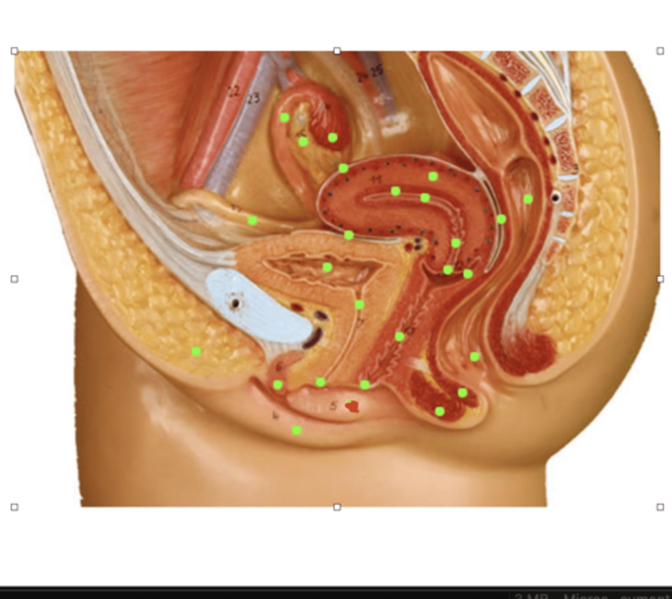
Clitoris
associated w/ female orgasm
-near apex of labia minora
Vagina
tube leading to the cervix
-rugae
-acidic environment !!!
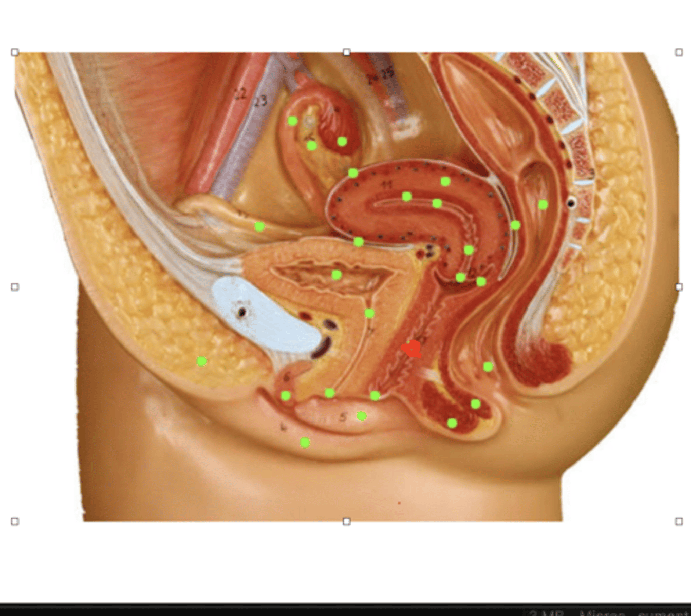
Perineum
between vulva & anus
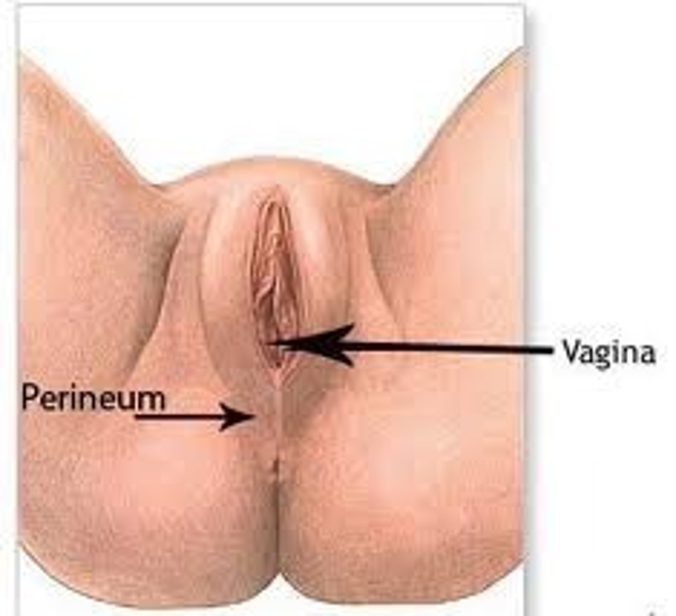
Uterus
site of implantation
- innermost layer sheds as menstrual cycle
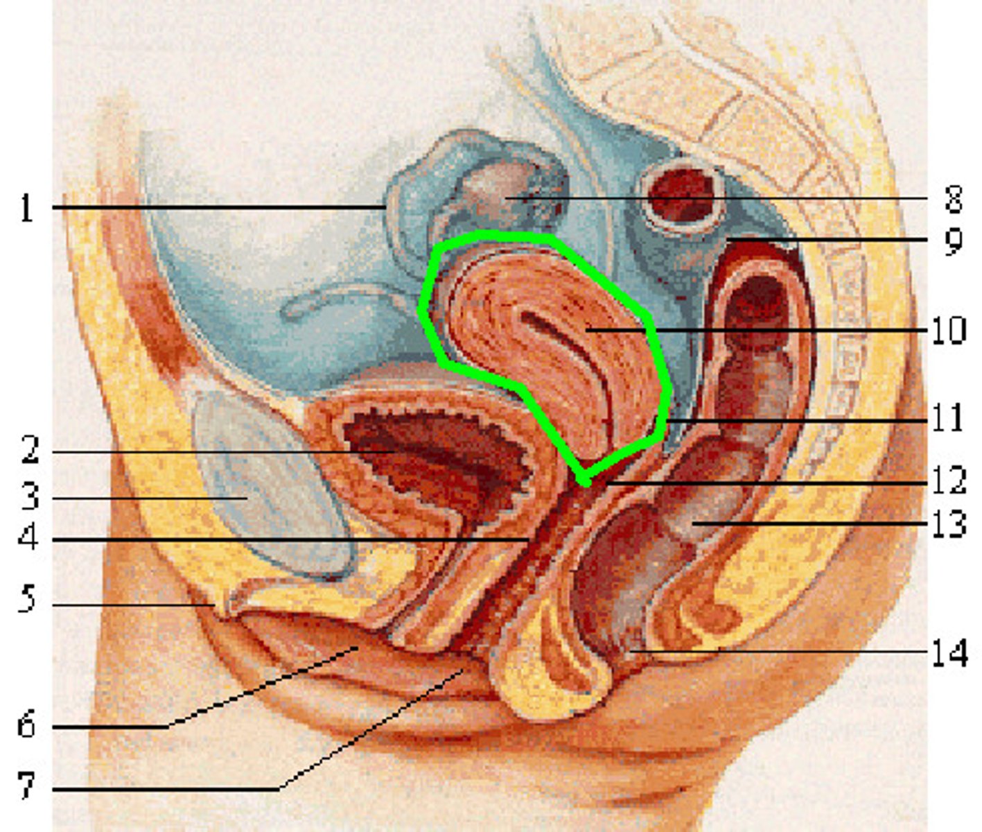
Cervix
narrow portion of the uterus
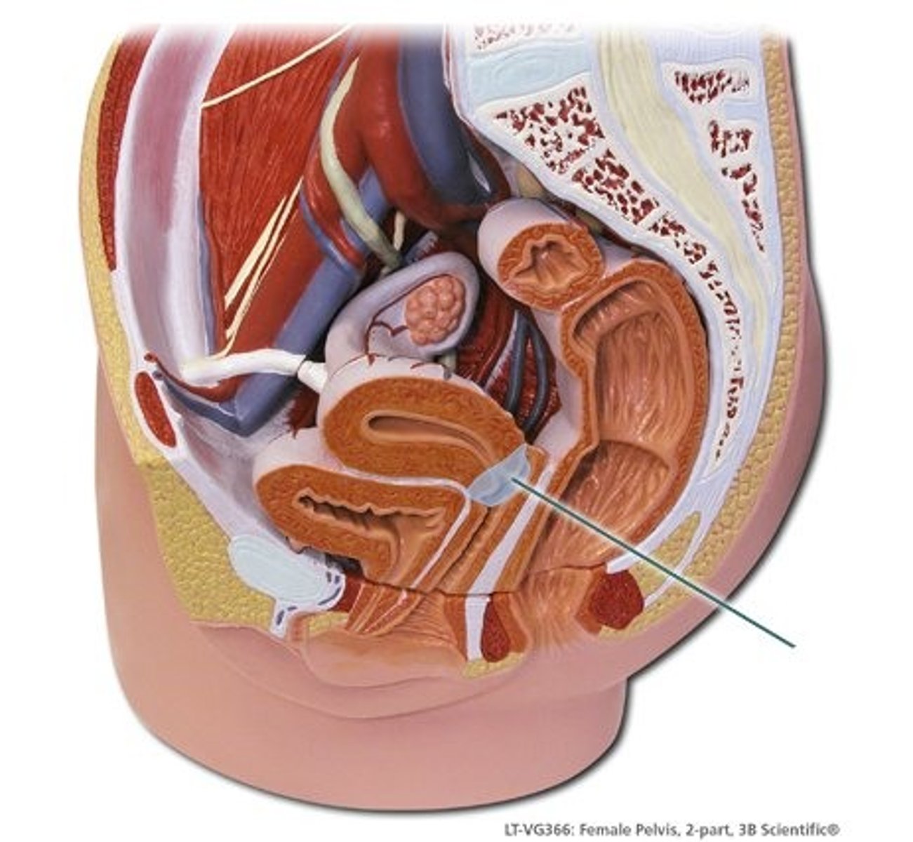
Oviducts
extend from the ovaries to the uterus
- site of fertilization NORMALLY
-transports fertilized oocyte → uterus
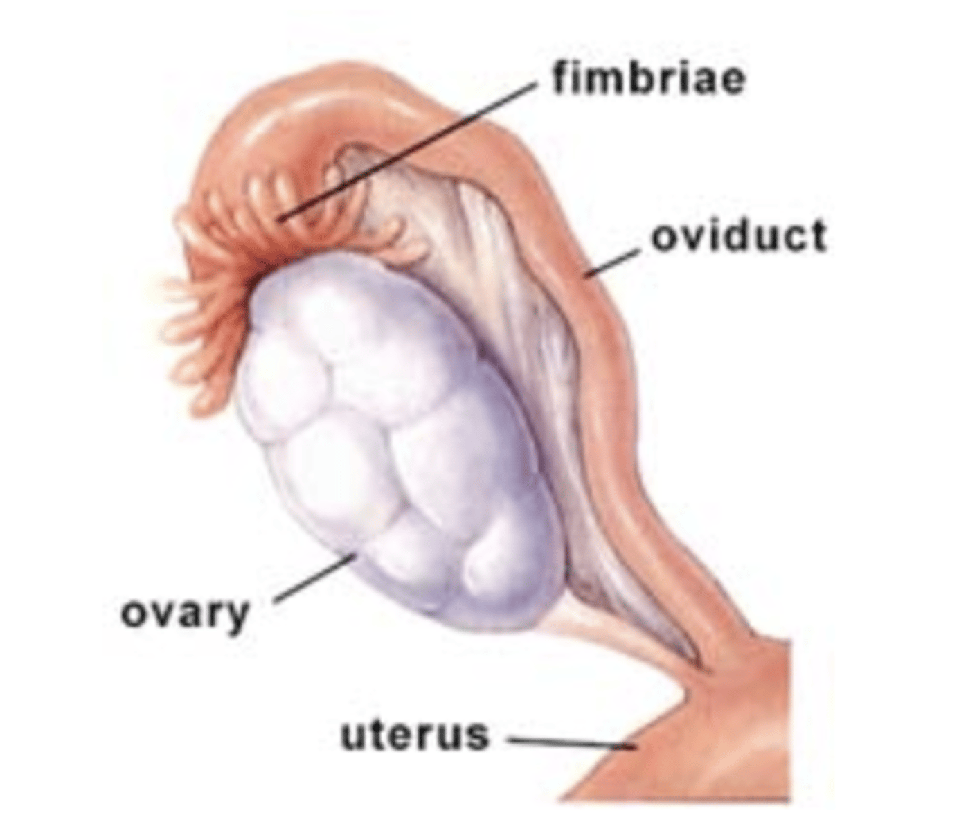
Fimbria
end of the oviduct
-finger-like fringe
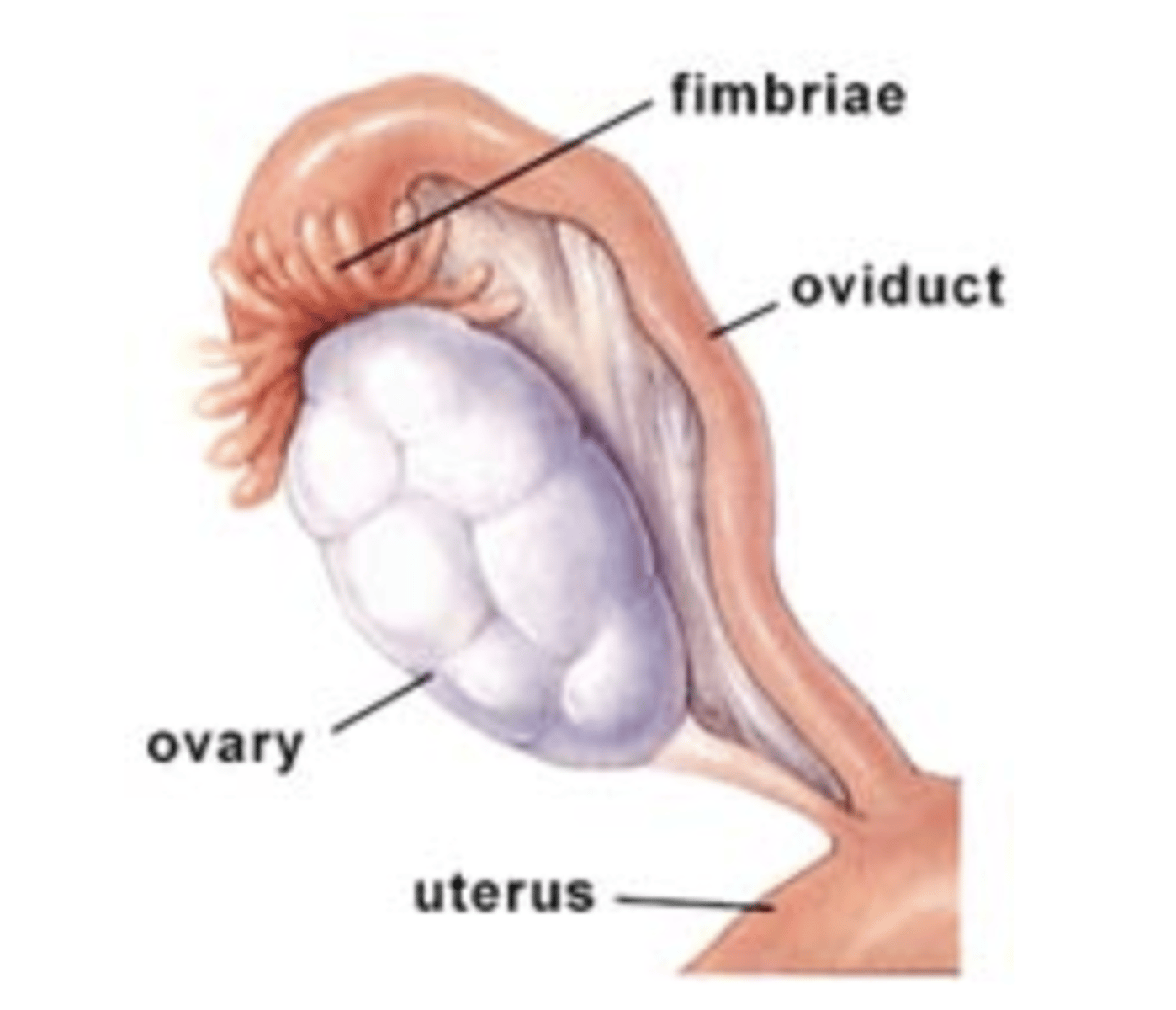
Ovary
gonad(sex organs), holds oocytes
-makes estrogen & progesterone
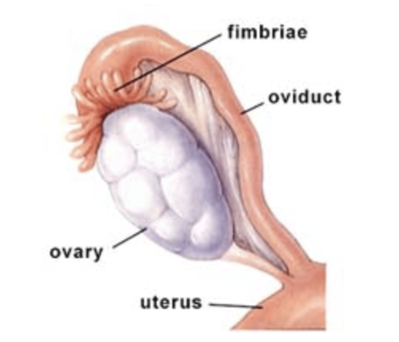
Oocyte
gamete; "egg cell"
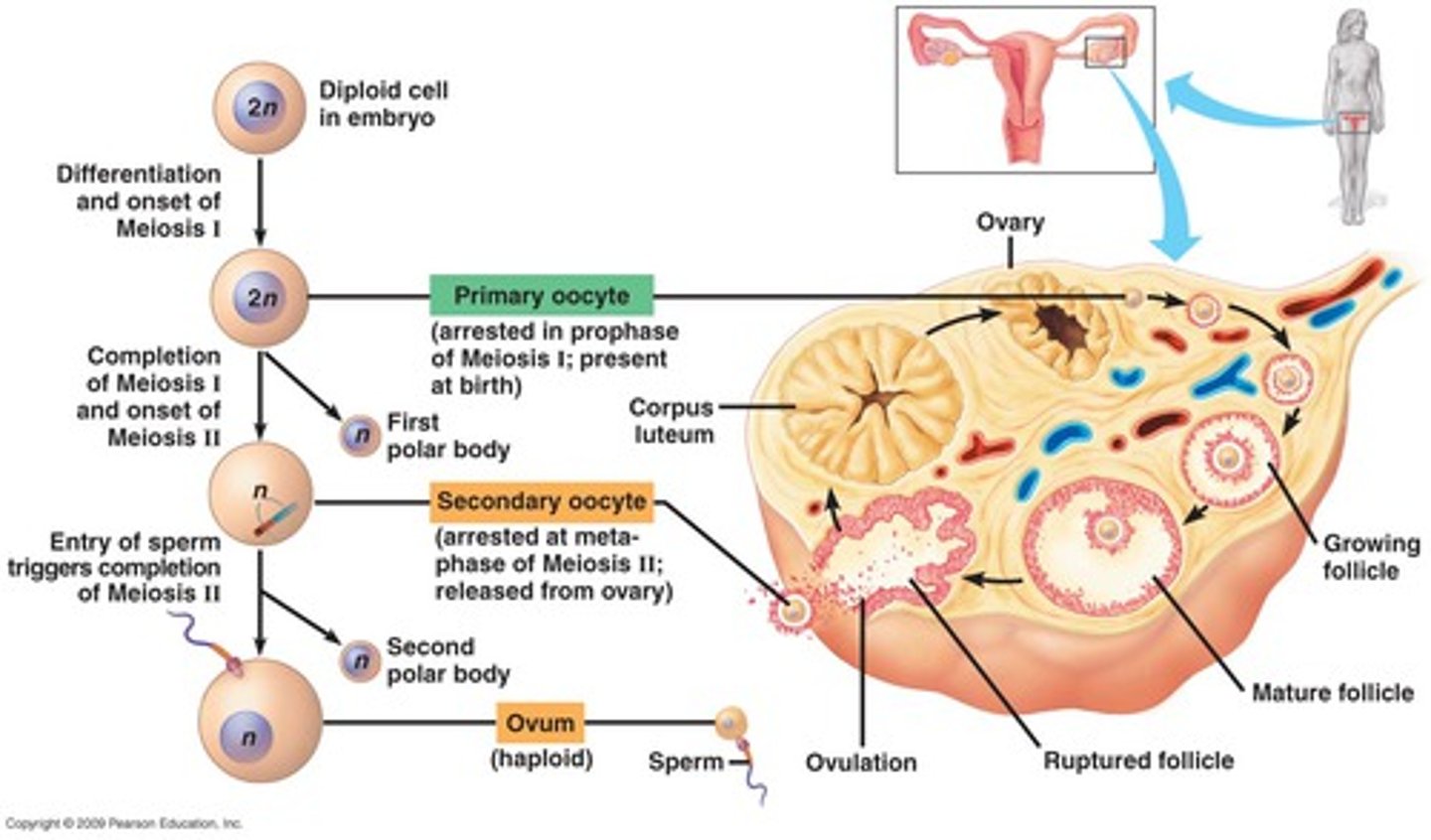
Follicle
oocyte + surrounding support cells
-makes estrogen
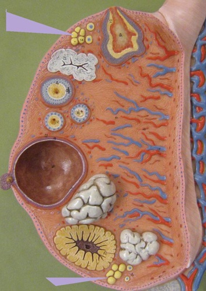
Corpus luteum (ovary)
yellow tissue that forms after ovulation
makes progesterone
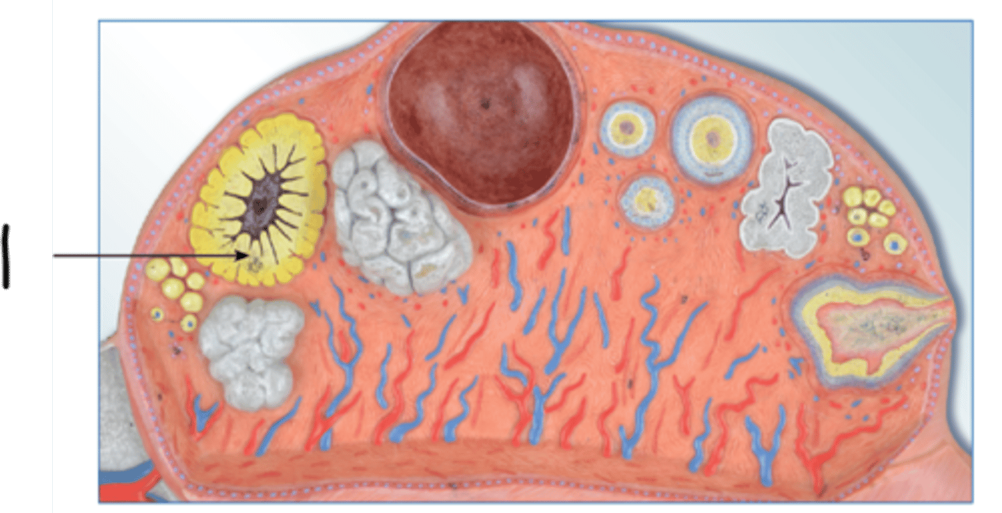
Corpus albicans
white tissue
corpus luteum
broken down into corpus albican
can scar!
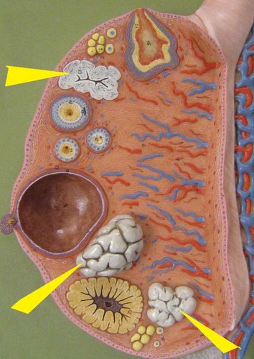
Nipple
a projection from the breast
delivers breast milk to young
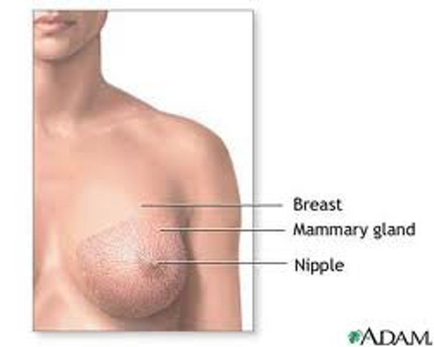
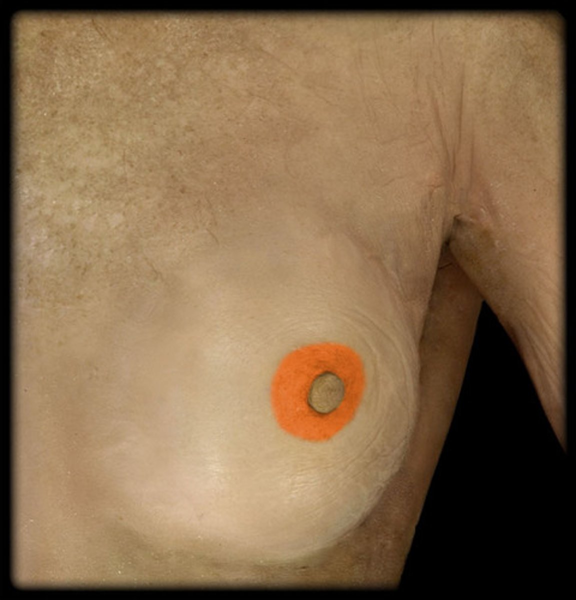
Areola
circular pigmented tissue
surrounds nipple