Lesson Material - Muscle Physiology
 Call Kai
 Learn
 Practice Test
 Spaced Repetition
 Match
 Flashcards
 Knowt Play
1/99

Earn XP
Description and Tags
In Lesson 5 we will learn about muscle physiology. The focus is on the structure and function of skeletal muscle tissue, how it is stimulated, and how it produces force (contraction). In lab, you will identify basic muscles of the body, forming a foundation to learn about actions later in the course (Unit 4).
100 Terms
Characteristics of muscle tissue
Excitable, elastic, extensible, and contractile
Excitability
Ability to spread electrical charge along the cell membrane
Elasticity
Ability to recoil and return to normal shape after being stretched
Extensibility
Ability to be stretched
Contractility
Ability to shorten (contract) and produce force

Skeletal muscle
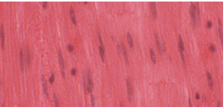
Smooth muscle

Cardiac muscle
Skeletal muscle
○Long, cylindrical cells, multinucleated
○Striated, voluntary
Smooth muscle
○Short, fusiform cells
○Non-striated, involuntary
○Lines hollow organs
Cardiac muscle
○Striated, involuntary
○Found in the heart
Functions of Skeletal Muscle
●Movement
●Stabilizes joints
●Control sphincters and reflexes for swallowing, urination, and defecation
●Protection (e.g. abdomen)
●Generates heat
Connective tissue coverings of skeletal muscle
Epimysium, perimysium, and endomysium
Muscle fiber
Another name for a skeletal muscle cell
Epimysium
Covers the entire muscle; blends with the tendon
Perimysium
Surrounds groups of muscle fibers called fascicles
Fascicle
A bundle of muscle fibers
Perimysium function
Provides passageways for blood vessels and nerves to reach muscle fibers
Endomysium
Surrounds each individual muscle fiber (muscle cell)
Tendon
Attaches muscle to bone
Muscle cell organelles
Sarcolemma, sarcoplasmic reticulum, sarcoplasm, myofibrils, and sarcomeres
Sarcolemma
Plasma membrane of a muscle cell
Sarcoplasmic reticulum
Extensive smooth endoplasmic reticulum that stores calcium
Sarcoplasm
Cytoplasm of a muscle cell
Myofibrils
Cylindrical structures made of actin and myosin that contract
Sarcomere
Smallest segment of a myofibril that contracts
Sarcomere
Smallest functional unit of muscle contraction; extends from one Z line to the next
Z lines
Mark the boundaries of each sarcomere
Thick filaments
Made of myosin
Thin filaments
Made of actin
Sliding filament theory
Muscle contraction occurs when actin and myosin slide past one another
Actin and myosin
Do not shorten during contraction
Sarcomere shortening
Occurs as actin and myosin slide over each other, bringing the Z lines closer together

Sarcomere
Sarcomere
Extends from one Z line to the next
Thick filaments
Formed by the protein myosin
Thin filaments
Made of actin, tropomyosin, and troponin
Actin
Globular protein arranged like beads on a string
Tropomyosin
Rope-like protein that covers myosin-binding sites on actin
Troponin
Binds calcium, moves tropomyosin, and exposes myosin-binding sites
A band
Dark band where thick (myosin) filaments are located
I band
Light band containing only thin (actin) filaments
M line
Center of the sarcomere; anchors thick (myosin) filaments
Myosin heads
Globular heads that reach up and attach to actin during contraction
Actin movement
Actin is pulled toward the M line (center of the sarcomere)
Z disc
Anchors thin (actin) filaments
Z disc movement
As actin slides toward the M line, the Z discs are pulled closer together, shortening the sarcomere

Relaxed
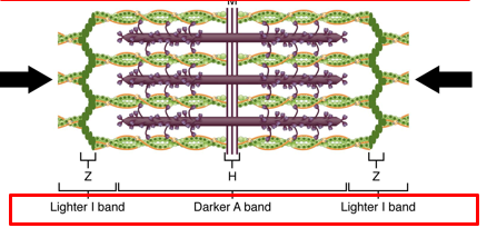
Contracted
Relaxed sarcomere
Myosin and actin are not actively interacting; I band and H zone are visible
Contracted sarcomere
Myosin heads contact actin and pull it toward the M line
I band
Becomes smaller during contraction
H zone
Becomes smaller during contraction
Sarcomere shortening
Z discs move closer together as actin slides toward the M line
Muscle contraction
Sarcomere shortens, but actin and myosin filaments do not become shorter
ATP role in muscle contraction
Provides energy for the repeating four-step cross-bridge cycle
Cocking
Myosin ATPase uses energy from ATP to cock (energize) the myosin head
Cross-bridge formation
Calcium binds troponin, moving tropomyosin and exposing myosin-binding sites on actin; myosin attaches to actin
Power stroke
Myosin pulls actin toward the center of the sarcomere (M line)
Detachment
ATP binds to myosin, causing it to release actin
Cross-bridge cycle
Repeats as long as ATP and calcium are present
Neuromuscular junction (NMJ)
The nervous system stimulates specific muscle fibers to contract
The end of the motor neuron
Expands to form the axon terminal.
Axon terminal
Contains vesicles with acetylcholine
Synaptic cleft
A small space between the motor neuron and the skeletal muscle fiber
Acetylcholine receptors
Bind acetylcholine and initiate an action potential (electrical current) in the skeletal muscle fiber
Step 1
Action potential arrives at axon terminal
Step 2
Voltage-gated Ca2+ channels open and Ca2+ enters the axon terminal
Step 3
Synaptic vesicles containing acetylcholine merge with the plasma membrane
Step 4
Acetylcholine is released into the synaptic cleft
Step 5
Acetylcholine diffuses across the synaptic cleft
Step 6
Acetylcholine binds to acetylcholine receptors on the muscle fiber
Step 7
The muscle fiber initiates an action potential
Steps of Excitation-Contraction Coupling
An action potential spreads from the NMJ down the sarcolemma. It dives down transverse-tubules(t-
tubule) into the center of the skeletal muscle fiber. Along the t-tubules, the sarcoplasmic reticulum releases calcium
Calcium
Binds to troponin
Troponin
Moves tropomyosin, revealing myosin binding sites on actin. Myosin can now form cross-bridges and move actin
Acetylcholinesterase
Degrades acetylcholine in the synapse
Relaxation
Motor neuron is not active, Calcium is pumped back into sarcoplasmic reticulum, tropomyosin moves to cover the myosin-binding sites
ATP
Also necessary for muscle to relax
ATP role in relaxation
ATP binds to myosin and allows myosin to detach from actin
Rigor mortis
ATP is depleted and myosin gets “stuck” to actin in a contracted state
ATP powers
Ca2+ pumps that move Ca2+ back into the sarcoplasmic reticulum
Sources of ATP
Creatine kinase, Glycolysis, Aerobic respiration
Creatine kinase
One of the first sources of ATP, transfers a phosphate group to ADP, forming ATP
Glycolysis
Second source, produces a small amount of ATP for about 1 minute of contraction
Aerobic respiration
Using mitochondria and oxygen is necessary for long-lasting contraction
Fast glycolytic (FG)
Rely on glycolysis, Produce a lot of energy, but short-term only. Fatigue quickly, White in color; do not have a large blood supply
Fast oxidative (FO)
More fatigue resistant than FG fibers, Intermediate color
Slow oxidative (SO)
Rely on aerobic metabolism, Fatigue resistant, Red in color; large blood supply and lots of myoglobin
Tension
The force produced by a contracting muscle
Isotonic contraction
Amount of force generated remains the same, length of the muscle changes
Concentric contraction
Produces force while shortening
Eccentric contraction
Produces force while lengthening
Isometric contraction
Amount of force generated increases, muscle length remains the same
Motor unit
One motor neuron and all of the muscle fibers it innervates
Motor unit recruitment
One way to increase the amount of force produced
Optimal overlap
Skeletal muscle produces maximal force
Passive length
There is already maximal overlap and no contraction is possible
Muscle is passively stretched
There is not enough overlap and cross-bridges can’t form; this also leads to low force generation
Optimal resting length
Muscles are under tension in our bodies