Practical 3
 Call Kai
 Learn
 Practice Test
 Spaced Repetition
 Match
 Flashcards
 Knowt Play
1/75

There's no tags or description
Looks like no tags are added yet.
76 Terms
Abdominal Aorta
This is the part of the aorta that is found in the abdomen
Afferent Arteriole
These blood vessels branch off of the cortical radiate arteries and enter the renal corpuscle to bring blood into them.
Second line on left

Albuminuria
This is urinary condition where excess protein is found in urine. It usually signifies that the glomerulus is damaged, allowing protein to leak into the urine. 10mg/100mL is the normal concentration of protein
Arcuate Arteries
These arteries branch off of the interlobar arteries in the kidneys, and they then branch into the cortical radiate arteries.
Bottom lines on right

Arcuate Veins
These veins are formed from multiple cortical radiate veins, and they then form arcuate veins.
Bottom line on right

Ascending Limb
This part of the nephron loop is going up. it is impermeable to water and permeable to solutes.
Top right line

Bowman’s Capsule
This is another name for the glomerular capsule, the thin, double-walled capsule surrounding the glomerulus.
Collecting Duct
This is a tube that receives fluid from several nephrons for final modification. Multiple of these merge to form papillary ducts.
Bottom right line
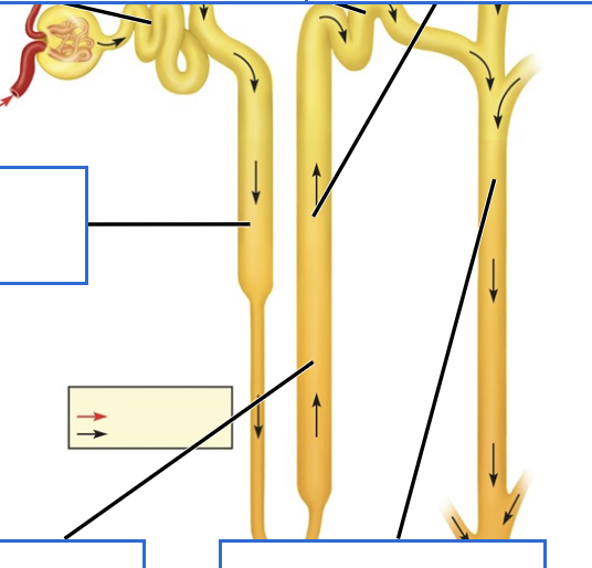
Cortical Radiate Artery
The arcuate arteries branch off into these arteries, which then form branch off into multiple afferent arterioles.
First line on right

Cortical Radiate Veins
These veins take blood from multiple peritubular capillaries and vasa recta. They empty into the arcuate veins.
First line on right

Descending Limb
This part of the nephron loop is going down, and it is permeable to water but not solutes.
Middle line on left

Detrusor
This muscle is part of the bladder, and it is made of collectively of the irregular crisscrossing bands of smooth muscle that make up the bladder.
Second line on left
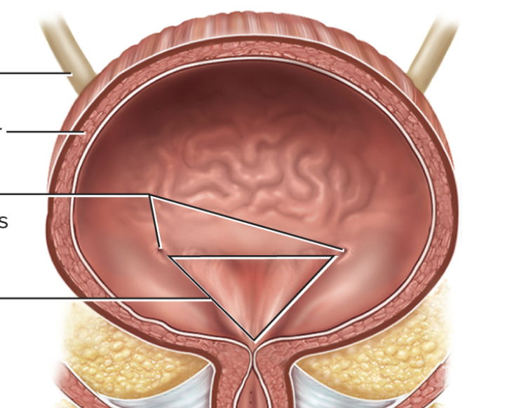
Distal Convoluted Tubule
This is where filtrate goes after the ascending loop, and it then directs fluid into the collecting duct.
Middle line on top

Efferent Arteriole
This is the blood vessel that takes filtered blood out of the renal corpuscle. They either branch into vasa recta or peritubular capillaries, which are both used for reabsorption and secretion into the nephron loop.
Top line on left

External Urethral Orifice
This is is the external opening of the urethra through which urine exits the body. It is much longer in males than females, which is why females get more UTI’s.
Middle bottom line

External Urethral Sphincter
This is a ring of striated skeletal muscle that surrounds the urethra and provides voluntary control over urination. It is innervated by the pudendal nerve.
Bottom line on right

Glomerular Capsule
This is the thin, double walled capsule that surrounds the glomerulus. It has a visceral inner layer and a parietal outer layer, which have the capsular space in between them.
Glomerulus
This is the name for the cluster of capillaries where filtration occurs. The afferent arteriole flows into it, and the efferent arteriole flows out.
Glycosuria
This is a condition where someone has high amounts of glucose appearing in their urine because there blood glucose levels exceed the number of available sodium-glucose transporters.
Hematuria
This is the condition of an abnormal presence of blood in urine, which causes urine to be pink or red in color and signifies a serious issue.
Hemoglobinuria
This is a medical condition where the protein hemoglobin appears in abnormal concentrations in the urine. It happens when red blood cells break down rapidly inside the blood vessels faster than the body's binding proteins can clear it
Renal Hilum
This is the medial entry and exit site for structures that service the kidney, including blood vessels, nerves, lymphatics, and ureters. It extends into the renal sinus.
(R/L)
Inferior Vena Cava
This is the major vein of the body, and it is where the renal veins empty into.
Interlobar Arteries
These arteries branch off of the segmental arteries, and when these reach the cortex they bend and branch to form the arcuate arteries.
Second line on right

Interlobar Veins
The arcuate veins empty into these veins, which then empty into the renal veins.
Second line on right

Internal urethral Sphincter
This is an involuntary, smooth muscle ring located at the junction of the urinary bladder and the proximal urethra. It stops semen from flowing back into the bladder.
Second line on right

Ketonuria
This condition is diagnosed by an excess amount of ketones in urine, which signifies diabetes or a severe deficiency of carbs or proteins. It is caused by the use of fats to produce energy in place of carbs.
Kidney
This pair of organs lies on either side of the spine between the peritoneum and the posterior abdominal wall. They filter the blood and produce urine. They consist of a cortex and medulla.
Loop of Henle
This is another name for the nephron loop, which consists of the ascending and descending limb.
Major Calyx
These structures conduct urine into the large renal pelvis, and they are formed by multiple minor calyces.
Fourth line on left

Membranous Urethra
This portion of the urethra comes after the prostatic urethra and before the spongy urethra. It is only found in the male urethra. It passes through the deep muscles of perineum, where it is invested by the overlying urethral sphincters.
Sixth line on left

Minor Calyx
This is where urine goes first after the collecting ducts of the nephrons, and they lead into the major calyces.
Third line on left

Nephron
These are the structures of the kidney that filter blood into urine. They include the renal corpsucle, the PCT, the nephron loop, and the DCT.
Nephron Loop
This structure of the nephron includes the descending and ascending limb
Papillary Duct
These ducts are combinations of multiple collecting ducts, and they empty into the minor calyces at a renal papillae
Parietal Layer
This is the outer layer of the glomerular capsule, and it is made of simple squamous epithelial tissue.
Top line on right

Peritubular Capillaries
The efferent arterioles flow into these, and they are part of where the reabsorption and secretion take place in the nephron tubules.
Prostatic Urethra
This is the first region of the urethra, and it passes through the prostate gland. This allows the urethra to receive sperm via the ejaculatory ducts.
Fifth line on left

Proximal Convoluted Tubule
This is the first part of the nephron tube, and it receives filtrate directly from the renal corpuscle.
Top line on left

Pyuria
This condition is the presence of leukocytes in urine, and it may indicate a UTI.
Renal Arteries
These are the first blood vessels to enter the kidney, and they stem from the abdominal aorta. It divides into the segmental arteries.
Sixth line on left

Renal Columns
These are connective tissue extensions that radiate downward from the cortex through the medulla to separate the renal pyramids. They provide a supportive framework for vessels, and divide the kidneys into 6-8 lobes.
Fifth line on left

Renal Corpuscle
This structure includes both the glomerulus and the glomerular capsule, and it makes up the proximal end of each nephron. It is where filtration takes place.
Renal Cortex
This is the outer region of the kidney, and parts of the nephron as well as some blood vessels are located here.
First line on left

Renal Medulla
This is the inner portion of the kidneys. The renal pyramids sit in this section of the kidney.
Renal Papilla
This is the blunt point that occurs at the end of a renal pyramid. They are bundles of collecting ducts that transport urine to the calyces of the kidney for extraction.
Renal Pelvis
This is where the major calyces conduct urine into, and this structure emerges out of the kidney at the hilum into the ureter.
(R/L)
Second from bottom on left

Renal Pyramid
These are the functional units of the kidney, and they each contain multiple complete nephrons. They each end in a renal papilla.
Second line on left

Renal Tubule
These are the tiny, microscopic tubes in the kidneys that process filtered fluid from the blood to form urine, reabsorbing vital nutrients and water while removing wastes. They are major parts of nephrons. They consist of the PCT, nephron loop, and DCT
Renal Veins
These are the two major veins that take blood from the segmental veins in the kidney to the inferior vena cava. They exit the kidney from the hilum.
(R/L)
Eighth line on left

Rugae
These are the wrinkles found in the urinary bladder when it isn’t full. They allow it two stretch when needed.
Second line on left

Segmental Artery
This is what the renal artery branches into, and they are located within the renal sinus. They branch into the interlobar arteries.
Third line on right

Spongy Urethra
This is the last portion of the urethra in males, and it exits a the tip of the penis after passing through the erectile tissue.
Third line from bottom on left

Thick Segment
These are the thicker portions of the descending and ascending loops. They are found at the top of each side of the U.
Thin Segment
These are the thin portions of the descending and ascending loops. It is found at the bottom of the U
Trigone
This is the term used to describe the triangular shaped area between the beginning of the urethra and the two ureteral openings.
Fourth line on left
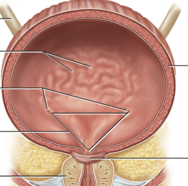
Ureter
This is what the renal pelvis turns into after exiting the kidney and slimming down. They are approximately 30cm long and consist of three tissue layers. They conduct peristalsis to move urine faster.
Bottom line on left

Ureteral Openings
These are where the ureters enter the urinary bladder.
(R/L)
Third line on left
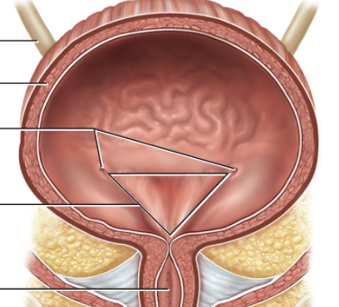
Urethra
This is the terminal organ of the urinary system, which transports urine from the bladder to the outside of the body for disposal. There is differences between the male and female organs.
Urinary Bladder
This is the primary organ that collects urine from both ureters. It empties into the urethra
Vasa Recta
These are the less common branches of the efferent arterioles that are important for producing concentrated urine.
Visceral Layer
This is the inner layer of the glomerular capsule, and it is composed of cells called podocytes (cells with foot-like processes)
Third line on right

Absorptive Cells
These are cells that line the small and large intestines, they absorb water and salts as well as vitamins produced by intestinal bacteria
Accessory Organs
These are the organs that aid the main organs of the digestive tract. They include the salivary glands, liver, pancreas, and gallbladder
Acinar Cells
These are cells found in the pancreas that secrete enzyme-rich pancreatic juice into tiny merging ducts
Adventitia
This is a dense sheath of collagen fibers found in the mouth, pharynx, and esophagus.
Alimentary Canal
This is another name for the GI tract, and it is a one way tube whose main function is to nourish the body via the digestion and absorption of nutrients.
Anal Canal
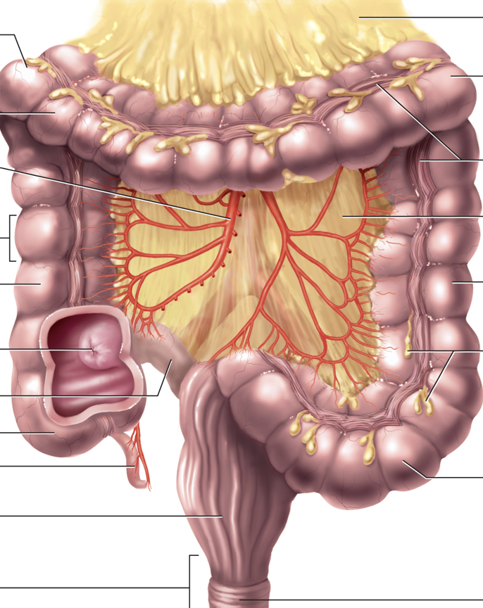
This is the last part of the large intestine, and it is located in the perineum completely outside of the abdominopelivc cavity. It opens to the exterior of the body at the anus.
Bottom line on left

Antrum
This is the wider, lower-most region of the stomach that lies just before the pyloric canal and the small intestine
Top line on left

Anus
This is where the large intestine opens to the outside of the body
Appendix
This is a winding tube that attaches to the cecum whose function is not fully known.
Third line from bottom on left

Ascending Colon
This is the first part of the large intestine after the cecum.
Fifth line on left
Bile Duct
This duct is where bile flows into the small intestine.
Fourth line on right

Body of Pancreas
Bottom right line

Brush Border
This is what the walls of the small intestine are called because of the bristle like appearance of the microvilli