BISC 101: Animal Biology
 Call Kai
 Learn
 Practice Test
 Spaced Repetition
 Match
 Flashcards
 Knowt Play
1/73

There's no tags or description
Looks like no tags are added yet.
74 Terms
Animal Physiology
How the physical structures in an organism works (to sustain life)
How they maintain homeostasis, respond to their environment, and perform essential functions (respiration, digestion, reproduction)
Main purposes of anatomy and physiology
Homeostasis and Reproduction
Which of these mammals have to eat more to support each gram of it’s tissue: a dog or a human?
The dog; it has a higher mass specific metabolic rate than humans (higher SA:V ratio than a human)
Surface Area to Volume Ratio
An organism’s size is linked to it’s metabolism
Smaller animals, with a higher SA:V ratio will lose more heat to their surroundings (higher metabolic rate)
Large animals, with lower SA:V ratio will lose less heat to their surroundings (lower metabolic rate)
4 strategies animals use to get food
Suspension feeders - capture tiny particles/organisms from water
Deposit feeders - feed on organic matter (bacteria, algae, etc.) buried within sediments
Fluid feeders - absorbs liquids, often by sucking on them (ex. butterflies, leeches, etc.)
Mass feeders - consume large pieces of food (meat, grass, etc.)
Major components of a homeostatic system
Maintains a homeostasis via a feedback loop
Sensor - monitors the environment for changes (thermoreceptors, chemoreceptors, pH changes) and sends a signal to the integrator
Integrator - control center that processes information for the sensor and compares the signal to a predetermined set point and decides if a response is needed
Effector - receives a command from the integrator and carries out a response to bring the variable back to a set point (ex. muscles contracting to produce heat)
4 ways which animals exchange heat with the environment
Conduction - heat transfer through direct contact; molecule to molecule
Convection - heat transfer through the movement of fluids
Radiation - heat transfer through electromagnetic waves
Evapouration - heat transfer from a liquid during phase changes (liquid to gas)
What would happen if the epithelial cells in the gills of a shark are treated with ouabain (molecule that poisons Na+/K+ ATPase)
Na+ and Cl- cannot move out of the epithelial cells into the surrounding seawater; salt would build up in the sharks tissues
Epithelial Cells
Cells that form a protective layer on surfaces of the body, they help with…
Protection - physical barrier against pathogens/environmental damage
Secretion - secrete substances like mucus, hormones, and sweat
Absorption - in the digestive tract, they absorb nutrients from the foods you eat
Filtration - in the kidneys, they are involved in filtering waste from the blood
Partial Pressure
Pressure of a specific gas in a mixture of gases; depends on…
Total pressure of the gas
Fractional pressure of the gas
Lymphatic System
A circulatory system made up of lymph vessels, which helps drain excess fluid (lymph) from the capillaries back to circulation
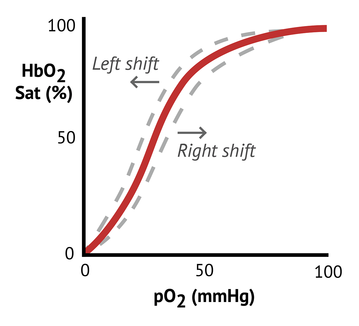
In the blood, DPG shifts the oxygen-hemoglobin equilibrium curve to the right, how does this shift affect oxygen loading and unloading?
Right shift = decreased affinity for oxygen, DPG promotes release of oxygen from hemoglobin to tissues
What would happen to the resting potential of an axon if the potassium leak channels were blocked?
The resting potential will be less negative as potassium would be unable to leak out of the cell
What would happen if feedback inhibition fails in a hormone signaling pathway?
Hormone production would not stop and the signal/response would continue
What would be the consequence of treating a person with drugs that inhibit trypsin?
Trypsin will inhibit protein digestion and reduce amino acid absorption - this could lead to weight loss and muscle atrophy due to protein deprivation
Myasthenia gravis is a diease that develops in humans when the immune ystem produces proteins that bind with Ach receptors in the muscles and causes muscle weakness. Why?
If Ach receptors are covered, they cannot bind to Ach and become activated - this means the muscles would have trouble contracting
Phenotypic Plasticity
The ability of an organism to change it’s appearance, behaviour, or physiology in response to a change in it’s environment (ex. elephants that grows up in hot climates developed strong muscles for flapping it’s ears)
Digestive System
Food is taken in (ingestion)
Food is broken down (digestion)
Nutrients are absorbed (reabsorption)
Waste is removed (excretion)
Ingestion
Different (evolutionary) adaptations allow animals to access food
Fluid feeder
Suspension feeder
Deposit feeder
Mass feeder
Digestion
Process of breaking down food into smaller molecules that the body can absorb for energy, growth, and repair
Absorption
Absorption of nutrients, vitamins, minerals, and water through the small intestine
Small diffusion distance (one cell width) between lumen and blood
Huge surface at organ and cell level
Transport proteins bring in nutrients from digestive tract and into blood
Excretion
Formation and elimination of feces
Large intestine: colon and rectum
Colon: form feces by absorbing additional water and compacting the wastes that remain
Rectum: storage location for feces until it can be eliminated
Mouth
Mechanical & chemical processing of food; break down of lipids & carbohydrates
Salivary amylase - breaks down starch into simpler sugars
Salivary (lingual) lipase - breaks down triglycerides (lipids)
Esophagus
Moves food from the mouth into the stomach through peristalsis; a series of muscle contractions that push the food downwards
Salivary amylase from the mouth can continue working until the stomach acids inactivates it
Lingual lipase is secreted by the tongue and continues working in the stomach
Stomach
Stores and breaks down food into a liquid mixture (chyme) before releasing it into the small intestine, the stomach acid helps kill microbes and denatures proteins for easier digestion
Pepsin - breaks down proteins into smaller peptides
Gastric lipase - breaks down fats (triglycerides) into fatty acids and monoglycerides
Lingual lipase - breaks down triglycerides
Small Intestine
Main site of digestion and nutrient (vitamins, minerals, water) absorption; chyme from the stomach mixes with bile (from liver) and pancreatic juices and enzymes are secreted to finish breaking down macromolecules. Nutrients are absorbed into the bloodstream and lymphatic system
Pancreatic amylase - breaks down starch into simpler sugars
Pancreatic lipase - breaks down fats into fatty acids and monoglycerides
Large Intestine
Absorbs water and electrolytes (Na+/Cl-) from the remaining indigestible food, forms and stores feces before elimination (excretion) in the rectum
gut bacteria helps break down some undigested material, which can be thought of as a form of enzymatic action; producing beneficial vitamins
Rectum
Stores feces until it’s ready to be excrete through the anus; when the rectum is full, stretch receptors signal the brain to trigger the defecation reflex
In mammals, where does most of the enzymatic breakdown of molecules occur?
Small intestine; enzymes secreted from the liver and pancreas (including digestive enzymes), plus any left-over enzymes from the mouth and stomach, are active in the small intestine
How are nutrients stored and used to fuel activity
Carbohydrates - stored mainly as glycogen in the liver and muscles
during short-term or high-intensity activity; glycogen is broken into glucose for ATP production
Lipids - stored as triglycerides in adipose tissues and muscle cells
during low to moderate intensity activity or rest; fats are broken into fatty acids and glycerol for aerobic ATP production
Proteins - doesn’t store protein specifically for energy
during prolonged exercise or fasting; amino acids can be converted into glucose or used directly for energy
How do the small intestines maximize the rate of transfer of nutrients (across a surface) into the body?
By enabling a continual gradient = (amount on one side - amount on the other side)
Between two things close together = distance across
Over a large area = surface area
Endotherm
Animals which create most of their own heat from metabolism (mammals and birds)
Ectotherms
Animals which receive heat primarily from external sources (reptiles, amphibians, and fish)
Adaptation used by endotherms living in cold environments
Circulatory system arranged in a countercurrent flow to minimize heat loss
ex. blood flowing in opposite directions (veins and arteries)
Countercurrent Exchange
Two fluids run in opposite directions in adjacent pipes
Useful because they maintain a gradient between two fluids along the length of adjacent pipes
Concurrent Exchange
Two fluids (ex. blood and water) run in the same direction in adjacent pipes
Countercurrent Exchange in Kidneys
Ensures the production of concentrated urine
Filtrate in the Loop of Henle runs in opposite directions of the blood capillaries directly beside it
Descending limb is permeable to water, but impermeable to solutes
Ascending limb is permeable to solutes, but impermeable to water
Allows for solutes and water to be transported back into blood from the Loop of Henle, resulting in more concentration of urine
Countercurrent Exchange in Limbs
Minimizes heat loss at the body’s extremities
Arteries and veins are located beside each other, but the blood flow is in opposite directions
Heat is transferred from the arteries to the veins
Osmoregulation
Process of maintaining a stable balance of water and salt concentration within an organism’s body fluids
Osmotic stress in water (osmoregulators)
Oceans & seas (saltwater/marine) - hypertonic
Fish would lose water by osmosis, and gain salts (electrolytes) by passive transport
Drinks water (also contains electrolytes) as a result; pumps ions out
Lakes, ponds, rivers, (freshwater) - hypotonic
Freshwater fish would gain water by osmosis, and lose salts (electrolytes) by passive transport
Doesn’t drink water; excretes lots of water; bring in extra electrolytes (active transport across gills, or through eating)
Getting Na+ and Cl- out of a shark (salt excretion)
Na+ from epithelial cell into blood; K+ from blood/ECF into cell-
ATP is required (primary active transport)
Establishes a sodium concentration gradient (high in blood/ECF, low in cell)
Na+, Cl-, and K+ are transported from blood/ECF into epithelial cells
ATP is required from concentration gradient (secondary active transport)
Brought Cl- into the cell - cell now have high [Cl-]
Cl- moves into ocean from cell, K+ moves into blood from cell
No ATP (passive facilitated diffusion)
Excreted Cl- which rebalanced K+ back into blood
Na+ diffuses from blood to ocean, between cells
No ATP (simple diffusion)
Na+ excreted
Kidneys
Where osmoregulation primarily occurs
Renal artery - brings blood (with nitrogenous wastes) into kidneys
Renal vein - carries blood (cleared of waste) away from kidneys
Kidney Pathway
Renal Corpuscle - filtration
Proximal Tubule - reabsorption of nutrients, vitamins, salts, water
Descending Loop of Henle - reabsorption of water (using osmotic gradient)
Ascending Loop of Henle - reabsorption of salts (making osmotic gradient)
Distal Tubule - reabsorption (when Na+ low)
Collecting Duct - combining from multiple nephrons; reabsorption of water (if dehydrated)
Renal Corpuscle
Blood enters and filtration occurs as it passes through
Proteins, cells, and other large components of blood do not fir through the pores and do not enter nephron (stay in blood)
Once filtered, the fluid is now known as pre-urine (filtrate)
Proximal Tubule
Filtrate contains water and small solutes (urea, glucose, amino acids, vitamins, electrolytes)
Almost all nutrients (& most of the NaCl and water filtered by renal corpuscle) are reabsorbed here
Loop of Henle
Reabsorption of water and salt (Na+/Cl-) using an osmotic gradient
Descending limb - water reabsorbed (aquaporins), using salt gradient
Ascending limb - Na+ and Cl- reabsorbed, using and making salt gradient
Distal Tubule
Reabsorption may continue (K+ and H+ from blood may move into distal tubule)
Filtrate that enters the distal tubule is always dilute
Collecting Duct
More water reabsorption, especially if dehydrated
ADH plays a role in the production of concentrated or dilute urine; part of a negative feedback loop (returning the system to a set point)
Respiration Pathway
Ventilation - Inhalation and Exhalation
Diffusion (gas exchange across lungs/gills/trachea) - exchange of O2 and CO2 between the air in lungs’ alveoli and the blood
Circulation (not all animals have this) - the circulatory system pumps oxygenated blood to the body’s tissues and carries CO2 back to the lungs
Diffusion (into cells) - exchange of gases between the blood and the body’s cells
Cellular Respiration - creates usable energy in the form of ATP using glucose and O2
Pulmonary and Systemic Circulation
Pulmonary:
Sends deoxygenated blood to the lungs to oxygenate it
From lungs to heart
Uses pulmonary arteries and veins
Systemic:
Sends oxygenated blood to organs and tissues
From heart to body
Uses aorta, arteries, veins, and capillaries
Movement of air in the body
Nose/Mouth - air enters the body
Pharynx (throat) - a passageway that connects the nasal and oral cavities to the larynx and esophagus
Larynx (voice box) - protects the airway during swallowing by using the epiglottis to block food from entering the trachea
Trachea (windpipe) - reinforced with cartilage; main function is to provide passage from the larynx to bronchi for air to pass
Bronchi - trachea divides into two main tubes, left and right bronchi; carries air into the lungs
Bronchioles - in the lungs, bronchi branch into smaller tubes called bronchioles that brings air through tiny ducts and into elastic air sacs (alveoli)
Alveoli - where gas exchange occurs; O2 inhaled from air diffuses into capillaries and attaches to red blood cells, while CO2 moves from the capillaries to alveoli to be exhaled (removed)
Inhalation
Diaphragm contracts and moves down
Intercostal muscles contracts - ribcage moves up and out
Increases chest cavity volume and decreases pressure to draw air into the lungs
Exhalation
Diaphragm relaxes and moves up
Intercostal muscles relax - ribcage moves down and in
Decreases the chest cavity volume and increases the pressure to push air ut of the lungs
Why would it be beneficial to breathe more heavily on a mountain top?
The partial pressure of oxygen in the air is lower than at sea level
Partial pressure is the pressure of a particular gas in a mixture of gases
Gas exchange in aquatic animals
Water flows in through the mouth, and out through the operculum (stiff flap of tissue covering the gills)
Gill filaments
Lamellae
Oxygenated blood
Deoxygenated blood
How is gas exchange of fish gills supported by it’s structures?
A - area for gas exchange
Should be maximized
Lots of surface area - capillaries and gills have huge surface area to volume ratio
D - diffusion distance
Should be minimized
Blood capillaries right next to the water, very short distance for diffusion
(P2-P1) - difference in partial pressure on either side of barrier
Is maximized
Blood and water are flowing in opposite directions; countercurrent exchange keeps a continual gradient over the whole gill
Do humans have respiratory structures for countercurrent exchange?
No; ventilation & gas exchange in our environment isn’t as challenging as under water
Water - low oxygen (low PO2) and the fluid is heavy - harder to move around
Air - lots of oxygen (high PO2) and the fluid is light - easier to move around
Gas Exchange - Movement of O2 and CO2
Depends on the partial pressure of O2 and CO2 in the body; gases will move via diffusion down their concentration gradients
Lungs (higher concentration of O2 in lungs, higher concentration of CO2 in blood)
Net diffusion of O2 out of alveoli into blood
Net diffusion of CO2 out of blood into alveoli
Tissues (higher concentration of O2 in blood, higher concentration of CO2 in tissues)
Net diffusion of O2 from the blood into tissues
Net diffusion of CO2 from tissues into blood
Role of Hemoglobin in Circulation
O2 is transported through the blood via Hemoglobin
Hb has iron that binds to O2 - binding causes a conformational change in Hb, increases affinity for O2, allowing Hb to be saturated with O2
Amino groups of Hb bind CO2
Hb reaches lungs - higher affinity for O2 due to high PO2
Hb reaches tissues - lower affinity for O2 due to low PO2
2,3 - DPG helps regulate release of O2
Other factors can affect Hb’s affinity for O2; resulting in shifts of the oxygen-hemoglobin dissociation curves
Oxygen-Hemoglobin Dissociation Curve
Left shift - less oxygen being released
Increased affinity for O2
Right shift - more oxygen being released
Decreased affinity for O2
How are O2 and CO2 carried in the blood?
Most CO2; dissolved in the plasma, in the form of bicarbonate ions
Most O2; bound to hemoglobin
Solutions:
Convert CO2 into an ion that dissolves easily using Carbonic Anhydrase
Bind O2 to a carrier protein, Hemoglobin
Hemoglobin carries & releases oxygen
Tissues are actively respiring using up O2, so they have a lower blood PO2
When O2 is bound to Hb, the O2 is stuck in the red blood cell - not accessible to the tissue that is actively respiring
At the tissues, Hb needs to release O2 into the blood, so that it can diffuse into the cells there (so the cells can use it)
Hb picks up O2 when high amounts are present
Hb drops off O2 when low amounts are present
What happens to an O2-Hb dissociation curve in response to pH?
Bohr Effect
Low pH deceases Hb’s ability to bind to oxygen
In low pH environments, Hb unloads more O2
Shift to the right
Provides the tissues with more oxygen so they can continue with cellular respiration
Bohr Effect vs. Root Effect
Bohr Effect (in mammals)
Reduces O2 affinity - harder for Hb and O2 to bind in response to an increase in CO2 or decreased pH (acidic)
Makes it easier to unload O2 into the tissues
Root Effect (in fish)
Reduces both affinity and O2 carrying capacity in response to decrease in pH
Builds up high O2 pressure in specific organs
2 main types of circulation
Closed Circulatory System
Keeps blood inside a continuous network of vessels; blood is always contained within them
Blood is separate from interstitial fluid
High pressure; rapid blood flow
Efficiently transports respiratory gases
Open Circulatory System
Pumps blood (hemolymph) into open cavities to directly bathe organs
Hemolymph mixes with interstitial fluid
Low pressure; slow blood flow
Does not transport respiratory gases efficiently
Directionalities
Veins - towards heart
Arteries - away from heart
Deoxygenated - towards lungs
Oxygenated - away from lungs
Pulmonary Circulation - to and from lungs (including right atrium/ventricle); lower pressure
Systemic Circulation - to and from body (including left atrium/ventricle); higher pressure
What would happen if one of the ventricles had thinner, less muscular walls?
It leads to a weakened ability to contract and pump blood effectively because it requires lots of pressure to move blood through the body
Why does the left ventricle have thicker walls?
It has to pump (oxygenated) blood to the entire body, which requires significantly more pressure than the right ventricle
Aldosterone
A lipid (steroid)
Hydrophobic and can cross the membrane
Receptor is inside the cell
ADH
A peptide
Hydrophilic/polar and cannot cross the membrane
Receptor is on the cell surface
Aldosterone and ADH effect on target cells in kidneys (osmoregulation)
Aldosterone saves sodium
Distal Tubule: aldosterone increases amount of sodium transporters in membrane; sodium moves into blood
ADH saves water
Collecting duct: ADH increases amount of aquaporins in membrane; more water can move passively into blood
Overall outcome
Negative feedback to achieve homeostasis
An adaptation in your body, with systems that respond to keep your enzymes and systems happy
Digestion & absorption in the physiological response
Sensor (signal) - presence of food in digestive tract
Integrator - causes production/secretion of hormones into bloodstream
Effector (response) in small intestine - increase in the amount of digestive enzymes & relevant transporters
Types of blood vessels
Arteries
Transports blood away from the heart under high pressure
Have thick, muscular, elastic walls to handle high pressure
Veins
Transports blood towards the heart under low pressure
Have thinner, less muscular walls than arteries
Capillaries
Allows for the exchange of gases and nutrients between blood and tissue
Have extremely thin, semipermeable walls for substances to diffuse in and out
Pathway of blood in heart
Deoxygenated blood enters from the superior/inferior vena cava into the right atrium
Deoxygenated blood moves from the right atrium, passing the tricuspid valve into the right ventricle
From the right ventricle, blood moves past the semilunar valve and out of the heart via pulmonary artery
Oxygenated blood comes back via pulmonary vein into the left atrium
From the left atrium, blood moves past the bicuspid valve into the left ventricle
Oxygenated blood flows into the aorta and blood is distributed to the rest of the body