Lecture 3: Interpretation of Genetic Variation
 Call Kai
 Learn
Learn Practice Test
 Spaced Repetition
 Match
Match Flashcards
 Knowt Play
1/46

There's no tags or description
Looks like no tags are added yet.
Name | Mastery | Learn | Test | Matching | Spaced | Call with Kai | Chat |
|---|
No analytics yet
Send a link to your students to track their progress
47 Terms

*****What is a major limitation of current global genomic databases?
hint: has to do with race/ethnic groups
Genomic databases are European centric, balancing input is being worked on.

Humans share 99.9% of their DNA (99.9% identical)
How does Geography affect our genomes and relatedness
what factors cause genetic variation among human populations
Geography (relatedness amongst groups mimics geographic location)
Local adaptations (to factors like altitude, diet, and microbial pathogens)
Admixture with extinct hominid groups (like Neanderthals and Denisovans)
Can influence disease risk eg. depression, severe COVID
***** What are the 3 IMPORTANT CAVEATS mentioned in this lecture?
IMPORTANT CAVEAT #1
The amount of variation between individual is so great, it dwarfs the amount of interracial variation.
Populations are a continuum. No support from genetics for saying that the members of any racial group are genetically better or not
There is an essential genetic equality of all human groups.
IMPORTANT CAVEAT #2
All humans carry deleterious variants, estimated 10-12 variants each.
No individual can claim to be genetically superior to another.
Variability in genes and their products can improve adaptability to environmental change, pathogen exposure
IMPORTANT CAVEAT #3
Genetic changes are typically considered “good” or “bad”.
Ascertainment bias.
Keep in mind temporal changes in the “value” of a mutation or allele. (value of a mutation (good vs. bad) is dependent on context)
The mutations which have helped us survive other times in human history may be “deleterious” now.
Eg. NIDDM, Type II diabetes, Hemochromatosis, Susceptibility to severe COVID
****If there is essential genetic equality of all human groups why do we ask for an individual’s ethnic background?
We use the variation present in human populations and its
frequency to help us evaluate whether a variant is clinically
significant. We ask for an individual’s ethnic background on our
requisitions for that purpose.
*****Genetic variants or alleles in the clinic can be:
what are the 7 possible results
Does not change the protein encoded or the change has no effect on protein function or gene expression. Eg. F9 gene polymorphism.
Neutral
Benign
Likely benign
Protein function or gene expression is changed and an individual’s health is compromised. Eg. BRCA1 and cancer.
Pathogenic
Likely pathogenic
Unclear what effect change has on protein function or gene expression
Variant of unknown clinical significance (VUS, VOUS)
Protein function or gene expression is changed and has a beneficial or protective effect on an individual’s health. Eg. CCR5 and HIV susceptibility.
Positive/Protective
Is there perfection in anyones genome?
NO such thing as perfection
Each of us carries variants in all classes of pathogenicity
*****How do we interpret the variation we see?
What are 6 things we need?

How do we orient ourselves so when we talk about genetic variation, so we all are looking at the same positions in the genome……
aka how To consistently identify and communicate genetic variations across research and clinical settings
Standardized genomic coordinates and nomenclature.
Look at chr + physical landmarks when looking through microscope eg. cytogenetic chromosome bands
Nomenclature
Genome build + exact nucleotide positions on the chr that we identify with molecular based techniques (eg. chr21:21,379,642–30,108,064del (hg18, NCBI build 36))
Commonly used to describe large CNVs
Accession # for genomes sequences for Chr 21 in NCBI genbank eg.(NC_000021.5:g.34,450,000T>A) = a T to A substitution at g.34,450,000
Commonly ysed to describe SNVs or SNPs
**Chromosome Structure Value: Why is chromosome architecture still important during high-resolution molecular testing?
Even with molecular techniques = high res (where we can focus on individual bp) knowledge of chr and their structure can still be key to interpreting how a variant occurred.
Architecture of genome, condensed + folded structure of genome and the orientation within the nucleus are relevant ad being investigated for their impact in health and disease
*****What type of nomenclature format is typically used to describe large copy number variants (CNVs) vs. small SNPs + SNVs?
GENOME BUILDS = CNV
Chromosome range notation based on a specific genome build
Accession Number = SNV + SNP
Limitation of Nucleotide Coordinates
Question: What is the limitation of using absolute nucleotide positions (like Genome builds and Accession #) alone?
This has value for some variants, but lacks info on what is happening at the level of the gene
They do not automatically provide information about what is happening at the gene or coding level.
using only genome builds and accession numbers provides the exact physical coordinates of a mutation, but fails to explain its functional impact on a specific gene.
Genome Builds (e.g., GRCh37, GRCh38): These act like a map of the entire human genome.
Accession Numbers (e.g., NC_000001.11): These identify a specific chromosome or sequence within that map.
*****When evaluating the clinical significance of a genetic variant, why is it essential to establish a single consensus reference transcript and how is this transcript typically selected?
Alternative splicing of mRNA occurs in most human genes
more than 1 version of RNA transcript = more than 1 product can be produced from the same gene
These may be expressed at diff times, diff conditions, tissues and time in development
This need to be considered when evaluating a variants significance
a variant's impact depends heavily on which transcript and protein isoform are affected
eg. a variant might fall in an exon or splice site that is skipped or in a tissue-specific transcripts, meaning the variant could be neutral in one isoform but pathogenic in another
Reference transcript = LONGEST mRNA

*****How do we describe what we see in a genetic variant?
How to number a gene location (hint: relative to what)
Numbering a gene location is always done relative to the REFERENCE transcript (NM#)
[Reference Sequence] : [Level & Location] [What Changed] -> [Protein Consequence].
uses Coding sequence (EXONS) of a gene as a reference (c.#)
Mentions what consequences there are on the protein (p.#) or mRNA (r.#)

****Explain how Introns and Exon positions are denoted in gene location nomenclature with HGVS
Since it uses the coding sequence as a reference, Nucleotides in EXONS (the parts that code for protein) receive a number all the way until the stop codon.
Coding DNA numbering (c.) only counts the exons.
When a mutation happens inside an INTRON, HGVS uses a + or - to show how many steps into the intron the mutation is located:
+ = 3’ of a splice site (aka. after the stop codon)
- = 5’ upstream of a transcription start
The “A” of the Start codon -→ always = 1
Example: c.77+1 = Go to exon position 77. Move 1 letter forward (+1) into the intron

****Common codes used in description of variants: Nomenclature rules
What do the HGVS prefixes c., g., r., and p. stand for
The Level (Prefix): Tells you what kind of molecule is being looked at:
c. = coding sequence on DNA
g. = genomic sequence
r. = RNA/transcript sequence
p. = Protein sequence
****Common codes used in description of variants: Nomenclature rules
What does * and > mean?
* indicates the presence of a stop codon
> = nucleotide substitution

***Please denote what this nomenclature means

*****Assessing Genetic variants for clinical significance: What is the DEFAULT classification of a genetic variance?
Pathogenic (P)
Likely Pathogenic (LP)
Variant of Uncertain Significance (VUS)
Likely Benign (LB)
Benign (B)
UNCERTAIN SIGNIFICANCE = default until you have evidence other wise
NO ANSWER IS BETTER THAN WRONG ANSWER
*****What type of Evidence Evaluated in the SCORING process for clinical significance?
Population Data
Variant Type
Clinical Observations
Functional Studies
Population Studies
In silico/ Computational Data
How has ACMG variant classification guidelines EVOLVED from 2008 → 2014? for GERMLINE variants
Germ line = inherited genetic changes in sperm or egg cells that affect every cell in the body
Somatic mutations = acquired genetic changes in non-reproductive cells that happen after conception and cannot be passed to children
Evolved from a rigid, outcome-based classification system → standardized, evidence-weighted framework

*****How does the different evidence looked affect if a variant is considered more benign or pathogenic
eg. how does de novo status influence if a variant is pathogenic vs. benign
for: Population data, Computational, Functional, Family (Segregation), De novo, allelic data (variant data)
Population data:
Many people in the population have the variant = strong indicator of Benign
Variant is absent in public genome data bases = moderate indicator of pathogenic
Variant is prevalent in affected populations is > than contol = strong pathogenic indicator
Computational and Predictive Data: Uses computer algorithms (e.g., SIFT, PolyPhen) to predict how a DNA change alters protein structure or function.
Majority of the data suggest variation will have no impact on gene product = Supports Benign
Much of the data suggest deleterious effect of gene product = support Pathogenic
Novel change that causes as change in different pathogenic mutation that is already known = Moderate pathogenic
Variant causes SAME change as an established pathogenic variant = Strong pathogenic
Variant causes LOF (loss of function) where it is a known mechanism of disease = Very strong pathogenic
Functional Data: real-world, laboratory-bench experiments
Experiments show variant does not disrupt cellular or protein function = strong Benign
Studies directly demonstrate a severe deleterious effect on protein function = Strong pathogenic
Family Data (segregation): Showing whether a specific variant is inherited together with a disease
Non-segregation. Affected family members do not carry the variant, or unaffected members carry it when they shouldn't = Benign
Increase in segregation (aka. disease phenotype is prevalent along side variant = inherited) = Pathogenic
De Novo Data
De novo = Indicates pathogenic
Allelic Data: Looks at the relationship between the variant and other variants in the same gene.
The variant is observed in trans (on the opposite chromosome copy) with a known dominant pathogenic variant, or in cis (on the same copy) with another pathogenic variant, meaning it doesn't cause disease on its own. = Benign
For recessive conditions, detecting this variant in trans with a known pathogenic variant on the other copy points toward pathogenicity = Pathogenic


Summarize this slide
Pop data: High incidence in data = benign. Low/ absent = Pathogenic
Variant Type: Mild or silent changes = lean benign. Severe protein-disrupting changes (nonsense, frameshift, splice site dinucleotides) lean pathogenic.
Clinical Observations: Patient and family case history (e.g., co-occurrence patterns, family segregation, or number of affected families). High number of family cases = pathogenic
Experimental Studies: reputable experimental results are only weak/support of pathogenicity or benign (do not strongly influence categorization)
Computational Data: In silico predictive tools act as indirect evidence, evaluating whether a change is likely completely neutral or highly deleterious.

****Interpretation of variants form NGS (nxt gen seq). What is the work flow? What tools + data bases are used?
Tools:
Databases: ClinGen, ClinVar, NCBI dbSNP, and gnomAD (Genome Aggregation Database).
In Silico (Computational) Tools: SIFT, PolyPhen, Mutation Taster, and REVEL.
Clinical/Family Analysis: Familial segregation, pedigree analysis, and cis/trans variant configuration.

Why must clinicians be cautious when relying on databases and in silico tools?
Because data in databases and predictions generated by computational tools can be wrong or inaccurate.

****What is the suggest classification of this assessment and WHY?
UNCERTAIN SIGNIFICANCE
The default unless enough evidence to classify as BENIGN, LIKELY BENIGN, LIKELY PATHOGENIC OR PATHOGENIC
All the categories that are in the yellow area (needed to be confident evidence) = 0 = no evidence

****True or false: the suggested classification of a variant can be OVERRIDED?
TRUE
******If the suggested classification can be overriden → what type of variant can be manually overridden to "likely benign"?
Synonymous variant (silent mutation) with in silico data that suggests no splice change can be called as likely benign
*****What evidence of Pathogenicity puts a varints in the following categories
PVS
PS
PM
PP
BA
BS
BP
PVS (Pathogenic Very Strong): null variant - nonsense, frameshift etc.- where LOF is a known mechanism
PS (Pathogenic Strong): confirmed de novo mutations, well-established functional studies, very high prevalence in affected individuals
PM (Pathogenic Moderate): located in a mutational hot spot, absent/ low in general population controls
PP (Pathogenic Supporting): co-segregation in family members, in silico computational evidence, reputable sources recently reports variant as pathogenic
BA (Benign Stand-Alone): allele frequency high in general populations, which automatically rules out rare disease causation
BS (Benign Strong): allele frequency is greater than expected for the disease, studies show no effect, lack of segregation in affected family members
BP (Benign Supporting): synonymous variants with no predicted splice alterations, nucleotide not highly conserved, reputable source recently report as benign
*****Above what % allele freq. in population data can pathogenicity of a variant be automatically ruled out? aka. labelled as BA (Benign Stand-Alone)
5% +

******is BS1: Above what % allele freq. is considered GREATER THAN EXPECTED for disease incidence and why?
0.1% and above
b/c that is the baseline probability
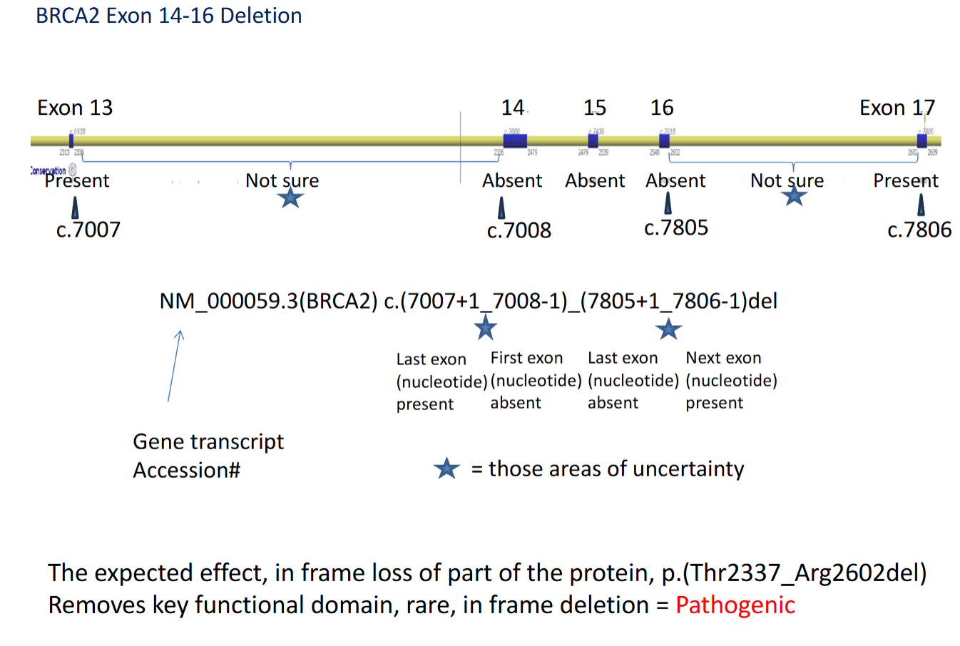
*****What are somethings we need to Consider when Assessing CNV’s in genes?
Truncating
In frame
Alternative splicing
Key functional domain
eg. In this BRCA2 case it was classified as PATHOGENIC despite being in frame because it deleted a key functional DNA-binding domain.
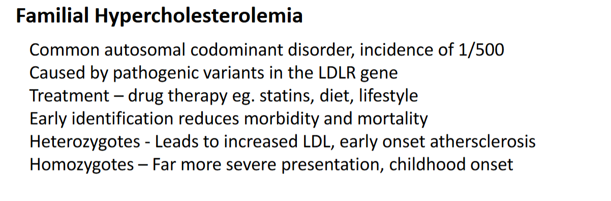
What is Familial Hypercholesteremia?
Autosomal vs. sex chromosome disorder
Incidence? 1/out of how many
What gene?
Treatment?
Heterozygotes vs. Homozygotes disease affect
How does early identification help?

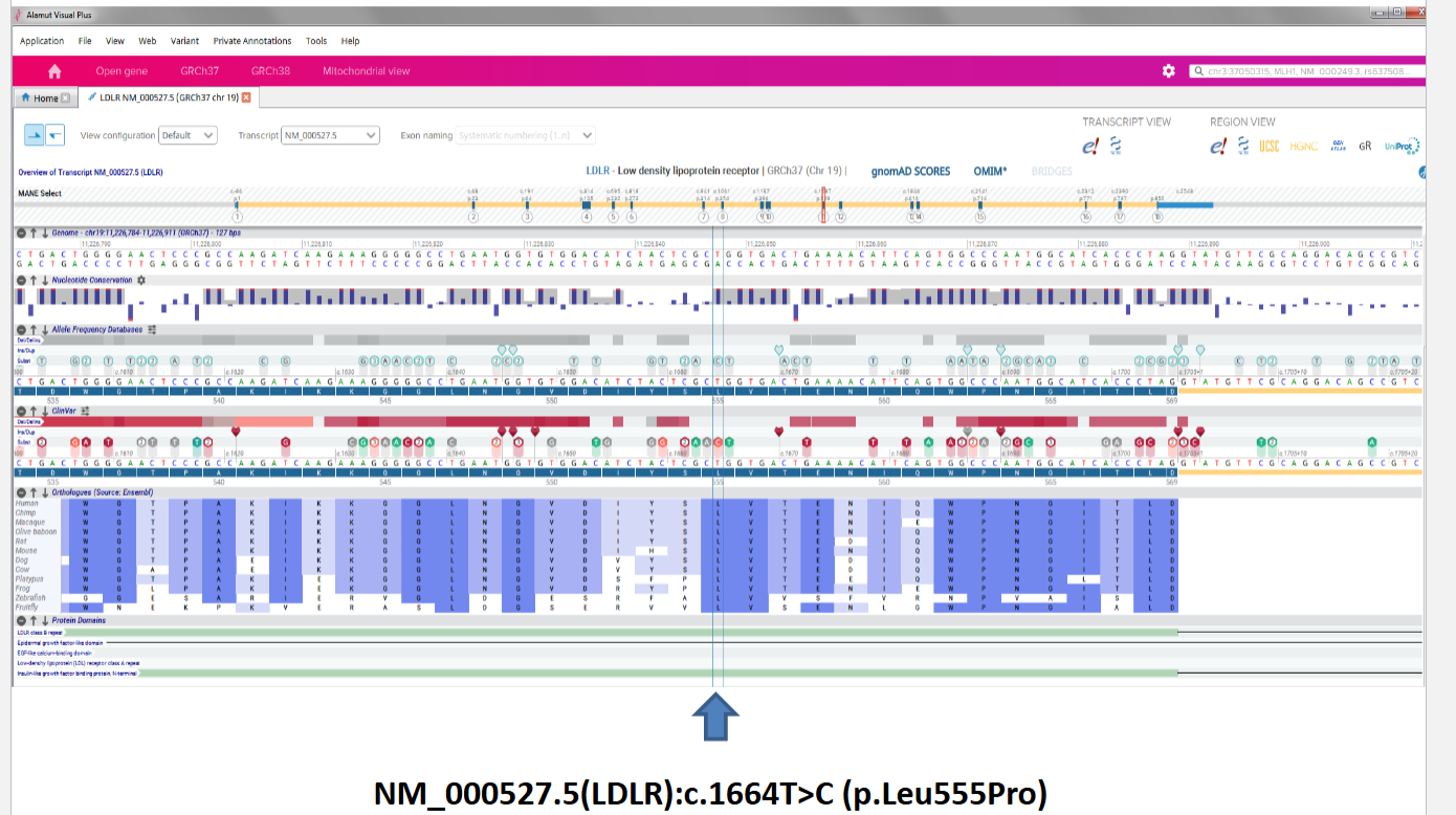
****Assessing missense vs. synonymous variation: How to begin assessing a variant aka. what are the first couple things you would do?
See what is known about the variant and location
compare the patient to data sets
In this data: green = common variant + red = pathogenic

*****The results for our patient with high LDL
HOW TO CLASSIFY? + WHAT ARE THE NEXT STEPS?
benign, likely benign
Uncertain
Pathogenic, likely pathogenic
LIKELY PATHOGENIC
Nxt steps = TREATMENT + potential Assessment of family members


*****The results for our patient If they had another variant of the a.a.
HOW TO CLASSIFY? + WHAT ARE THE NEXT STEPS?
benign, likely benign
Uncertain
Pathogenic, likely pathogenic

What is the diagnostic odyssey?
A set of steps for suspected mitochondrial DNA (mtDNA) disorders
structured, multi-tiered approach that progresses starting from non-invasive clinical INVESTIGATIONS to invasive tissue biopsies and specialized genetic sequencing.

*****How are Mito variants classified
What are 2 things taken into consideration and WHY?
are they part of a Haplotype?
A haplotype (or sub-haplogroup) represents a specific cluster of mutations shared by individuals who belong to the same maternal lineage. Thus if a haplotype is present it means that many people have this variant and it is likely benign (ancestral marker)
disease-causing mutations are frequently heteroplasmic (a mix of healthy and mutated mitochondrial DNA within the same cell),
is the variant Disease associated
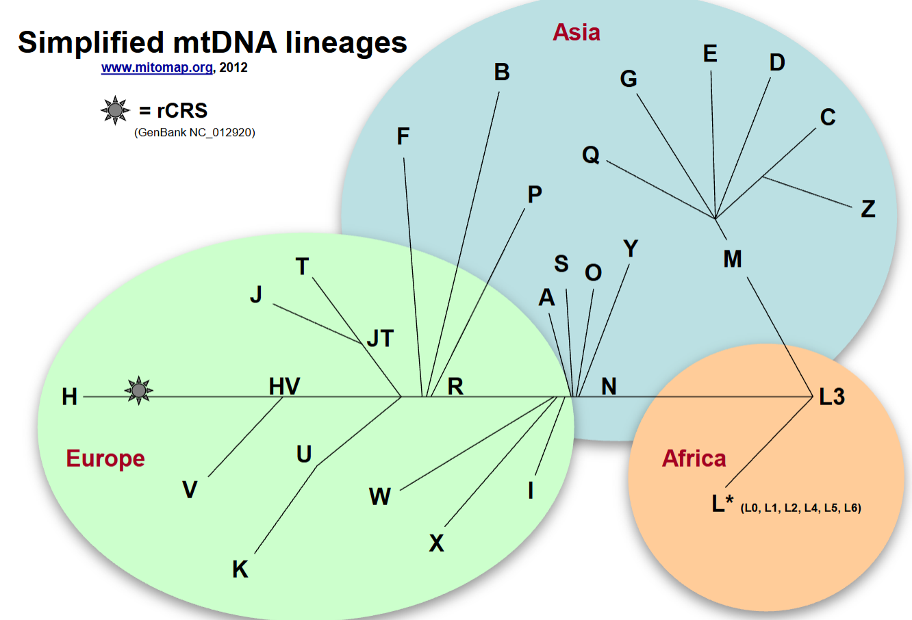
What is the key take away?
human mitochondrial DNA (mtDNA) is distinct in different ethnic groups
evolves along clear, non-recombining maternal lineages (haplogroups) that map directly to global geographic ancestries, tracing back to a common African origin
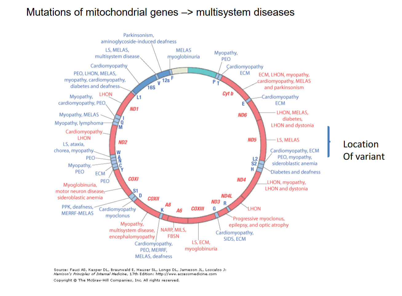
What is the main message?
Mut of mito genes ==> multisystem diseases
Mutations within the mitochondrial genome lead to complex, overlapping multi-system diseases because mitochondria provide essential cellular energy (ATP) to tissues all across the body.
Mitochondrial mutations affect many organs and systems
******Mitochondria INHERITENCE
why is there variable penetrance and expressivity?
MATERNAL inheritance
only mom passes it on
If dad has variant non of children are affected because he does not pass on
Penetrance and expressivity is dependent on the amount of affected mitochondria in the patient

*****What are some considerations for CLINICAL REPORTS
what is the frame work to make a good clinical report
What do the clinicians want to know?
How do we make it concise and meaningful?
Start of with:
Clinical Lab Interpretation: start with clinically relevant interpretation so physician can immediately grasp what the results mean for their patient.
Clinical Report Generation: what to include and not include depends on the clinical request
Accurate test methodologies and limitations
explicitly categorizing pathogenic variants vs. Variants of Uncertain Significance (VUS)
Report incidental findings
genetic counseling consideration
Recommendations for subsequent testing.
Educational Support for End Users: Provide consultation on reports and implications of variants identified and need for additional testing (help clinician interpret the report if needed)

*****What type of results can you expect:
How are the following variants categorized/ referred to?
what affects if the disease is observed
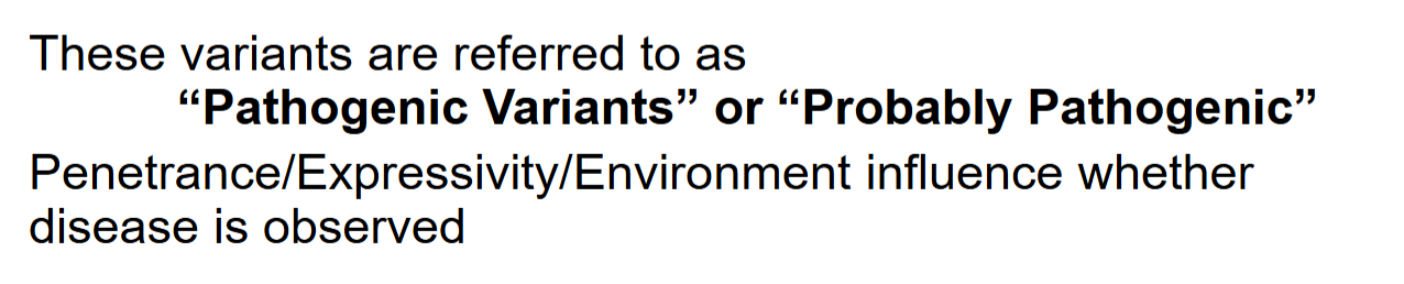

*****What type of results can you expect:
In this patient what can these results mean?

*****What type of results can you expect:
what are the possible results When a Patient is said to carry a VUS
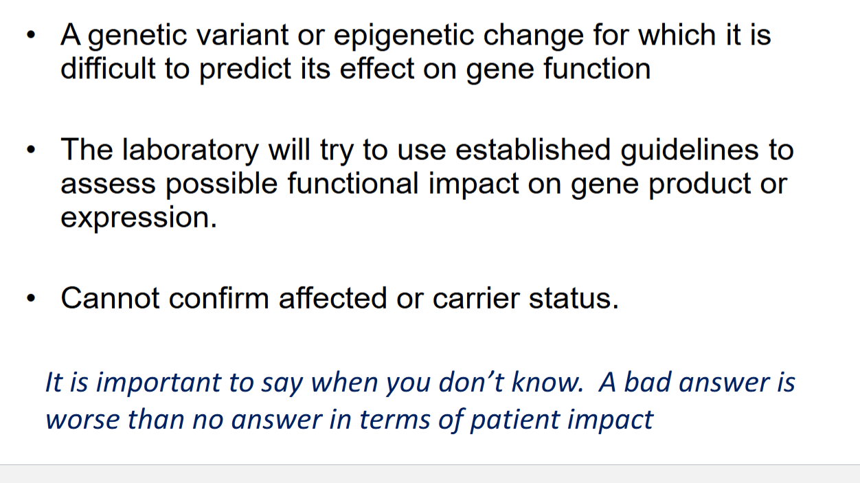
WHY? what is an example of where this went wrong/ wrong person was tested?
A boy had sudden cardiac death. Since the boy died he could not be used to test for the cause thus they tested the boys brother.
They detected a LQT variant in the brother → Doctors assumed this genetic change was the deadly killer of the boy. Because of this guess, they surgically put a heart-shocking device (a defibrillator) inside the healthy brother.
The mistake spread: They tested the rest of the family. Over 20 relatives had the exact same genetic spelling change. Doctors told all of them that they had a deadly heart condition.
The truth came out later: Scientists realized that the genetic change was completely harmless. It was just a normal family trait, not a disease.
Testing the wrong family member first can cause a cascade of false diagnoses and unnecessary, invasive medical procedures across an entire family.
Review of ECGs in those family members not consistent with long QT
Review of variant showed it had not been definitively linked to long QT
DNA Test repeated on blood sample from affected collected at autopsy
Pathogenic variant found in another gene linked to sudden cardiac death
THE DOCTORS SHOULD HAVE TESTED THE DEAD BOY INSTEAD OF THE BROTHER TO DETERMINE THE CAUSE OF DEATH

****Work flow for molecular autopsy
aka called postmortem genetic testing
