Topic V: Recombination, Repair and Mutations W10-12
 Call Kai
 Learn
Learn Practice Test
 Spaced Repetition
 Match
Match Flashcards
 Knowt Play
1/119

There's no tags or description
Looks like no tags are added yet.
Name | Mastery | Learn | Test | Matching | Spaced | Call with Kai | Chat |
|---|
No analytics yet
Send a link to your students to track their progress
120 Terms
What are the different types of mutations?
*Point mutation
Replace on base with another
Leads to a change in one aa
*Frame shift
Add/delete bases
Changes to reading frame
Changes more than one aa
More significant effects
^Non-sense mutation
RNA sequence becomes a stop codon
Very bad - more significant
^Missense mutation
Changes amino acids from one to another
^Silent mutation
No affect on the protein (due to redundant nature of aa)
^Conservative mutations
Produces amino acids of the same nature
E.g. Glu mutates to an Asp but they are both acidic
^Non-conservative
Produces amino acids of different nature from original
E.g. Ser (polar) replaces with Phe (non-polar)
*Named after how they affect the DNA
^Named after how they affect the protein

What are the types of DNA damage?
Double-stranded break
Caused by cellular activity, ionising radiation, chemotherapeutic drugs, DNA repair of other types of damage
We can potentially loose a part of the chromosome
Chemical bound between neighbouring nucleotides
Can be caused by UV light
This is INTRAstrand crosslinking between nucleotides (aka the same strand)
Repair mechanism: Nucleotide Excision Repair
Chemical linkage of two strands
Can be caused by reactive oxygen species, chemotherapeutic drugs or other cellular and environmental chemicals
INTERstrand crosslinking between strands
Repair mechanism: Homologous Recombination
Chemical modification of a nucleotide
Can be caused by reactive oxygen species, chemotherapeutic drugs or other cellular and environmental chemical, as well as normal modification that regulate what genes are active
These occur very often but the cell if very good at repairing this
Methylation, Depurination and Deamination

What are the way that DNA can repair itself?
Direct repair
Fix DNA molecules that carry nucleotides damaged by mutagens
Find a DNA that has been changed, place that change onto an enzyme
Search/fix abnormal chemical structures in DNA
Excision repair
Fix DNA molecules that carry nucleotides damaged by mutagens
Damaged base/ is excised from a nucleotide by a specific DNA glycosylase and phosphodiesterase and then fixed via a DNA polymerase and sealed by a DNA ligase
Search/fix abnormal chemical structures in DNA
Mismatch repair
Fix mismatched but otherwise normal nucleotides that result from errors in replication
Detects absence of proper base pairing between parent and daughter strands (MutS/MutH)
Nonhomologous end joining
Use to mend double-strand breaks in DNA
Ku binds to each end and helps bring the 2 fragments close together and then they are then ligase together, but this is hard to do
Very low fidelity - very common to errors


What are inherited (above) and non-inherited mutations?
Inherited (germline mutations)
Present in all nucleated cells of the body (including germ cells)
Acquired from parents (Predisposition (familial traits))
May be transmitted to offspring
Responsible for single gene disorders: cystic fibrosis
May or may not be present in multifactorial disorders: cancer, heart disease, diabetes, Alzheimer’s (aka diseases caused by multiple mutations)
Non-inherited (somatic mutations)
Affect only the mutant somatic cell and its descendants
Will not be transmitted to offspring
Could occur due to environmental factors and build up over time (at the start we can fight back and fix these mutations but over time as these mutations accumulate we can no longer deal with them and the consequences of the disease can present (e.g. cancer))
Contribute to multifactorial disorders: cancer, heart disease, diabetes, Alzheimer’s etc. (sporadic cases, also act as triggers for those with pre-disposition)
Cumulative → Accumulate in age related processes, UV and mutagen exposure...

What are the major classes of mutations?
One impact includes the grain of function, this is where a mutation causes over activity in the mutated cell, this is the case with cancers, where mutations lead to overgrowth and over proliferation of tumours etc.
Conditional → certain enzymes are only active under specific conditions, these mutations changes those conditions that enzymes act in


Point mutations
Single base substitution (missense, nonsense, silent) or single base deletion/insertion (frame shift)
Base substitution → no change in the overall number of bases
Missense → Results in an alternate amino acid (e.g. sickle cell)
This can be very impactful depending on the location of mutation (e.g. active site)
Nonsense → Results in a premature stop codon (e.g. Dopa-responsive dystonia and Sepiapterin reductase
deficiency)
If this mutation occurs very downstream of the active site (tail end) then it may not necessarily impact function
Silent → Does not affect the Amino Acid Sequence (due to redundant nature of aa)
Base deletion/insertion
Results in a changes in the number of bases (e.g. Duchenne (DMD) and Becker Muscular Dystrophies (BMD))
**If a base is substituted and results in a STOP codon → Missense
If a base is inserted/removed and results in a STOP codon → Frame shift

Sickle Cell Anaemia case
Sickle Cell Anaemia → Point mutation results in clumped haemoglobin, instead of globular
Gene: HBB encoding beta globin protein
Associated Disease: Sickle Cell Anaemia
Mutation: Point mutation – base substitution: A>T
Effect on translated protein: Missense Glu>Val
Symptoms
Fatigue and anaemia
Bacterial infections
Sudden pooling of blood in the spleen and liver congestion
Lung and heart injury
Leg ulcers

Dopa-responsive dystonia and Sepiapterin reductase deficiency
Gene: sepiapterin reductase gene (SPR)
Associated Diseases: Dopa-responsive dystonia and Sepiapterin reductase deficiency
Mutation: Point mutation – base substitution (A>T)
Effect on translated protein: premature stop – nonsense (Lys>Stop)
Sepiapterin reductase is an enzyme required for the synthesis of key neurotransmitters dopamine and serotonin
Deficiency: Symptoms include involuntary muscle contractions (dystonia), muscle stiffness, tremors, problems with coordination and balance (ataxia), and involuntary jerking movements
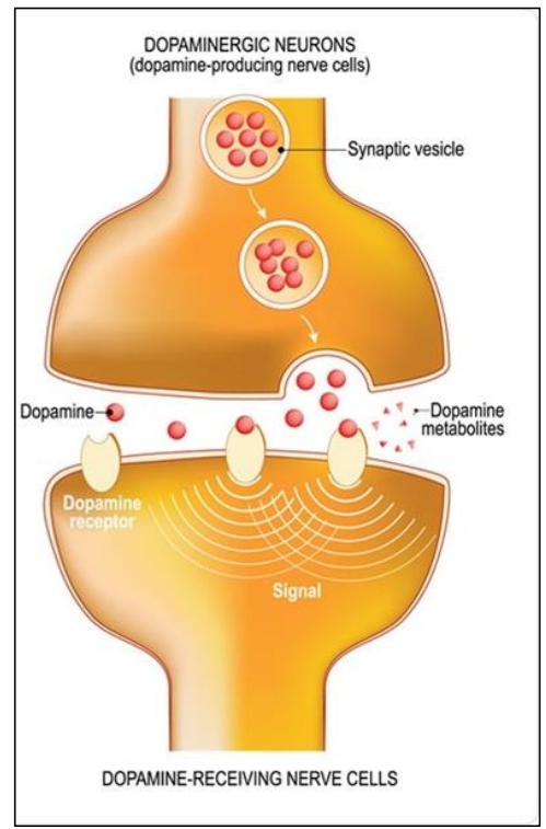

Duchenne (DMD) and Becker Muscular Dystrophies (BMD)
Gene: dystrophin
Associated Diseases: Duchenne (DMD) and Becker Muscular Dystrophies (BMD)
DMD: severe, BMD: mild
Progressive muscle wasting diseases due to mutations in the 79-exon dystrophin gene
Dystrophin connects the muscle cytoskeleton to the ECM
Duchenne muscular dystrophy (DMD)
Often results in frameshift or Nonsense mutation: Truncated protein
Becker Muscular dystrophy (BMD)
In-frame mutation (deletion) (entire codons removed i.e. nucleotide multiple of 3 are removed)
Does not affect the frame downstream: smaller protein but retains some function and very similar to the wild type

What are some larger scale mutations?
DID IT (acronym)
This occur at chromosomal level and affects MULTIPLE GENES
very important for evolution
Deletion
DiGeorge syndrome, large scale deletion of part of chromosome 22
Delayed development, congenital heart defects, reduced immune function, cleft palate
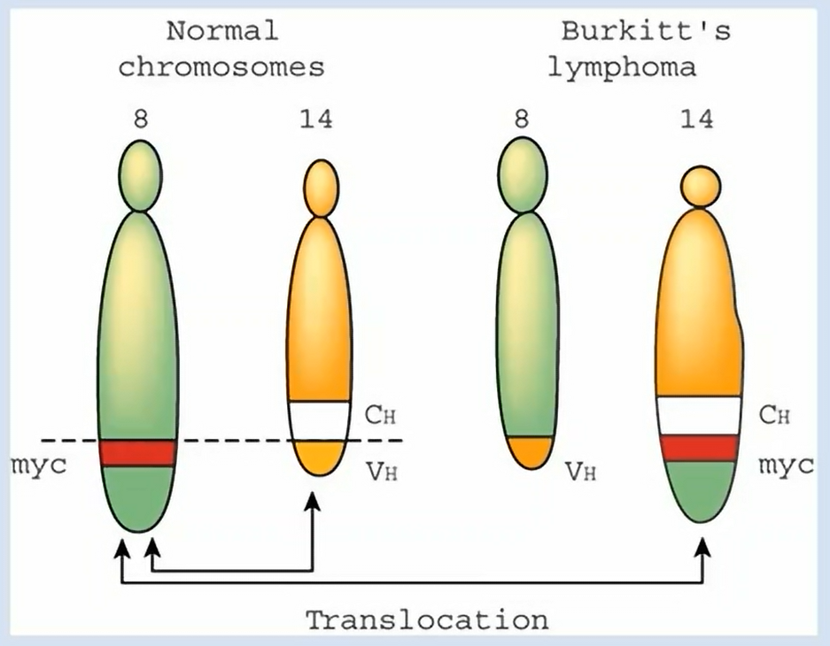
Translocation (image above)
Burkitt’s Lymphoma, translocation from chromosome 8
Results in increased activity of c-myc (proto-oncogene)
A proto-oncogene can the potential of becoming an oncogene which leads to cancer
Transcription factor involved in regulation of proliferative genes
Results in lymph node tumours (cancer)
Cause? → commonly associated with Epstein-Barr Virus

What causes mutations?
Spontaneous - occur naturally
Arise in all cells at low frequency
Errors in DNA replication (e.g. due to tautomers)
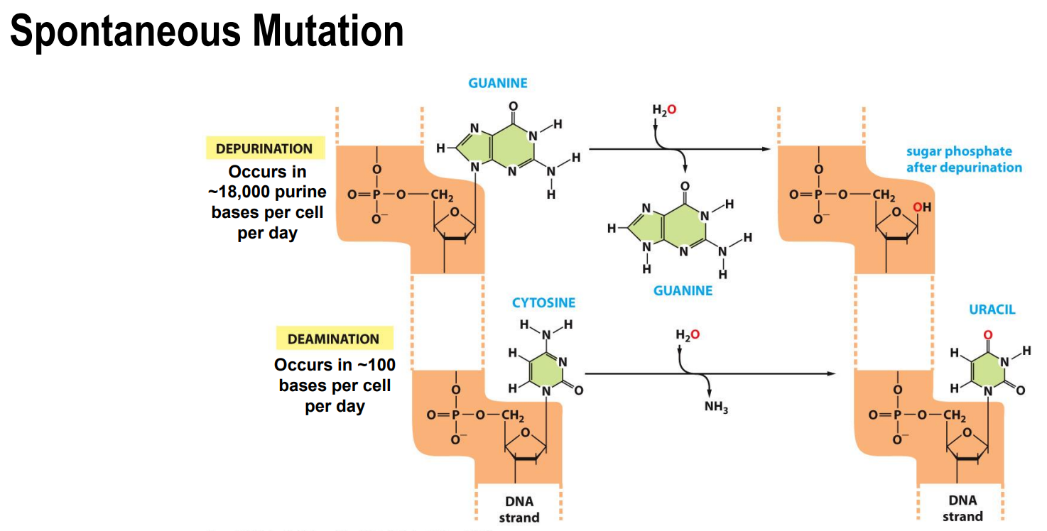
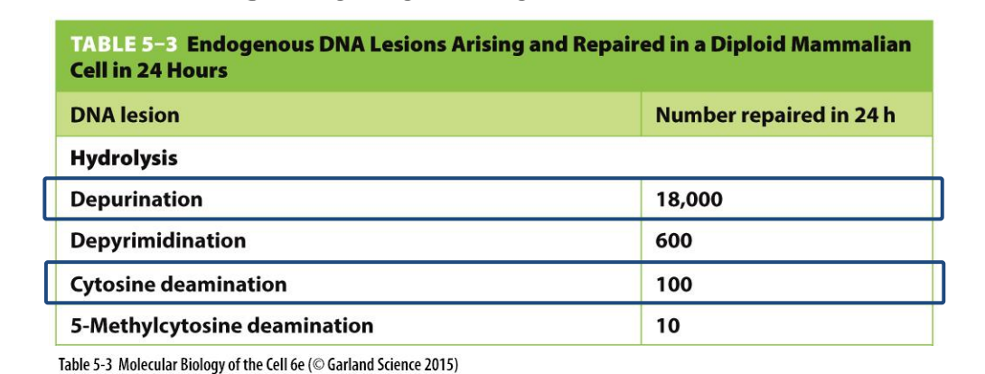
Spontaneous lesions or damage (e.g. depurination, deamination of cytosine) (image)
Sites on each nucleotide known to be modified by spontaneous: (below)
Oxidative damage (red arrows): METABOLIC guanine is more susceptible
Hydrolytic attack (blue): CLEAVES chemical bonds in DNA (this causes a removal of a base or deamination)
Uncontrolled methylation: (green): ALKYLATION of bases (this changes the base pairing)
Induced - require a ‘mutagen’
Chemical → Base analogues, intercalating agents, base modifiers
E.g. Aflatoxins, benzopyrenes, nitrites
Radiation → UV light, electromagnetic ionizing agents (free radicals)
UVB damage causes pyrimidine dimers that results in a lesion or a kink in the DNA this can lead to incorrect replication/base pairing thus can lead to a permanent mutation (e.g. melanoma)
Inherited - also known as germline
Present in all nucleated cells of the body (including germ cells)
Acquired from parents
May be transmitted to offspring


Induced DNA damage: UV Irradiation
Covalent linkage between two adjacent pyrimidine bases
Caused by UVB radiation from the sun
Thymine dimers: covalent linkages on the C-C bonds form lesions
Can occur between any two neighbouring pyrimidine bases – (T or C)
UV irradiation leads to: Sunburn, increased melanin production, If left unrepaired: Can lead to melanoma (cancer)


Spontaneous DNA damage by hydrolysis (damage to the bonds)
Depurination = spontaneous loss of purine bases (adenine and guanine) by hydrolysis
Depurination leads to loss of a nucleotide pair (deletion)
The backbone remains intact but the base is lost
When replication machinery encounters missing purine on template, it skips to next nucleotide resulting in a deletion: frameshift
Can be repaired well
Deamination = spontaneous conversion of cytosine to uracil by hydrolysis
Deaminated cytosine becomes uracil and mutation propagated as uracil pairs with adenine: base substitution
As a result of a hydrolytic event the amine group of a cytosine is cleaved, which then converts the base to uracil (the base still remain intact, its just the base changes)
If deamination remains we end up in a change to the complementary base and a mutation will be introduced into the 2nd strand that will then be incorporated into the genome following replication of that
*Note that one copy of the DNA remains unchanged as these mutations only occur to one strand, during replication both the normal and mutated strands separate and are replicated
Alkylation Damage: Methylated Guanine
Guanine is particularly susceptible to this type of damage
Results in an altered base that doesn’t follow base pairing rules
Alteration to the base – Methyl group attached to oxygen of guanine
Methyl Guanine pairs with thymine, not cytosine – Base substitution

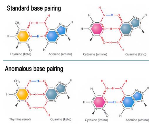
Spontaneous mutations - Tautomeric forms of DNA bases
Bases can be incorporated into DNA during replication in their rare tautomeric forms
Base pairing will follow different rules → Base substitution

What are the effects of mutations within genes?
Reduced quantity and efficiency and prevention of protein forming would all be a loss of function mutations
Change in proteins function → could be a gain/loss of function


What are CFTR Mutations?
Large gene with many known mutations
Not all these mutations will cause CF, some can go unnoticed
So many people can have different CFTR genes but not necessarily present with actual CF
CFTR encodes for the Cystic Fibrosis Transmembrane Conductance Regulator
Membrane protein
Chloride channel
Very important for epithelium fluid transport between the cell and the cytosol
Cystic Fibrosis: dysregulation of epithelial fluid transport
F508del results in low numbers of misfolded protein in the cell membrane (this is an example of the very common deletion mutation)

Complex mutations: CFTR gene associated with cystic fibrosis in humans
Large variation in the CFTG gene in the population
Remember not all of them lead to CF (come can be silent/small)

What is the Human Variation in the genes?
We are different in large part because the sequence of our DNA is different
On average <1 difference in every 1,000 bp
This is still ~ 1 x 10⁶ differences between individuals
The majority of these differences occur in the non-coding regions of our DNA
However, even amongst our 25,000 or so genes, at least one third are polymorphic within the healthy population
Each different form of a gene is called an allele and has arisen as a result of mutation
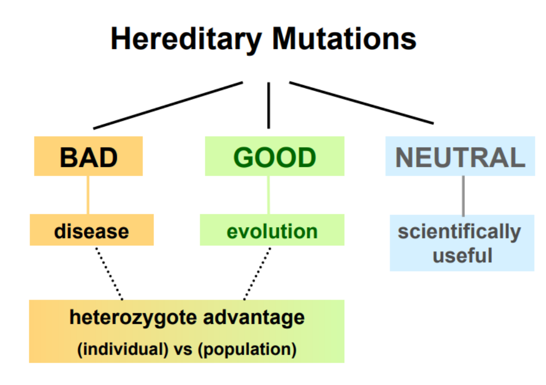

Mutation in Evolution: Principle
Some mutations that provide a selective advantage are perpetuated: natural selection
These mutations give a higher chance of surviving and thus can be passed one (i.e. selected)
High Altitude adaptation: Tibetans and Nepalese live altitude 4,000m
Air contains 40% less oxygen
Compensate with bigger chests: greater lung capacity
Evolution of few red blood cells with better blood flow in brains
These mutations have lead them to thrive in these environments


Example: CCR5 receptor helps HIV-1 enter cells
CCR5 receptor is a type of chemokine receptor present on immune cells and is what HIV particles will bind to in order to infect an individual
Therefore people who have the mutation where they do not have those receptors are protected against HIV
Beneficial mutation up to 14% in Northern Europe
So we see that the 32 allele is common where HIV is rare and 32 allele is rare/absent where HIV is rampant

What are Neutral Mutations?
Neither advantageous nor disadvantageous
They don’t cause disease but they also do not provide any evolutionary advantage because they don’t actually affect the proteins or any coding regions of DNA (aka silent mutation), but they are very scientifically useful in forensics, paternity testing etc.
The majority of mutations are neutral and inherited
Can also spread in population
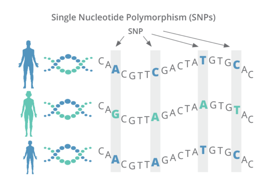
In humans in the form of single-nucleotide polymorphisms (SNPs)
99.5% similarity
Why do many disease-causing mutations persist in the population?
There are a multitude of conditions/diseases (> 4000) which result from the ‘bad’ or detrimental effects of mutations that are passed on through the generations → so why do they persist in the population
Many genetic diseases only manifest when the individual has inherited 2 mutated copies of the same gene (homozygous)
Recessive inheritance: the affected person must be homozygous for a disease mutation or compound heterozygous (mean 2 faults genes) for two different disease-causing mutations for the condition to manifest
No working copy of the gene to provide sufficient function
In these cases, heterozygotes with one normal gene copy and one mutant gene copy are healthy (most of population?)
Their normal gene provides sufficient normal protein/function to compensate for this mutated allele → so disease does no present
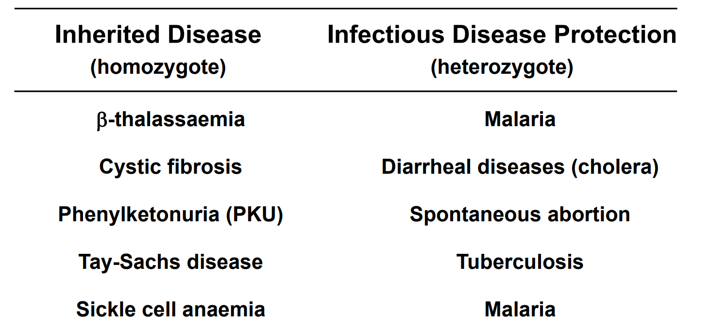
Heterozygote Advantage: If the mutation confers some selective advantage to heterozygous individuals while not causing diease, it may be maintained in the population by natural selection (image)
E.g. Homozygous to CF causes disease but being a heterozygote causes protection against cholera
Sickle Cell Allele and Malaria


New genes and genome evolution
DNA is not made in random sequences, it is generated from pre-existing DNA
Changes in DNA sequence lead to changes in structure and the development of new functions for proteins
Intragenic mutation
Could be simple point mutations, deletions, insertions
So the original gene picks up mutations and begins to evolve, particularly if the particular mutation is advantageous
So new genes are created through mutation
Gene duplication
New genes introduced via duplication
This can happen during replication
We then have 2 copies of the gene that can evolve at different rates (e.g. they can acquire mutations etc. at different locations at different rates) and essentially become completely different genes
DNA segment shuffling
Happens during replication
This is where translocations and stuff occurs (during recombination)
This can cause new combinations of genes
Gene mixing/ Horizontal gene transfer
This uptake can occur from: Bacterial transformation, Conjugation, Transduction, Viral reassortment (above)


Evolution through expansion of Gene Families
These DNA duplications, mutations and shuffling then give rise to different gene families
These genes that are related are called gene families
Example
The alpha and beta subunits are only slightly different, and thus comes from an initial duplication, translocation and then subsequent mutations of the ancestral gene
Genes evolve as families: members encode proteins with related structure and function
Globin gene family:
Derived from common ancestral gene
Encode globular proteins transporting oxygen
Common to many species
Essential for multicellular animals to grow to large size
Occurred over millions of years
At 400 million years there was a translocation event where the alpha section split off from chromosome 11 and translocated onto chromosome 6
*Note that not all genes evolve at the same rate

Why is Genetic Stability important?
For survival, organisms not only need accurate mechanisms for replicating DNA but also mechanisms for repairing spontaneously occurring damage in DNA
We do not want long lasting permanent mutation occurring that can cause ongoing or permanent damage to our proteins and subsequently cause disease
DNA damage can be caused by heat, metabolic accidents, radiation, exposure to the environment, different substances
Fewer than one in 1000 accidental base changes results in permanent mutation thanks to DNA repair
This is very important for genetic stability, if our cells are acquiring mutations at a really high rate then they are not going to be able to replicate successfully
Why is DNA repair critical?
DNA repair occurs through a range of coding genes involved in repair
Inactivation of DNA repair genes causes an increased rate of mutation (as other mutations start to build up) → this can lead to disease
Many of these were originally identified in bacteria
Many serious human diseases are linked to decreased DNA repair enzymes/genes
Cancers, UV sensitivity, leukaemia, growth and development

Recap from Theme I: Errors during replication
If errors not fixed during replication: mutations arise
Errors such as:
Tautomeric bases
Mismatch
Mechanisms to prevent replication errors
Proofreading polymerase (fixing majority of errors)
Errors occur usually in 1:100,000 to 1:1,000,000 bases
With proofreading: 1:100,000,000 bases
Mismatch repair system
Identifies errors in the secondary structure
Mismatch repair enzymes recognize this and remove/replace the nucleotide
Recap from Theme I: Proofreading Polymerase
If errors not fixed during replication: mutations arise
Proofreading polymerase (fixes majority of errors: up to 99%)
Incorrect base paired, elongation pauses
3’-5’ exonuclease activity of the polymerase removes several bases, including the incorrect one
Replication resumes 5’-3’
Occurs during replication (S phase)

Recap from Theme I: Mismatch Repair
If errors not fixed during replication: mutations arise
Mismatch repair system identifies errors in the secondary structure (e.g. tautomeric bases)
Mismatch repair enzymes recognize this and bind to the base (MutS)
MutL scans downstream of the DNA to find a nick
Region between mismatched base and nick excised by exonucleases
DNA polymerase fills the gap and DNA backbone sealed via DNA ligase
Occurs mostly in S phase of the cell cycle, follows behind replication

How can the DNA double helix be repaired?
We have two separate copies of all genetic information (double helical structure of DNA)
When one strand is damaged, the complementary strand (copy of same information) remains intact
This is used to restore correct nucleotides to damaged strand
Overview of DNA damage repair pathways
Base lesions: single (methylation, depurination) or double (pyrimidine dimers)
Base excision repair (BER)
Nucleotide excision repair (NER)
For BER & NER
Damage is excised (removed)
Original sequence restored by using undamaged strand as template
Remaining break sealed with ligase
Direct reversal repair (DR)
Direct removal of lesion
No cleavage or ligation of a base, instead we remove the lesion (e.g. methylation - we remove the methyl group)

Base Excision Repair
Repairs damage to a single base (deamination or depurination - damage to the base as opposed to the entire nucleotide)
A set of enzymes acting sequentially
Specific DNA glycosylases
Recognise specific type of altered base by ‘flipping out’ (rotate) from helix
Excise/remove base via hydrolysis (breaking the bonds b/w sugar and base NOT the phosphate backbone)
AP endonucleases (AP for apurinic or apyrimidinic)
Recognise this phosphodiester backbone with a missing base in helix
Then cuts the phosphodiester backbone
DNA polymerase adds new nucleotides into the gap
DNA ligase seals the nick

Nucleotide Excision Repair
Repairs larger changes to DNA helix: 2 or more bases and entire nucleotides need to be removed
Multi-enzyme complex scans DNA for distortion
This complex will then cleaves phosphodiester backbone of abnormal strand on both sides of distortion
DNA helicase (DNA unwinding enzyme) peels away single-stranded oligonucleotide containing lesion
Gap filled by DNA polymerase
Sealed with DNA ligase

Xeroderma pigmentosum - Nucleotide Excision Repair
Autosomal recessive genetic defect: nucleotide excision repair enzymes are mutated (inherited)
Prevalence 1 in 250,000
Symptoms: severe sunburn after minutes of sun exposure, freckle
UV light can cause mutations and without proper nucleotide excision repair mechanisms due to accumulation of unrepaired DNA
High risk of developing skin cancers: tumour suppressor genes are affected, melanoma
Life expectancy shorter by ~30 years
Transcription-coupled DNA repair
Occurs during transcription
Ensures cell’s most important DNA is efficiently repaired
Links excision repair systems with RNA polymerase (enzyme that transcribes DNA into RNA)
RNA polymerase stalls at DNA lesions and falls off at these points and this then directs the nucleotide excision proteins (DNA glycosylases and AP endonucleases) to these sites
Targets repair to genes that are actively being transcribed into mRNA
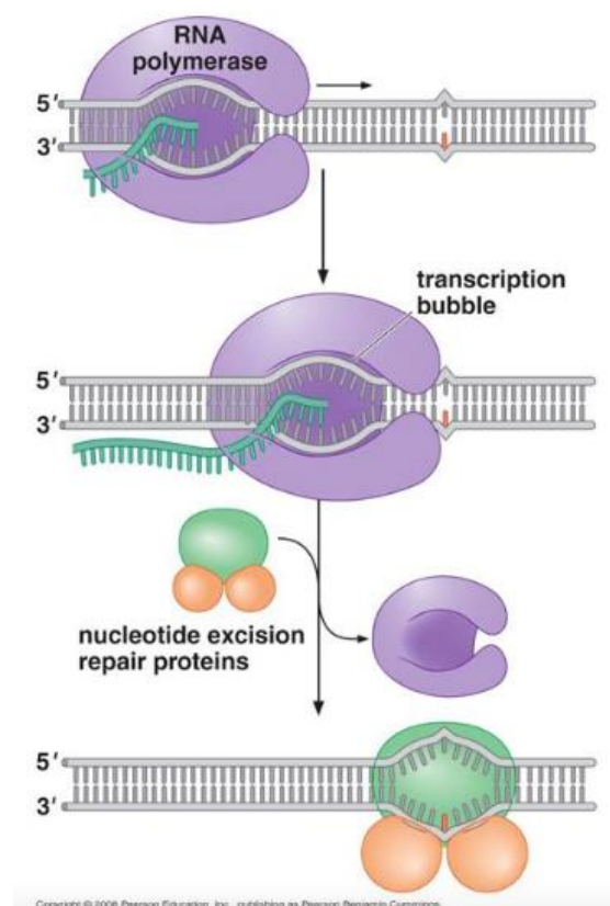
Cockayne syndrome - Transcription-coupled repair and human disease
The particular RNA polymerase that could stall or detect the lesion is permanently stalled or is unable to return to polymerisation and this then results in a high level of apoptosis as they encounter high levels of DNA damage as cells age
Autosomal recessive congenital disorder with a high level of apoptosis
Prevalence: 1 in 200,000
Symptoms: growth retardation, skeletal abnormalities, progressive neural degeneration and retardation, severe sensitivity to sunlight
Defect in transcription-coupled repair
RNA polymerase molecules become permanently stalled at sites of DNA damage in important genes
Causes cell apoptosis (programmed cell death)
Life expectancy 10-20 years

Direct Reversal Repair
Most efficient form of DNA repair
As it does not require the removal of bases or nucleotides
Rapid removal of certain highly mutagenic or cytotoxic lesions
E.g. Alkylation lesion 6-O-methylguanine
Methyltransferase (MTase) protein accepts methyl group (CH3) on cysteine residue from alkylated guanine nucleotide
Restores normal guanine
MTase inactivated
No DNA cleavage or ligation required

Emergency repair of heavily damaged DNA
Highly accurate replicative DNA polymerase (Pol III) stalls and falls of when it encounters damaged DNA
In emergencies, they employ less accurate back-up polymerases to replicate through the DNA damage - translesion polymerases (Pol V) from where the Pol III has left off
Pol V is able to replicate DNA and continue polymerisation despite damage being identified (usually they be falling off/stalling), hence transcribed through the lesion thus called translesion polymerase
The back-up polymerases lack exonucleolytic proofreading activity
These polymerases only add one or a few nucleotides before it falls off and the replicative polymerase (Pol III) continues from there
Risky for the cell: responsible for many mutations as the mutations become incorporated into the DNA

DNA damage can delay progression of cell cycle
When does repair occur?
In most cells, DNA damage causes a delay in cell cycle
Ensure that all damaged is repaired before a cell divides
G2/M checkpoint → does not enter mitosis until this is happed
Intra S phase checkpoint → S phase is slowed down when DNA damage is detected

Cell Cycle involves critical check points
Cell cycle will not progress pass these check points until damage is repaired
Orderly progression of cell cycle maintained through use of checkpoints to ensure completion of one step before next step begins
Cell cycle stops if damaged DNA is detected
In mammalian cells, the presence of DNA damage can:
Block entry from G1 to S phase (checkpoint)
Slow S phase (replication) once it has begun
Block transition from G2 phase to M phase (checkpoint)
Delays facilitate DNA repair by providing time needed for repair to reach completion

Integrating DNA repair and cell cycle progression
DNA damage results in increased synthesis of some DNA repair enzymes
Special signalling mechanisms that arrest the cell cycle and respond to DNA damage
ATM protein: large kinase (phosphorylate proteins) that signals intracellularly to delay the cell cycle in response to DNA damage
Individuals with ataxia telangiectasia (AT) (defects in ATM protein) suffer from effects of unrepaired DNA lesions (neurodegeneration, genome instability etc)
p53: ‘Guardian of the genome’
Arrests the cell cycle at G1/S checkpoints until damage repaired
Activates DNA repair enzymes
Can initiate apoptosis if damage too great (if cell is held at rest for too long)
Huge implication in cancer: tumour suppressor
Chk1: kinase
Cycle arrest at S and G2/M checkpoints
DNA repair or cell death
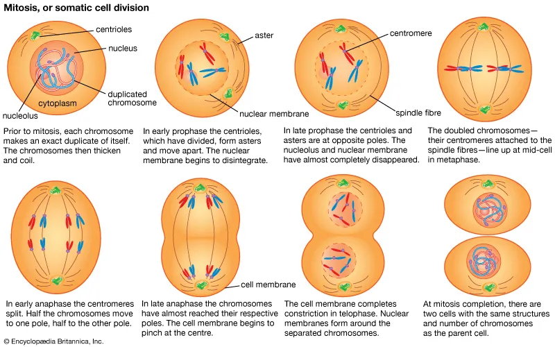
Mitosis Revision
Prophase: chromosomes condense and become visible, the nuclear envelope breaks down, and spindle fibers form.
Metaphase: chromosomes line up along the middle of the cell (the equator).
Anaphase: sister chromatids are pulled apart to opposite poles.
Telophase: nuclear envelopes re-form around each set of chromosomes, and the chromosomes uncoil.
Chromosomal Crossover (homologous cross over) Revision
When: Prophase I of meiosis
What happens: Homologous chromosomes (one from each parent) pair up as a tetrad, which is 4 chromatids in total. Non-sister chromatids break and swap matching sections
Chiasma (plural: chiasmata): the point where the swap happens
Result: Each chromosome ends up with a mix of maternal and paternal DNA. The genes are the same, but the alleles (variants) can differ
Why it matters: It creates genetic variation in gametes, on top of the variation from different sperm meeting different eggs
Meiosis Revision
Before it starts: Interphase, where DNA replicates, so each chromosome has 2 sister chromatids
Meiosis I (homologous pairs separate, so the cell becomes haploid)
Prophase I: Chromosomes condense and the nuclear envelope breaks down. Homologous chromosomes pair up as tetrads, and crossover occurs.
Metaphase I: Homologous pairs line up at the middle in random orientation (independent assortment).
Anaphase I: Homologous chromosomes are pulled to opposite poles. Sister chromatids stay together.
Telophase I and cytokinesis: The cell splits into 2 haploid cells, each with chromosomes that still have 2 chromatids
Meiosis II (sister chromatids separate, similar to mitosis)
Prophase II: The spindle forms in each of the 2 cells, and there is no DNA replication.
Metaphase II: Chromosomes line up at the middle.
Anaphase II: Sister chromatids are pulled apart.
Telophase II and cytokinesis: Each cell splits again
Result: 4 genetically unique haploid cells (gametes).
Key contrast: Meiosis I separates homologous chromosomes, and meiosis II separates sister chromatids.
Humoral response
The humoral response is commonly activated to fight bacterial infections or toxins, and relies on using antibodies to destroy the foreign antigens
The response is tightly regulated, and involves several steps of clonal selection: specific cells are selected and cloned to fight a specific target
Cells that are clonally selected will then clonally select other immune cells, creating a rapid expansion in the number of lymphocytes targeted to a specific pathogen
This rapid expansion of lymphocytes is one reason an infected person may feel fatigue.

What are the function of antibodies?
Antibodies are small Y shaped proteins that are secreted by Plasma B cells
They destroy or deactivate the pathogen when they bind to it
They are highly specific to a particular antigen
The level of antibodies in your blood can indicate the strength of your immunity to that specific pathogen
Diagram
Antibody
All antibodies have a constant region (a region that is the same for all antibodies). However, there is a region at the two tips of the antibody that can be different, called the variable region
Agglutination is when pathogens become trapped in a network of antibodies, making it easier for them to be phagocytosed (engulfed) by other lymphocytes, for example macrophages
Antibodies can also bind to toxins that are either produced by a pathogen, or directly introduced into a person (e.g. through a snake bite). In neutralisation, the antibodies deactivate these toxins by binding to them, thereby stopping these toxins from affecting other cells
The final way antibodies function is through boosting the innate response of opsonisation. Antibodies coat the pathogen’s surface to ‘tag’ the pathogen. This makes it easier for immune cells to identify the pathogen, and also prevents the pathogen from binding to host tissues and helps phagocytes to phagocytose them
Complement activation is a process where antibodies, when bound to a pathogen, trigger a cascade of other proteins to be activated. These complement proteins will have other functions, e.g. physically ‘punching’ a hole in the pathogen’s membrane, making a pathogen more easily identified, or deactivating a toxin. Complement proteins work as part of the innate immune response and while their action is boosted by working with antibodies, complement proteins can work independently of antibodies

What is the Cell-mediated response
It is commonly activated to fight cancerous cells or cells that have been infected by viruses
The cell-mediated response relies on using cells (e.g. cytotoxic T cells) to destroy other diseased cells, hence the name ‘cell-mediated’
This differs from the humoral response that uses antibodies to fight an infection
Like the humoral response, the cell-mediated response involves a cascade of cells being activated and dividing. This large consumption of resources contributes to the symptoms a sick person may feel, such as fatigue.
Diagram
If a cell becomes diseased, for example cancerous or infected by a virus, it will present this information on the MHC I class protein, beginning the cell-mediated immune response (Tip: MHC I class proteins are for endogenous antigen)
Antigen presenting cells such as dendritic cells or macrophages patrol through the body’s tissues. If one encounters a foreign antigen from a diseased cell, it will phagocytose it and then present a fragment, or antigen on its MHC II class protein. It will then travel to the lymph node via the lymphatic system to get back-up
When an antigen presenting cell arrives in the lymph node, it will activate a specific T cell by presenting the antigen fragment on a MHC II class protein. This process is called selection
The selected T cell will then begin to divide into memory T cells and helper T cells which subsequently differentiate into either effector or memory T cells. This process is called clonal selection

Overview of the Adaptive immune response

What are Double stranded breaks?
Both strands are broken
Problem: no template for repair
Causes:
Environmental damage:
Double stranded breaks can occur from ionising radiation (e.g. x-rays) and reactive chemicals (e.g. anti tumour-agents)
DNA replication:
Most of the time they arise from DNA replication forks that become stalled or broken (accidents occur during nearly every round of DNA replication)
If lesions not repaired:
Breakdown of chromosome into smaller fragments
Catastrophic loss of genes when cell divides
Double stranded break during replication
These unrepaired lesions could be a result of a deamination or depurination → can be fixed via emergency repair
These unrepaired breaks leads to a double stranded break

What are the two mechanisms of Double stranded break repair
Non-Homologous End Joining (NHEJ)
Re-joining of broken ends by DNA ligase'
Generally loss of nucleotides at site of joining
Common in mammalian somatic cells
Quite error prone
Essentially just ligates the 2 ends back together
Homologous Recombination (HR)
More accurate
Uses sister chromatid as template
We process the blunt ends, such that the 5’ ends are resected (which is they are digested back a little), then the identical sister chromatid can be used as a template to repair this DSB (aka strand invasion)

What are the steps in Non-homologous end joining?
Ku protein: a heterodimer that recognises and grasps the broken chromosome ends
Protein complex forms, holding the two ends together
These proteins, including nucleases process the ends of the DNA (though not as much as HEJ)
Then the Ku proteins on either end pull the two ends together
DNA polymerase may be required to fill any gaps
DNA ligase completes the process resulting in the end being covalently joined
Results in a loss of nucleotides at the site of joining
This can lead to a deletion → mutations

What is Homologous recombination?
Homologous Recombination involves the exchange of DNA strands between two homologous duplexes of DNA
Duplexes: double helix DNA
Homologous: identical or in this case, highly similar sequences
Sister chromatids (repair)
Homologous chromosomes (meiosis)
Very similar but have different alleles → still homologous


How is DNA base pairing critical for recombination?
Homologous recombination only between DNA duplexes that have extensive regions of sequence similarity
Two DNA duplexes engage in extensive base-pairing between a single strand from one DNA duplex and the complementary single strand from the other duplex
A perfect match is not necessary but it must be close for homologous recombination to happen

Homologous recombination: functions
Accurate repair of double stranded breaks: most widespread use of homologous recombination
Genetic exchange between two homologous DNA sequences (DNA sequences similar or identical in nucleotide sequence)
Mechanical role in assuring accurate chromosome segregation during meiosis in eukaryotes
Double stranded break repair via HR (homologous recombination)
1) Exonuclease chews/resects 5’ ends of the DS break
2) Strand exchange/invasion by homologous base pairing
One of the 3’ overhands will invade the intact sister chromatid/duplex → pairs with complementary bases → DNA synthesis using the 2nd duplex as a template
3) Extension of invading strand by DNA polymerase
4) Invading strand released, original helices reformed
Once released the extended region can then bind to the 2nd overhanging 3’ region (this is why it is important that those exonucleases at the start resect a large region either side of the break)
5) Gap filling by DNA polymerase
6) Ligation
*No loss of nucleotides

Double stranded break repair during replication
1) Nick or gap in parental DNA helix ahead of the replication fork
2) When fork reaches lesion, it falls apart: causing a DS break in one duplex, replication halts
3) Homologous recombination (strand invasion) restores DNA sequence using undamaged duplex as template
4) Replication can resume

How does a strand invade?
Invading strand needs a way of disrupting a stable DNA helix of the other duplex (because the sister chromatid otherwise has no reason to open up), therefore we need protein it help this process
The 3’ end of the invading strand is acted upon by RecA (E. coli) or Rad 51 (eukaryotes)
ATP bound RecA binds tightly to ssDNA forming a DNA-protein filament on the 3’ overhang that is going to invade → this actually stretches the DNA out a little bit exposing the nucleotides so that they can scan the duplex for complementary bases (in a sequence independent manner)
RecA also binds to the intact dsDNA helix and stretches (destabilise) it out, so that the ssDNA can invade
Once homologous sequence located (extended stretch of at least 15 nucleotides), strand invasion occurs
Single strand displaces one strand of the duplex and forms base pairs with the other strand resulting in a heteroduplex (pairing of two DNA strands from two different DNA molecules
Other proteins help in this process
Destabilises the duplex DNA
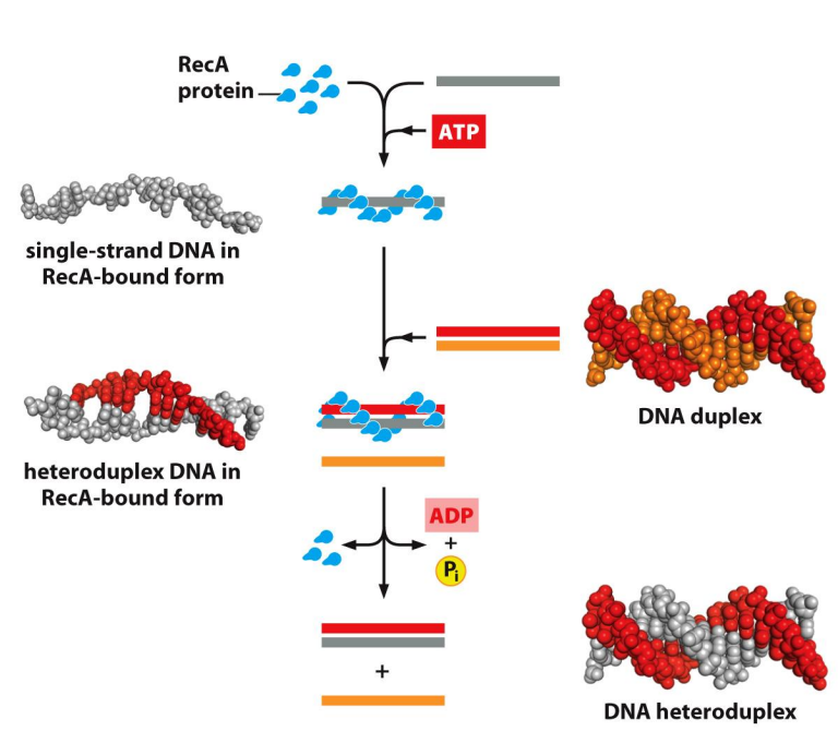
What are Heteroduplexes?
Strand invasion requires the pairing of a region of single-stranded DNA with a complementary strand in a different DNA double helix
DNA hybridisation occurs between strands that are highly similar, but not necessarily identical
Creates a region of DNA helix formed from strands that originate from two different DNA molecules – heteroduplex
Some mismatch may occur
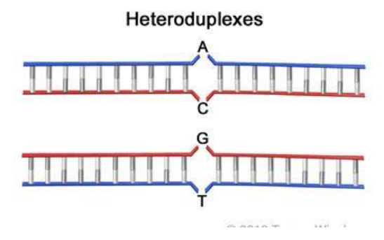
What can go wrong in Homologous Repair?
Wrong template used (other chromosome homolog, not sister chromatid) lead to loss of heterozygosity
Mutations can become homozygous
if the mutant gene is used as a template, therefore we lose the functioning copy of our normal gene and we not are homozygous for a mutant
Can lead to cancer (e.g. tumour suppressor genes)
Too little homologous recombination = increased mutation rate → can lead to cancer
Results from damage to portions in homologous recombination
Mutations in Brca1 and Brca2 cause breast cancer due to inefficient repair by homologous recombination
Brca2 helps to bring Rad51 protein to sites of damage and releases it in its active form onto ssDNA

Difference between mitosis and meiosis?
dsDNA repair occurs shortly after DNA replication in the S and G2 cell cycle phases - when a daughter duplex can act as the repair template, (i.e. Before Mitosis)
Meiosis differs from mitosis in that:
Two divisions,
Homologous chromosomes separate
Sister chromatids separate
Chromosomes in meiosis undergo recombination
Shuffles the genes
Different genetic combination in each gamete
Chromosomes in mitosis are identical
Outcome of meiosis is four (genetically unique) haploid cells
Outcome of mitosis is two (genetically identical) diploid cells
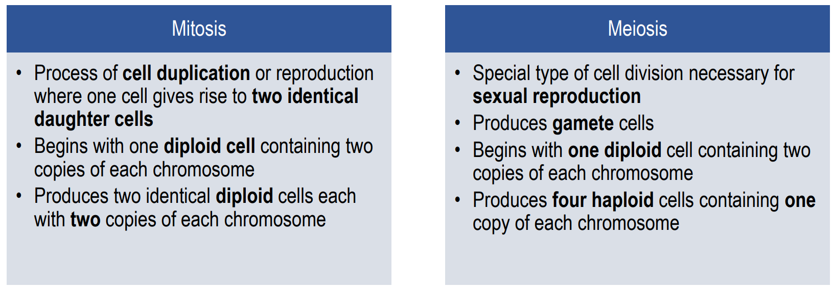
Steps in meiosis vs. mitosis

How can Recombination generate chromosome crossovers?


Recombination during meiosis
Process differs slightly from Homologous Repair
Begins with enzymes Spo11 and Mre11 creating a ‘double stranded break’ in one of the homologous chromosome
Nucleotides cleaved/resected from the 5’ ends either side of the break
Rad51 initiates strand invasion
Second strand from nicked chromosome ‘captured’
DNA polymerase and ligase synthesise new DNA and seal the gaps
Results in a double Holliday junction structure
Branch migration extends the cross over region or region of heteroduplicity
Endonucleases cleave strands to resolve junctions
*Cross over is an exchange of genetic material between non-sister chromatids of homologous chromosomes


Key stages of Homologous Recombination: 1) Double stranded break either spontaneous or induced in one of the homologous duplexes
In repair, this may begin as a nick, collapsing the replication fork
During meiosis, enzymes Spo11 and Mre11 create a break

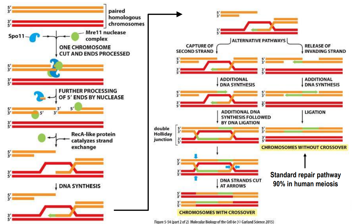
Key stages of Homologous Recombination: 2) 5’ ends of break processed leaving 3’ overhangs


Key stages of Homologous Recombination: 3) One of the two 3’ overhangs invades the undamaged duplex at the point where the sequence is complementary
Rad51 in Eukaryotes
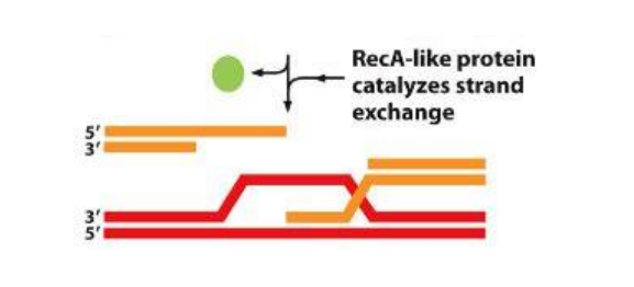

Key stages of Homologous Recombination: 4) DNA synthesised from the 3’ overhang, using the complementary strand as a template replacing the gap from the break


Key stages of Homologous Recombination: 5) Alternative pathways
Invading strand release → what happens during repair
Second Strand Capture → DNA recombination


Key stages of Homologous Recombination: 6) Invading strand release
Broken double helix re-forms
DNA polymerase fills any remaining gaps
Ligation
No crossover
This mechanism used for repair


Key stages of Homologous Recombination: 6) Second Strand Capture
Additional DNA synthesised to fill gap
DNA ligated back together
3’ end ligated back to the 5’ end of it’s original strand
BUT with a region now complementary paired to the homologous duplex
Double Holliday junction
Points where single strands separate from double helix: Branch Points
Points where strands crossover and join 4 DNA strands: Holliday junction
Branch points can move along the DNA strand and extend those regions of exchanged bases: branch migration

Second Strand Capture: Branch Migration
An unpaired region of one of the single strands displaces a paired region of the other single strand, moving the branch point
Increasing the regions of cross over/DNA exchange and heteroduplicity
Specialised proteins and ATP hydrolysis ensure the branch moves in one direction
This image only shows one branch point, IRL we have 2 branch points migrating in different directions
*Key stages of Homologous Recombination


Resolution of Holliday junctions
Depending on how nucleases cut DNA, different product can arise, with or without crossover
Crossover: exchange of chromatid segments
Important to be able to identify crossover in the products
(image bottom)
2) The bottom strand has now rotated around
3a → vertical resolution results in a cross-over event
3b → horizontal resolution results in a non-cross over event

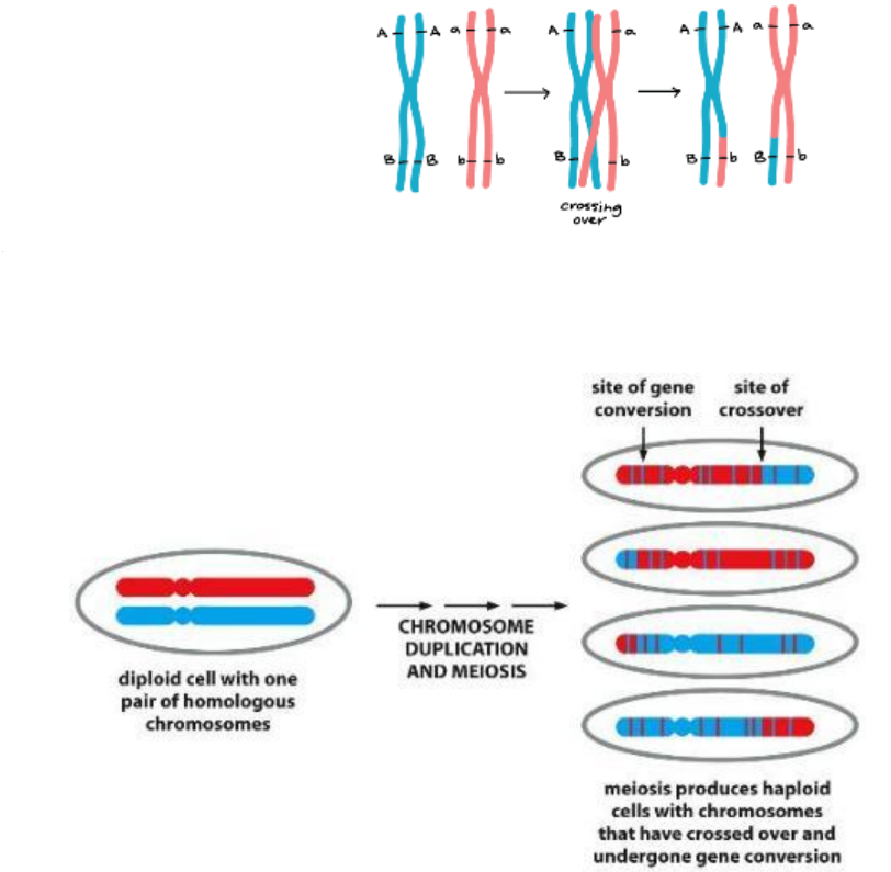
Recombination during meiosis
Recombination definition: The natural formation in offspring of genetic combinations not present in parents, by the processes of crossing over of homologous sequences
Repair-like event preferentially between maternal/paternal chromosome homologues
Acts to ensure no two daughter cells are identical nor identical to parent
Recombination can lead to gene conversion (bottom)
Heteroduplexes resulting from mismatched bases following recombination
Which is then detected by DNA repair systems
But these mechanisms cannot tell paternal/maternal apart
Therefore it will randomly choose a template strand and what strand will be corrected
Results in ‘conversion’ of one allele to another
Typically limited to small sections of DNA (part of genes)

Innate immune cells activate adaptive immunity
B and T cells develop in the bone marrow and thymus respectively
Naïve mature lymphocytes (B & T cells) constantly surveil the body for infection
Threats detected by B cell receptor (BCR) and T cell receptor (TCR)
Importantly adaptive immunity has:
Immunological memory
Antigen specificity (focus here)

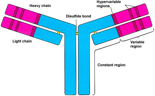
What are the common structural features shared by BCRs (antibodies) and TCRs?
The antigen binding site is in the variable portion of the receptors
V = variable
C = constant
The antigen binding site is contributed to by two different polypeptide chains
The constant portion of the receptors confers functional attributes unrelated to antigen specificity

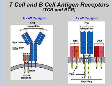
What are the differences in structural features between BCRs (antibodies) and TCRs?
The BCR (membrane-bound antibody) consists of two heavy (H) and two light (L) chains linked by disulphide bonds
The BCR can be secreted as an antibody by the B cell after it has been fully activated
The TCR consists of one alpha (α) and one beta (β) chain
The TCR is membrane bound and never secreted
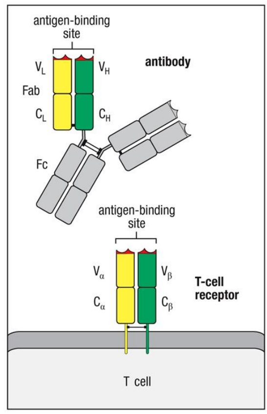

BCR (antibodies) and TCR bind different antigenic determinants
BCR → WHOLE antibody
TCR → epitope is a peptide fragment buried within the antigen presented in the contex of MHC
Antigen = molecule an adaptive immune response is directed against
Epitope = part of antigen bound by a lymphocyte receptor (can have multiple epitopes)

Each lymphocyte receptor chain has three hypervariable regions
The specificity of each BCR or TCR is unique
The antigen binding site contains the most sequence variability
These regions are known as hypervariable (HV) regions or complementarity determining regions (CDR)
There are three HV regions on each chain (heavy, light, alpha, beta)
Therefore, for the antibody in the lecture we would have 12 HV regions
The most variable of the three HV regions is HV3 in each chain


Where are Hypervariable regions located?
The HV region isn't encoded by one gene. It's assembled by DNA recombination from smaller segments:
Heavy chain (and TCR β): V + D + J segments
Light chain (and TCR α): V + J segments only
The hypervariable regions are found at the same point in each polypeptide chain
HV3 has the greatest variability of aa which means… (image below)
Amino acid sequence determines protein structure
Folding of loops determines antigen binding
HV1 and HV2 are both encoded within the V gene segment itself. The V segment is long and contains the sequences for CDR1 and CDR2. So these two loops are determined by which V gene was picked. Diversity here comes from having many different V genes to choose from, but there's no extra junctional variation’
HV3 sits right at the junction where the segments are stitched together. It includes the end of V, the whole D (heavy chain only), and the start of J. This is why it's the most diverse:
Many possible V, D and J combinations can be joined
During joining, nucleotides are randomly trimmed off and added at the junctions (junctional diversity, including N and P nucleotides)

So what do antigen receptors have to do with DNA recombination?
We now know where lymphocyte receptor specificity is determined by hypervariable regions of antigen binding site
But how is it generated?
There are approximately 1012 different B cells and 1012 different T cells (i.e. lymphocytes with different receptors)
With each receptor being encoded by 2 chains (H/L or α/β) this would mean we need approximately 4×10¹² genes
But only 19,000–20,000 protein-coding genes in human genome! (not enough)
So how does this work?
Somatic DNA recombination!
The DNA in each of our somatic cells is the same?
DNA recombination occurs during meiosis in the formation of ‘germ cells’ or gametes
Recombination does not occur in our somatic cells (non-reproductive cells)
DNA is preserved in an un-recombined form as the genetic blueprint for transcribing to mRNA and translation into required protein…..
Thus each cell in your body has the same DNA sequence
EXCEPT LYMPHOCYTES!!

Lymphocyte receptor diversity is generated through somatic recombination
Lymphocytes are an exception to the “no recombination” rule (in mitosis)
DNA recombination (somatic recombination) does occur in lymphocytes
Involves physically cutting out small regions of DNA and recombining these to create unique truncated sequences
Your genome doesn't contain a ready-made gene for every possible antibody. It contains a library of short gene segments, labelled V, D and J, and each B or T cell picks one of each and stitches them together. The hypervariable regions (HV1, HV2, HV3, the loops that touch the antigen) are built from pieces of these segments
The unique DNA sequences encode unique polypeptide chains that create unique lymphocyte receptors
So which sequences are being recombined?
Lymphocyte receptor loci composed of multiple gene segments
Lymphocyte receptor chain loci composed of multiple, distinct, V(D)J gene segments
V = variable
D = diversity (look how the light and alpha chains doesn’t have this)
J = joining
C = constant
Similar germline sequences in all loci

Receptor loci vary in somatic cells versus mature lymphocytes
We can see that only one section from each region has been selected in the mature B cell and have been combined into a unique receptor

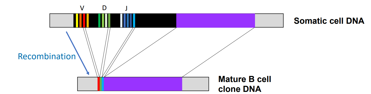
DNA sequence differs in a B cell clone versus a somatic cell
Somatic cells each carry the DNA ‘blueprint’ inherited from the parents
DNA in each mature B cell clone has been rearranged (“recombined”) with loss of a large amount of the genetic information
Same recombination process occurs in B and T cells
Lymphocyte receptor DNA sequence differs between clones
All progenitor lymphocytes share the same ‘blueprint’ DNA
In each B or T cell clone V(D)J gene segments are selected at random
Therefore each clone has a different rearrangement of their lymphocyte receptor locus

Steps in lymphocyte recombination
BCR heavy chain gene locus in germline DNA
Recombination first occurs between Diversity (D) and Joining (J) regions
The first recombination event joins one D segment to one J segment. The DNA between them is cut out and discarded, along with the other D and J segments in that stretch. The cell now has a DJ unit.
Recombination next occurs between Variable (V) and DJ regions
One V segment is joined to the DJ unit, again deleting the DNA in between. Now the cell has a complete VDJ exon
The recombined gene is transcribed to RNA
At this point the RNA contains the VDJ unit, then any leftover J segments that weren't used, then the C region sequences
The VDJ complex is spliced onto the Constant (C) region RNA
Extra J regions are spliced out
mRNA is translated to produce the BCR heavy chain polypeptide
*Light chain is also being transcribed/translated at the same time just without the D segments

umber of possible receptors depends upon number of combinations of V(D)J gene segments possible
Immunoglobin → BCR (Heavy chain + 2 diff types of light chains)
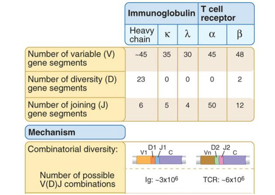
So how does recombination relate to antigen binding site specificity?
Hypervariable regions are encoded by the Variable (V), Diversity (D), and Joining (J) gene segments
Hypervariable regions 1 and 2 are encoded within V gene segments
Hypervariable region 3 is encoded by a combination of V, D and J for
BCR heavy chain
TCR beta chain
Hypervariable region 3 is encoded by a combination of V and J for
BCR light chains
TCR alpha chain
Each cell randomly picks different V, D and J segments, and since HV1, HV2 and HV3 are built from them, each cell ends up with a different set of loops
The loops together form the antigen-binding site, so a different shape means a different antigen recognized
Thus somatic recombination impacts protein function (i.e. antigen binding site specificity)
What mechanisms contribute to Lymphocyte receptor diversity?
1) Recombination of lymphocyte receptor gene segments (VDJ)
2) Nucleotide deletion at junctions
3) Addition of nucleotides at junctions
Somatic recombination occurs during B and T cell development
B cells rearrange their BCR in the bone marrow
T cells rearrange their TCR in the thymus
This process occurs before the lymphocytes have encountered the antigen that their receptor binds to
Recombination of the BCR and TCR genes involves coordinated activity of several enzymes
Recombination activating gene (RAG) 1 and 2 enzymes
Expressed only in developing B and T cells
DNA repair enzymes
Found in all cells and involved in DNA damage repair
The mechanism of V(D)J rearrangement
RAG enzymes recognise specific Recombination Signal Sequences (RSS) in lymphocyte receptor genes to initiate recombination
RSS are found…
3' of each V gene segment
5' of each J gene segment
Both sides of each D gene segment
RSS sites consist of a…
Conserved 7 nucleotide heptamer
12 or 23 non-conserved nucleotide spacer
Conserved 9 nucleotide nonamer (to allow enzyme binding)


Recombination only occurs between RSS sites that have 12 bp and 23 bp spacers
Using BCR heavy chain as an example:
A DH gene segment can be joined to a JH gene segment
A VH gene segment to a DH gene segment
VH gene segments CANNOT be joined to JH gene segments directly (heavy chain)
As both VH and JH gene segments are flanked by 23 bp spacers
12/23 rule (opposites 12 OR 23 attract - 23 and 23 will not attract/bind)
Recombination only occurs between segments flanked by 12 bp spacer and 23 bp spacer
Ensures that recombination will not occur between only V elements (or only J elements)

Rag 1 and Rag 2 enzymes mediate the synapse of two RSS sites with 12 bp and 23 bp spacers and cleave
Recognition of RSS sites by RAG1:2 recombinase enzymes
Binding of RAG enzymes and bringing V(D)J gene elements together
Cleavage of DNA fragments
Binding accessory proteins Ku70:Ku80 to cleaved ends
DNA ligase IV and XRCC4 bind to Ku accessory proteins (ends of the gene segments) to form the coding joint
Ligation of DNA ends to form the new V(D)J segment (coding joint)
Excess DNA in signal joint lost upon subsequent cell division

Junctional diversity introduces further diversity into HV3
1) Recombination of lymphocyte receptor gene segments (VDJ)
2) Nucleotide deletion at junctions
3) Addition of nucleotides at junctions
Combinatorial diversity can generate ~1.6 × 10⁶ different BCRs
Combinatorial diversity comes from which segments get picked. You choose one V, one D and one J from the library, and different choices give different antibodies
HV1 and HV2 sit inside the V segment, away from the joints, so they get combinatorial diversity only
Junctional diversity can increase this by a factor of ~3 x 10⁷
Junctional diversity comes from how the segments are joined. At each joint (D to J, then V to DJ), the cell randomly alters the DNA ends before sealing them. So even if two B cells pick the exact same V, D and J, their junctions can still differ
Introduces diversity into HV3
Combined, the number of unique lymphocyte receptors approaches ~4.8 × 10¹³ (48 trillion)
Changing the number of bases can also shift the reading frame. Roughly two thirds of rearrangements end up out of frame and non-functional, so the cell has to try again or die. This is the cost of the system
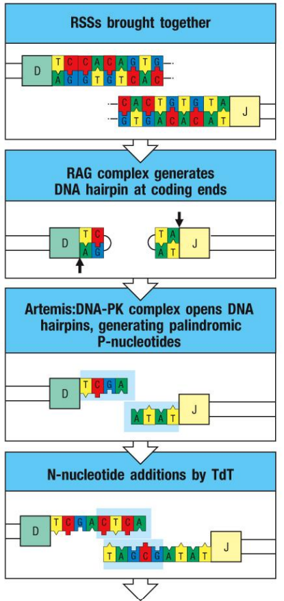
Junctional diversity is created by the addition and subtraction of nucleotides to the coding joint
RAG1/2 binds RSS creates DNA double stranded breaks
Artemis: DNA phosphokinase (DNA-PK) opens hairpins to form random overhangs
Palindromic or P-nucleotides
Terminal deoxynucleotidyl transferase (TdT) adds random nucleotides
Non-templated or N-nucleotides
Exonuclease removes random nucleotides
The removing and addition of nucleotides introduced variability
They also change reading frame - encoding novel amino acids
(bottom) Pairing of overhanging strands, DNA ligase IV and XRCC4 ligate strands back together
Exonuclease removes unpaired nucleotides
DNA polymerase “fills in” overhangs
New “DJ” segment!
Not always functional
Introduce frame shifts with stop codons → thus producing non-functional/truncated proteins
Encode molecules with different amino acid composition and thus structure
