Lecture 5: Signal Propagation and Transmission
 Call Kai
 Learn
Learn Practice Test
 Spaced Repetition
 Match
Match Flashcards
 Knowt Play
1/50

There's no tags or description
Looks like no tags are added yet.
Name | Mastery | Learn | Test | Matching | Spaced | Call with Kai | Chat |
|---|
No analytics yet
Send a link to your students to track their progress
51 Terms
Propagation within a neuron
The way an electrical signal travels along an axon by triggering the same voltage change in each neighbouring part of the cell membrane.
Trigger zone of axon hillock → axon terminal.
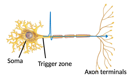
Two methods of action potential propagation
Continuous conduction
Saltatory conduction
Continuous conduction involves:
Step-by-step depolarization and repolarization of adjacent sections of the plasma membrane.
Where does continuous conduction occur in?
Unmyelinated axons and muscle fibers
Steps of continuous conduction
Stimulus moves the RMP to threshold, causing voltage-gated Na+ channels to open to generate an action potential.
Na+ influx causes the adjacent membrane segment to depolarize until threshold, propagating the action potential along the membrane as a wave of depolarization.
Repolarization occurs, caused by inactivation of voltage-gated Na+ channels and opening of voltage-gated K+ channels.
The area behind the depolarized membrane is briefly refractory.

What causes repolarization after the depolarization wave?
Closing of voltage-gated Na+ channels.
Opening of voltage-gated K+ channels.
Saltatory conduction involves:
The action potential leaping from one unmyelinated segment (myelin sheath gaps/nodes of Ranvier) of the axon to the next.
Why is saltatory conduction more efficient?
The entire membrane does not need to depolarize (skips myelinated sections since its insulating nature blocks charged ions).
Voltage-gated ion channels are concentrated at the nodes.
Steps of saltatory conduction
Stimulus move RMP to threshold, opening voltage-gated Na+ channels to generate an action potential.
Na+ influx creates ionic currents that travel from one node to the next. Each nose reaches threshold and generates another action potential.
Depolarization of each node is followed by repolarization.
Each node becomes briefly refractory.

Refractory period
Short period after action potential when a neuron/muscle cell cannot easily generate another electrical signal.
Absolute refractory period
First phase of refractory period; no new action potential can occur no matter how strong the stimulus is.
Relative refractory period
Second phase of refractory period; a neuron/muscle cell can fire again but only in response to a stronger-than-normal stimulus.
What is the importance of the refractory period?
Ensures one-way propagation → prevents unorganized communication).
Keeps action potentials as separate events → allows info to be coded by action potential frequency rather than amplitude, since all action potentials have about the same amplitude.
Allows time for recovery → re-establish ionic gradients while limit amount of ATP required to restore these gradient.
Refractory membrane
Segment of neuron/muscle cell that temporarily cannot respond to a new stimulus while recovering from a previous action potential.
What is greater stimulus intensity measured by in action potentials?
Higher action potential frequency (amplitude is irrelevant, unlike graded potentials).
Three factors that increase nerve conduction speed
More myelination
Larger axon diameter (less internal resistance to ion flow)
Higher temperature
What is saltatory conduction used for?
Fast actions and long pathways
Ex: motor reflexes; somatic senses like sharp pain, touch, pressure, body position.
What is continuous conduction used for?
Slow, generalized processes and internal regulation.
Ex: GI tract movement during digestion, dull pain, temperature sensations.
Synapse
Region where neuron-neuron or neuron-effector cell (muscle/gland) communication occurs.
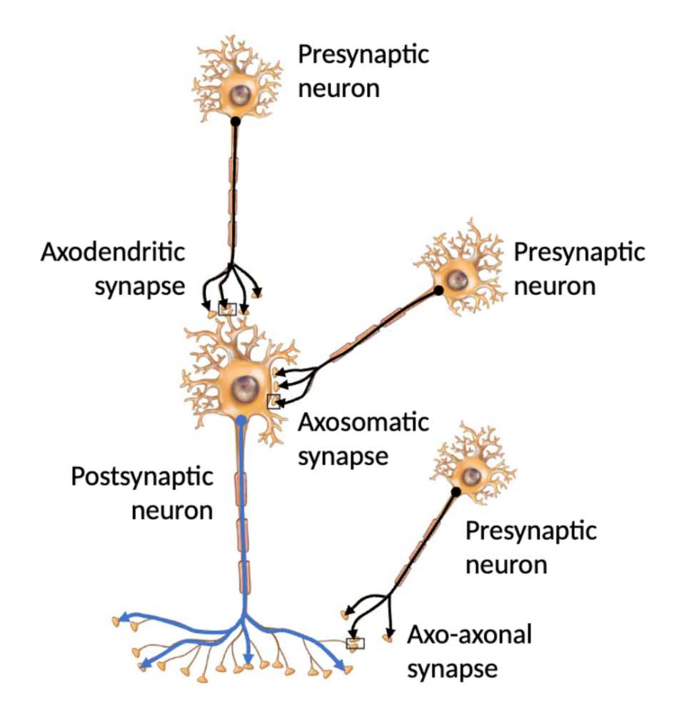
Presynaptic cell
Carries the impulse toward the synapse.

Post synpatic cell
Receives the signal from the presynpatic cell and carries the impulse away from the synapse.
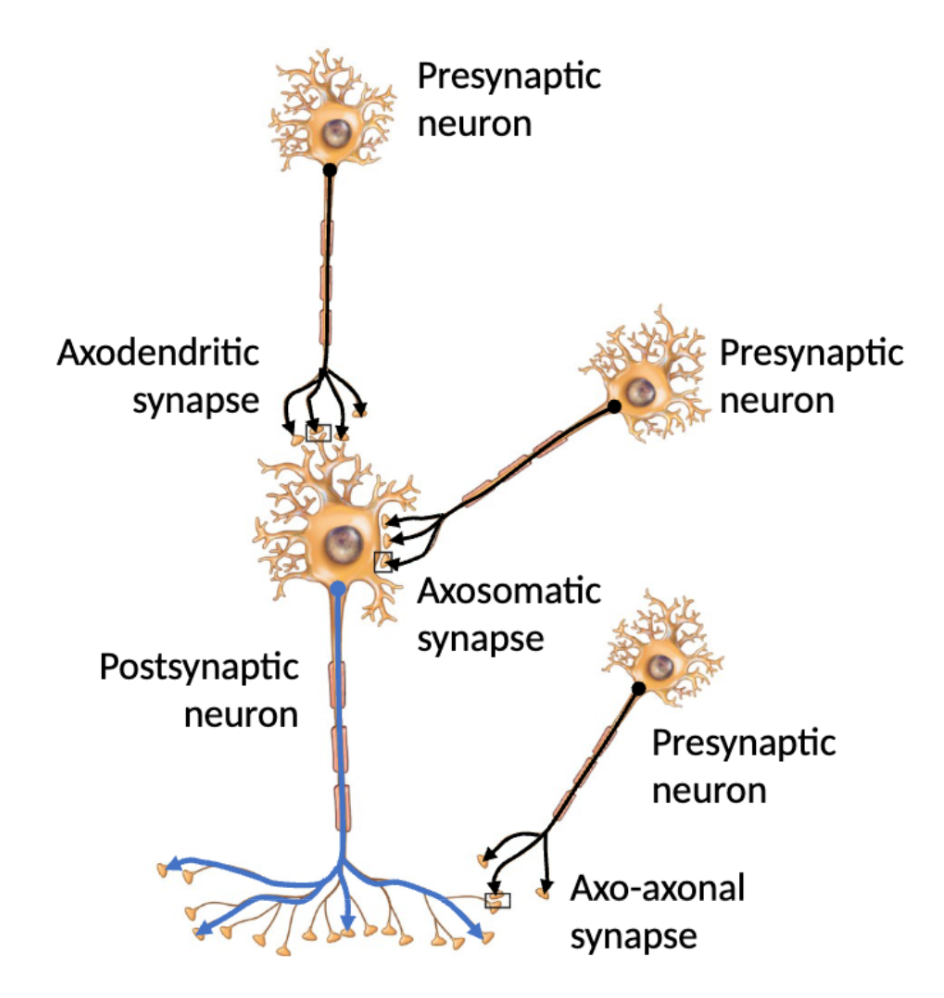
Three types of neuron-to-neuron synapses
Axodendritic (axon-dendrite); most common
Axosomatic: (axon-soma)
Axo-axonal (axon-another axon)
Two types of synpases
Electrical
Chemical
Electrical synapses
Action potentials are conducted directly between adjacent cells through gap junctions.
Faster than chemical synapses.

Gap junctions
Specialized intercellular channels that allow ions to flow directly from one cell to another, providing almost instantaneous transmission of signal.
Present in electrical synapses.
Where in the body do electrical synapses occur?
Visceral smooth muscle (GI tract), cardiac muscle (myocardium), nervous system (brain, spinal cord)
Synaptic cleft
Small space separating cells.
Present in chemical synapses.
Chemical synapses
Electrical impulses in the presynaptic cell release a neurotransmitter (chemical messenger) that travels across the synaptic cleft to the postsynaptic cell, where the NT binds to a receptor on its membrane to produce a graded potential.
If the postsynaptic potential reaches threshold → action potential.

Why is there a chemical synaptic delay?
Neurotransmitter release and receptor activation take time, so there is a delay of ~0.5 ms between arrival of the impulse at the presynaptic cell and generation of an action potential in the postsynaptic cell.
Steps for chemical synaptic transmission
Action potential arrives at axon terminal in the presynaptic cell.
Voltage-gated Ca2+ channels open, allowing Ca2+ to enter.
Ca2+ causes fusion of NT-containing vesicles with the synaptic end bulb.
NT is released into the synaptic cleft and diffuses across it.
NT binds to ligand-gated ion channels on the postsynaptic membrane, opening the channels.
Ions flow and create a postsynaptic potential (graded potential). If it reaches threshold → action potential.

Excitatory postsynaptic potential (EPSP)
Graded potential that moves the cell closer to threshold through a small depolarization. If enough EPSPs are summed → action potential.
Caused by Na+ channels opening → Na+ enters the cell.

Inhibitory postsynaptic potential (IPSP)
Graded potential that moves the cell further from threshold through a small hyperpolarization. Can cancel out an EPSP.
Caused by K+ channels opening → K+ leaves the cell; Cl- channels opening → Cl- enters the cell.

Two types of neurotransmitter receptors
Ionotropic
Metabotropic
Ionototropic receptor
Type of ligand-gated ion channel that opens when an NT binds to it.

Metabotropic receptor
Have an NT binding site but are not ion channels themselves.
Instead, they are connected to an ion channel through a G-protein, which opens/closes the channel through second messengers.

G-protein
Opens/closes ion channels connected to metabotropic receptors through second messengers.

Second messenger
Small intracellular molecules that relay/amplify signals from cell-surface receptors to target proteins inside the cell.

Three methods of neurotransmitter removal
Diffusion → NT diffuses away from synapse.
Enzymatic degradation → enzymes break down or inactivate the NT
Ex: acetylcholinesterase breaks down acetylcholine; monoamine oxidase degrades epinephrine, norepinephrine, and dopamine into inactive forms.
Uptake by cells → NT is actively transported back into the cell, where they can be recycled and released again.
SSRIs block serotonin re-uptake → increase serotoning in synaptic cleft.
Neurotransmitter
Substance that transmits signals across a synapse.
Two major classes of neurotransmitters
Small-molecule neurotransmitters
Neuropeptides
Small-molecule neurotransmitters
Fast-acting chemical messengers synthesized locally in the nerve terminal.
Neuropeptides
NTs made of amino acids that bind to metabotropic receptors, and can also function as endocrine hormones.
Acetylcholine
Small-molecule neurotransmitter
Released by many PNS neurons for muscle contraction and autonomic regulation.
Can also be released by some CNS neurons for memory/cognition.
Amino acids (as neurotransmitters)
Small-molecule neurotransmitters in the CNS
Aspartate → excitatory
Glutamate → excitatory
GABA → inhibitory
Glycine → inhibitory
Biogenic amines
Small-molecule neurotransmitters containing an amino functional group (R—NH2).
Epinephrine (adrenaline) → fight or flight, SNS activity
Norepinephrine (noradrenaline) → arousal, wakefulness, SNS activity
Dopamine → pleasure/reward, addiction
Serotonin → sleep, mood, appetite
Histamine → wakefulness, attention
Purines
Small molecule neurotransmitters in the CNS and PNS.
Adenosine, ATP, ADP, and AMP are excitatory.
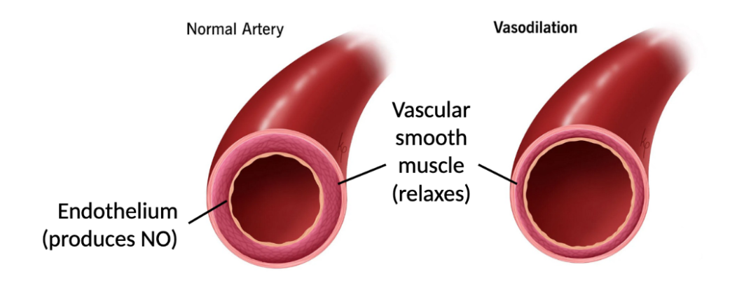
Nitric oxide (NO)
Lipid-soluble, excitatory small molecule neurotransmitters produced in brain and spinal cord.
Learning/memory; promotes vasodilation and inhibits blood clotting.
Substance P
Neuropeptide involved in pain signalling.
Enkephalins and endorphins
Neuropeptide that functions as an analgesic (painkiller) by blocking the release of Substance P.
Cholecystokinin (CCK)
Neuropeptide found in brain and small intestine. Regulates feeding and GI function → “stop eating” signal
Oxytocin
Neuropeptide released from the pituitary gland.
Bonding, sexual pleasure, uterine contractions, milk let-down.