KA Unit 6: Foundation 1: Biomolecules qs
 Call Kai
 Learn
Learn Practice Test
 Spaced Repetition
 Match
Match Flashcards
 Knowt Play
1/18

There's no tags or description
Looks like no tags are added yet.
Name | Mastery | Learn | Test | Matching | Spaced | Call with Kai | Chat |
|---|
No analytics yet
Send a link to your students to track their progress
19 Terms
The amino acids in hemoglobin (or any protein) uniformly have which of the following configurations?
All biologically produced amino acids are in the L configuration. (amine group on left)
Why is SDS (sodium dodecyl sulfate) used in SDS-PAGE?
to give protens a negative charge (so they will go toward positive end of gel)
During SDS-PAGE, DTT (dithiothreitol) or β-mercaptoethanol are sometimes applied. what are they and why are they used
reducing agents
used to break disulfide bonds (tertiary and quarternary sctructure) between cysteine residues in proteins.
Which of the following peptides is likely to have the greatest isoelectric point (pI)?
has the most basic aas
how does gas chromatography work
Gas chromatography uses a long tube to separate volatile organic compounds.
The two phases in gas chromatography are a liquid phase and a gas phase.
This gas, often referred to as the carrier gas, is the mobile phase - transports vaporized sample through the column where separation occurs based on the interaction with the stationary phase.
similar to liquid chrom, where liquid flows thru column and seperation is based on interaction with solid stationary phase
Native vs. Denaturing vs. Reducing Gel
NATIVE: protein retains its 3D structure. Banding pattern is determined by size and compactness
DENATURING: protein loses 3D structure. Banding pattern is also determined by size. If there are multiple subunits held together by non-disulfide interactions (such as by hydrophobic effects), then multiple bands will appear because those interactions are lost from the protein becoming negatively charged.
REDUCING: Pattern loses 3D structure, and disulfide bonds are broken. REDUCING gel patterns only differentiate from DENATURING if disulfide bonds are present. The solution used in REDUCING is the same as DENATURING (mainly SDS) except a reducing agent is added. This disrupts the quaternary structure of proteins that consist of more than one subunit. The bands visualized under reducing conditions reflect the size of the individual subunits.
centrifugation: which proteins sediment first, and what is their sedimentation coeff?
Larger proteins generally have higher sedimentation coefficients, meaning they sediment faster.

gel A = denaturing, as the mutated proteins are all similar molecular weight.
gel b = native, bc mutations can change shape and structure of protein based on noncovalent interactions


mutation leads to monomer instead of homo-trimer (noncovalent bonds cant happen)
*not denatured bc that would lead to weird/inconsistent results, but all three concentrations of the mutation show same thing and higher conc shows more oligomers (make sense, more protein = more association likely)


ligase? hydrolase?
isomerase: catalyzes changing one isomer to another
oxidoreductase: catalyzes oxidation or reduction rxn
lyase: breaks or make a bond
kinase: adds a phosphate
ligase: forms a bond by eliminating water
hydrolase: breaks bond by adding water
cofactor
coenzyme
a nonorganic molecule whose presence is necessary for the proper function of an enzyme.
a small, non-protein organic molecule that binds to an enzyme to help it catalyze a chemical reaction
induced fit model/theory of enzyme binding states:
the substrate itself alters the enzyme active site to better match its shape
Enzymes and substrates do not fit together like puzzle pieces immediately upon binding.
Enzymes and substrates will change their structure slightly upon binding to increase the affinity between the two during the reaction’s transition state.
The induced fit theory states that binding between enzymes and substrates is strongest at the corresponding reaction’s transition state.
The rate of a reaction
rate = k[reactant 1][reactant 2]
for enzymes: k[unbound enzyme][substrate]
E + S → ← ES
michaleis menten eq
y = mx + b
V0 = (Vmax[S]) / (Km + [S])
![<p>y = mx + b</p><p>V0 = (Vmax[S]) / (Km + [S])</p>](https://assets.knowt.com/user-attachments/66c93991-21ed-4299-be94-2b34203d7768.png)
x and y intercept of lineweaver burk plot


Km vs kcat

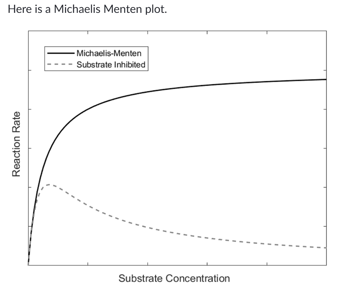
Enzymes following Michaelis Menten kinetics display first order kinetics, with regards to substrate concentration, at which substrate level?
Notice that the plot is nearly linear at low substrate concentrations, becomes more curved around Km and flattens near Vmax.
This is because MM kinetics exhibit first order kinetics at low substrate concentrations, where the rate or velocity of the reaction is proportional to the substrate concentration. At high substrate concentration, MM kinetics transition to zero order kinetics, where the rate is approximately constant and approximately equals Vmax.
Hill coefficient (n)
a measure of cooperativity in ligand binding to a multi-subunit protein or enzyme. It indicates how the binding of one ligand affects the binding affinity of additional ligands to the same protein.
1 = no cooperativity
>1 = ligand binding leads to more ligand binding
<1 = ligand bindnig leads to less ligand bidning