BIOS 1300 Exam #3 Review (Chapters 8-11)
 Call Kai
 Learn
 Practice Test
 Spaced Repetition
 Match
 Flashcards
 Knowt Play
1/215

There's no tags or description
Looks like no tags are added yet.
216 Terms
Regions of the spine
Cervical, Thoracic, lumbar, Sacrum, and Coccyx


Intervertebral discs
discs of connective tissue between vertebral bodies that cushion the spine and allow flexibility.
Nucleus pulposus
soft/squishy, gel-like inner portion of an intervertebral disc, cushioning part of disc, made of many polysaccharides and water

Annulus fibrosus
The tough outer ring of an intervertebral disc, made of fibrocartilage

Herniated disc
A condition in which the nucleus pulposus bulges outward through the annulus fibrosis
About how any adults have a herniated disk?
80%
Osteopenia
Reduced bone mineral density (BMD), bones are weaker but do not fracture as easily as in osteoporosis, happens due to aging
Osteoporosis
Severe loss of bone mineral density that makes bones fragile and increases fracture risk.
Vertebral compression fracture
most common type of injury in patients with osteoporosis; 25% of postmenopausal women and 40% of women over 80 will get one of these
Osteogenesis imperfecta
a group of genetic disorders involving imperfect bone formation, bone deformities, and brittle bones.
Brittle Bone Disease
genetic disease where bones break with very little force
Articulation/Joint
the site where two or more bones meet, providing attachment and sometimes mobility.
Joint naming
usually named after the two bones that join; exceptions listed include sutures, the knee, and the elbow.
Structural classification of joints
Bony Joints, Fibrous Joints, Cartilaginous Joints, and Synovial Joints
Bony joint
A joint with no movement, formed when bones fuse.
Synostosis
A bony joint in which bones fuse to become one bone, happens during development.
Examples of Synostoses
epiphyseal line, mandibular bones, frontal bones, pubic bone
Fibrous joint
A joint in which bones are joined by dense irregular connective tissue; little to no movement
Types of fibrous joints
Gomphoses, Syndesmoses, and Sutures
Gomphosis
fibrous joint connecting a tooth to its socket; slightly movable.
Syndesmosis
A fibrous joint with longer connective-tissue fibers than a suture, most moveable fibrous joint, not many in the body
Example of syndesmosis
intermediate radioulnar joint; allows forearm to pronate and supinate
Suture
A fibrous joint found only in the skull, where bones are held together by short connective-tissue fibers, small amount of movement, skull bones are less likely to break because of these
Fontanelle
A region of fibrous connective tissue between an infant's cranial bones, allows head to pass through birth canal more easily
Cartilaginous joint
A joint in which bones are connected by a disc or plate of cartilage; it permits more movement than a fibrous joint
Types of cartilaginous joints
synchondrosis and symphysis
Synchondrosis
A cartilaginous joint in which bones are united by hyaline cartilage; examples include the first sternocostal articulation and an epiphyseal plate during endochondral ossification.
Symphysis
A cartilaginous joint in which bones are united by a disc of fibrocartilage and hyaline cartilage; examples include intervertebral discs and the pubic symphysis.
Synovial joint
A highly mobile joint in which bones are separated by a fluid-filled joint cavity.
6 basic features of a synovial joint
articular cartilage, joint cavity, synovial fluid, articular capsule, reinforcing ligaments, and rich supply of nerve fibers and capillaries
Articular cartilage
less than a mm thick, smooth hyaline cartilage covering the ends of bones in synovial joints, slightly absorbs shock, remains of endochondral ossification
Joint cavity
The small space within a synovial joint that contains synovial fluid.
Synovial fluid
A viscous (thick) fluid in the joint cavity that lubricates the joint and supports weight-bearing.
Articular capsule
The structure enclosing a synovial joint cavity; it has a fibrous outer layer (continuous with periosteum) and an inner synovial membrane.
Synoviocytes
cells that secrete synovial fluid, maintain joint homeostasis, highly studied for the cause of Rheumatoid arthritis
Synovial membrane
The inner lining of the articular capsule layer of synoviocytes
Reinforcing ligaments
Ligaments that help hold a synovial joint together.
Purpose of sensory nerve fibers in synovial joints
allow us to feel pain, body position and stretches
Purpose of capillaries in synovial joints
nourishment and waste removal, supply things to make synovial fluid
Proprioception
sensation of knowing body position and feeling stretch, gives coordination
Functional classifications
Synarthrosis, Amphiarthosis, and Diarthrosis
Synarthrosis
A functional joint classification meaning immovable, ex) sutures
Amphiarthrosis
A functional joint classification meaning slightly or partly movable, ex) pubic symphysis
Diarthrosis
A functional joint classification meaning freely movable.
Intra-articular Fibrocartilages
extend from a fibrous layer of capsule, help cushion and stabilize joints
Types of Intra-articular Fibrocartilages
Articular Disc and Meniscus
Articular disc
An intra-articular fibrocartilage that extends all the way across a joint cavity, dividing it into two cavities; has a higher degree of motion.
Meniscus
An intra-articular fibrocartilage that protrudes only partway into a joint; in the knee, it cushions and contains the knee by preventing the femur from slipping off the tibia and helps provide a smooth surface.

Bursa
A flat, synovial-membrane-lined sac containing a thin film of synovial fluid that helps prevent friction. Found outside of the joint cavity.
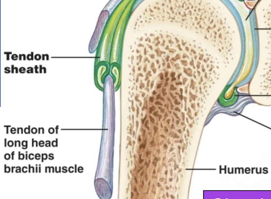
Tendon sheath
special type of bursae that is elongated, commonly found in the wrist and hand. Wrapped around the tendon
Bursitis
Inflammation of a bursa; caused by repetitive or excessive strain, infection, and rheumatoid arthritis. Can cause pain; increased synovial fluid is produced.
Prepatellar bursitis
Bursitis at the front of the knee, also called housemaid's knee or clergyman's knee

Ischial bursitis
Bursitis also called weaver's bottom.
Olecranon bursitis
Bursitis also called student's elbow.
Six types of synovial joints
Plane (gliding), hinge, pivot, condylar, saddle, and ball-and-socket.
Plane (gliding) joint
A synovial joint type where flat-ish surfaces slide past each other, 2-dimensional
Examples of Planar Joints
Intercarpal joints, intertarsal joints, facet joints (articular processes of vertebrae), costovertebral joints
Skull Joint
joint made of cranial and facial bones, fibrous and suture (structural) and synarthrotic; no movement (functional type)
Temporal Mandibular Joint (TMJ)
joint made of temporal bone of skull and mandible, synovial and modified hinge (structural) and diarthrotic; gliding and uniaxial rotation (functional)
Atlanto-occipital Joint
joint made of occipital bone of skull and atlas, synovial and condylar (structural) and diarthrotic; biaxial (functional)
Atlantoaxial Joint
joint made of atlas (C1) and axis (C2), synovial and pivot (structural) and diarthrotic; uniaxial (functional)
Intervertebral Joint
joint between adjacent vertebral bodies, cartilaginous and symphysis (structural) and amphiarthrotic (functional)
Sternoclavicular Joint
joint made of sternum and clavicle, synovial and saddle (structural) and diarthrotic; multiaxial (functional)
First Sternocostal Joint
joint between sternum and first rib, cartilaginous and syncondrosis (structural) and synarthrotic; no movement (functional)
Glenohumeral Joint (Shoulder)
joint between scapula and humerus; synovial and ball and socket (structural) and diarthrotic; multiaxial (functional)
Elbow Joint
joint between ulna (and radius) and humerus, synovial and hinge (structural) and diarthrotic; uniaxial (functional)
Proximal Radioulnar Joint
joint between radius and ulna, synovial and pivot (structural) and diarthrotic; uniaxial (functional)
Intercarpal Joint
joint between adjacent carpals, synovial and plane (structural) and diarthrotic; gliding (functional)
First Carpometacarpal Joint
joint between carpal and metacarpal 1, synovial and saddle (structural) and diarthrotic; biaxial (functional)
Metacarpophlangeal Joint (knuckle)
joint between metacarpal and proximal phalanx, synovial and condylar (structural) and diarthrotic; biaxial (functional)
Interphlangeal Joint (fingers and toes)
joint between adjacent phlanges, synovial and hinge (structural) and diarthrotic; uniaxial (functional)
Pubic Symphysis Joint
joint between pubic bones, cartilaginous and symphysis (structural) and amphiarthrotic; slight movement (functional)
Hip (coxal) Joint
joint between acetabulum of coxal bone and head of femur, synovial and ball and socket (structural) and diarthrotic; multiaxial (functional)
Knee (tibiofemoral) Joint
joint between femur and tibia, synovial and ball and socket (structural) and diarthrotic; multiaxial (functional)
Distal tibiofibular Joint
joint between the tibia and fibula (distally), fibrous and syndesmosis (structural) and synarthrotic; slight movement (functional)
Ankle Joint
joint between tibia and fibula with the talus, synovial and hinge (structural) and diarthrotic; uniaxial (functional)
Intertarsal Joint
joint between adjacent tarsals, synovial and plane (structural) and diarthrotic; gliding (functional)
Metatarsophalangeal Joint
joint between metatarsal and proximal phalanx, synovial and condylar (structural) and diarthrotic; biaxial (functional)
How is motion limited in the hip joint?
by the deep socket (acetabulum)
Acetabulum
The socket of the hip joint that receives the head of the femur.
Acetabular labrum
A lip of circular fibrocartilage attached to the rim of the acetabulum; it protects the rim, deepens the socket, and reduces dislocation risk.
Ligaments of Coxal Joint
Iliofemoral ligament, Pubofemoral Ligament, and Ischiofemoral Ligament

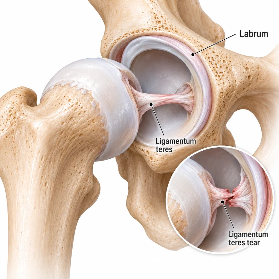
Round ligament (ligamentum teres)
An intracapsular hip ligament connecting the fovea capitis to the edge of the acetabulum; it contains/protects the artery that goes into the femoral head.
Arthritis
Inflammation of a joint; main examples include osteoarthritis and rheumatoid arthritis.
Osteoarthritis
The most common type of arthritis; degeneration of joints with loss of articular cartilage and growth of exposed bone, often associated with pain, inflammation, and stiffness. Linked to agining but not always, there is a genetic component
Rheumatoid arthritis (RA)
A chronic, relapsing autoimmune disease that causes joint degeneration; scarring and ossification can lead to ankylosis or possible joint fusion, no known cure
Non-synovial symptoms of rheumatoid arthritis
rash, rheumatoid lung disease, pericarditis, enodcarditis, osteoporosis, neuropathy
What groups of people are more likely to get rheumatoid arthritis?
women and smokers are both 3 times more likely
Myology
The study of muscles.
Skeletal muscle functions
Move the skeleton and skin; maintain posture and body position; support and protect soft tissues; control voluntary sphincters; generate heat; provide an amino-acid reserve; and help with glycemic control.
Skeletal muscle as an organ
Includes muscle tissue, connective tissues, blood vessels, and nerves.
Muscle fiber
A skeletal muscle cell; it is multinucleated and surrounded by endomysium.
Endomysium
Fine areolar connective tissue surrounding each muscle fiber.
Fascicle
A bundle of muscle fibers.
Perimysium
A fibrous connective-tissue sheath surrounding a fascicle.
Epimysium
Dense irregular connective tissue forming a sheath around the whole muscle.
Connective-tissue continuity in muscle
Endomysium, perimysium, and epimysium are connected to one another.
Direct muscle attachment
An attachment in which the epimysium is fused to the attachment site by fibrous collagen.
Tendon
A cord of dense regular connective tissue that attaches skeletal muscle to bone.
Aponeurosis
A broad, tendinous sheet of dense regular connective tissue that attaches muscles to other muscles or bones over a wide area.