locomotion lecture
 Call Kai
 Learn
Learn Practice Test
 Spaced Repetition
 Match
Match Flashcards
 Knowt Play
1/226

Earn XP
Description and Tags
VET30370
Name | Mastery | Learn | Test | Matching | Spaced | Call with Kai | Chat |
|---|
No analytics yet
Send a link to your students to track their progress
227 Terms
what is physical therapy?
the treatment of physical dysfunction/injury by the use of therapeutic exercise & the application of modalities, intended to restore/facilitate normal function/development
why is physical therapy necessary?
missing link in veterinary care for a long time
well recognized profession in the medical field
benefits in animals only recognized in recent times
area where vets & nurses can play a large role
carrying it out themselves on inpatient
educating owners when patient is discharged
should only be carried out under the direction of the vet and the physical therapist in charge of the case
why choose physical therapy?
concentrates in rehabilitation of the musculoskeletal system & nervous system
evidence based benefits
techniques based on physiological principles
benefits of PT
improve circulation
relax muscle spasm
prevent & relax of muscle contracture
dispersal of edema in muscle, joints, soft tissue
mobilize of adherent tissues
restore/maintain normal ROM in joints/limbs
contributes to analgesia (decrease muscle spasm & edema)
more comfortable & confident patient
quicker return to normal activity
neuromuscular re-education
what method is used for PT?
symptoms + precautions + prognosis + further contributing factors → hypothesis & working diagnosis
clinical reasoning
knowledge of:
anatomy & physiology
biomechanics
conditions
diseases
animal behavior
skeletal muscle functions
producing body movement
stabilizing body positions
storing & moving substances within the body
generating heat
skeletal muscle tissue
40% of total BW (640 muscles)
largest cells in the body & bound with connective tissue
abilities:
contractility
excitability
extensibility
elasticity
connective tissue
surrounds & protects muscular tissue
separates muscle from skin
provides pathway for nerves, blood vessels, and lymphatic vessels to enter and exit muscles
composed of:
areolar tissue- store fibroblasts & collagen
adipose tissue- stores triglycerides, insulates, reduces heat loss, protects muscles from trauma
fascia
dense sheet/broad band of dense irregular connective tissue that lines the body wall and limbs
support & surrounds muscles and all other organs in the body
functions:
hold muscles with similar functions together
allows free movement of muscles
carries nerves, blood vessels, and lymphatic vessels
fills space between muscles
all 3 connective tissue elements connect to structures (bone/other muscle) and may form tendon
epimysium, perimysium, endomysium
types of NS receptors
chemoreceptors
photoreceptors
thermoreceptors
mechanoreceptors
mechanoreceptors
sense change in joint movement, muscle tension, muscle length
feed info to CNS to respond by muscle contraction or inhibition
muscle spindles
small, elongated structures scattered among/arranged parallel to contractile extrafusal muscle fiber
respond to muscle stretch
responsible for muscle tone
rehab appilcations
repetitive & assisted exercises
massage
increase load of exercises
increase resistance
stretches to increase muscle tone
early mobilization
PROM & AROM
proprioception rehabilitation
body's ability to sense its location, movements, and actions in space without conscious thought
joint receptors
found in the capsule & ligaments around joints
stimulated by mechanical distortion that accompanies changes in the relative positioning of bones linked by flexible joints
movement
requires proper timing so antagonistic & synergistic muscle groups contract in the appropriate sequence and to the appropriate level
types of movement
reflex movement
postural reflexes
voluntary movement
rhythmic movement
reflex movements
least complex
integrated primarily in the spinal cord
stretch & reciprocal inhibition- control movement around the joint
flexion- pull limbs away from painful stimuli
postural reflexes
integrated in the brainstem
require continuous sensory input from visual & vestibular sensory systems
voluntary movements
most complex
integrated at cerebral cortex
initiated at will without external stimuli
rhythmic movement
combination of reflex & voluntary movements
initiated & terminated by input of cerebral cortex
once initiated can be sustiained without further input from the brain
golgi tendon organ
responds to muscle tension
found at the junction of tendons & muscle fibers
respond to the tension a muscle develops during an isometric contraction
cause relaxation reflex
methods of hydrotherapy
manual therapy (massage)
therapeutic exercise
hydrotherapy
thermotherapy
electrotherapy
manual therapy
breaks vicious circle of pain & muscle tension
increase blood flow
improves oxygenation to tissue & aids metabolic waste removal
improves venous & lymphatic return
breaks down & mobilizes adhesions
releases natural endorphins
provides pain relief
improves cutaneous perception in neurological patients
accelerates muscle recovery
promotes mental & physical relaxation
improves pet-owner bonding
cons to manual therapy
local inflammatory process
systemic/ local infection
neoplasia
hyperthermia
coagulopathy
bruising
cardiac compensation
48hrs post acute injury
disloaction/fracture/severe instability
edema of unknown origin
types of therapeutic exercies
passive range of motion (PROM)
stretching
assisted exercise
active assisted exercise
passive range of motion
helps contain muscle atrophy
prevents adhesions
improves proprioception
reduces stiffness from inactivity
stretching shrunken joint capsules, ligaments, tendons
should be carried out after massage & heat application
cons to PROM
joint dislocation
fractures involving joints
osteosarcoma
joint with restricted movement following trauma
stretching
can be combined with PROM
stretch & realign soft tissue structures without tearing them
when the joint has reached the end of range during PROM repitiotion
never stretch beyond the patients comfort level
2-3 reps 1-3x day
assisted exercise
bridge the gap between passive & active exercise
enhance strength & endurance
enhances proprioceptive training and prepares for more active exercise
gradually increase he length of each session as the patient gets stronger
10-15 reps 2-3x day
how to deliver assisted exercise
position patient with feet squarely underneath body
support the patient in this position using a towel/sling
allow them to bear as much weight as possible
as patient weakens gently reposition them
active assisted exercise
slow walks
weight shifting
withdrawal reflex
stair climbing/inclines
sit-stand exercises
wheel barrowing
pole weaving
cavatelli rails
dancing
hydrotherapy
the use of water to relieve discomfort and promote physical well-being
allows unloading of painful joints
early weight bearing on very weak or painful limbs
benefits to hydrotherapy
musculoskeletal system
muscle
joint
ligament
tendons
bones
cardiovascular system
circulation
edema
nervous system
proprioception
well being
cons of hydrotherapy
open & infected wounds
unhealed surgical incisions
surgical incisions without waterproof cover
fever
systemic compromise
cardiac/ liver/ kidney disease, hypertension, severe hypotension
respiratory compormise/distress
edema of unknown origin
advance debility
uncontrolled epilepsia
infectious disease
hydrotherapy precautions
fecal/urinary incontinence
older age patients
mild systemic compromised
extremely obese patients
external fixator
skin/ear/vestibular problems
epileptic
thermotherapy
use of physical agents/means to heat/cool the body
superficial heat
cryotherapy
superficial heat therapy
cause vasodilation to increase blood flow
reduce muscle spasm
increase tissue stretching ability
only effective is tissue temp raised by minimum 3ºC
harmful is raised above 45ºC
rapidly decreases once heat is removed to stretching & PROM should be done during or right after
15-20 mins 2-3x day
indications for heat therapy
chronic arthritis
muscle tightness/tension
in preparation for exercise
to warm muscle prior to stretching/ ROM
start 2-3 days post-op
cons for heat therapy
acute inflammation
bleeding disorders
open wounds
acute injuries
cryotherapy
vasoconstriction of blood vessels to decrease blood flow
reduce swelling
temporary pain relief
reduce muscle spasm
increase proprioception by increasing nerve impulse speed
decrease cellular metabolism
reduce tissue activity and the need for oxygen
allow cells to remain viable in reduced oxygen environment
induce hunting’s response if left for >20 mins
reflex dilation of deep arteries to cause increased blood flow to area
aid in natural debridement
15 mins 3-4x day
indications for cryotherapy
arthritis (+ acute flare up)
acute trauma
post-op pain relief in first 2-3 days
post exercise inflammation
cons to cryotherapy
hypersensitivity to cold
circulatory problems
near open wounds
care with metal prothesis
tissue healing phases
acute phase
tissue formation
remodeling
phase 1- acute phase
early (acute)- inflammatory response lasting 2-4 days
PRICE= protection, rest, ice, compression, elevation
manage initial swelling & pain
late (sub-acute)- continue inflammatory phase which is usually complete in 2 weeks
phase 2- tissue formation
tissue rebuilding 2-3 weeks (does not include chronic inflammation)
as pain decreases add:
cardiorespiratory fitness
increase ROM
restore/increase strength
re-establish neuromuscular control
continued use of modalities for pain control
phase 3- remodeling phase
adapt to original tissue & continues up to 1 yr post injury
continued collagen realignment
conformation
physical appearance & outline of a horse as dictated primarily by bone & muscle structures
varies according to purpose for which the horse is intended
poor conformation is not unsoundness but it can lead to lameness
good conformation does not guarantee good performance
soundness
horse is free of lameness & is suitable for a particular purpose
blemishes
scars/irregularities that do not affect the serviceability of the horse
unsoundness
causes a horse to be lame or otherwise unservicable
contributions to conformation
dam may contribute more to faults that sire
stallions are proven performers with exceptionally good conformation
fillies with faulty conformation may be retired from breeding if they develop lameness
heritable conditions
toeing in/out
back at the knee
offset kneee
tied in below the knee
sickle hock
straight behind
conformation analysis
balance
assessment of lengths, angles, heights
muscling
limb conformation
balance
relationship between:
forehand and hindquarter
short back- may have shorter strides
long back- may have trouble engaging hind limbs
limbs & body
left & right halves
body length (shoulders to tuber ischii) = withers height
withers height = rump height
horse divisions
front= muzzle to girth
middle= girth to rear flank
hind= rear flank to tuber ischii

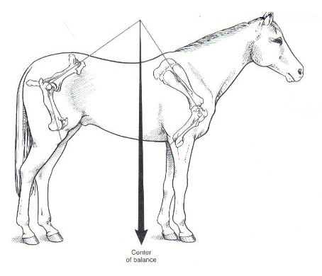
center of gravity
located at the intersection of
vertical line dropped from the highest point of the withers
horizontal line drawn from the point of the shoulder to tuber ischii

length, angles, heights
shoulder length & angle
related to stride length
long shoulders- long strides
short shoulders- short strides
line from scapula should be at same angle as pasterns (45º)
upright pasterns
shorter angle = shorter strides

what produces long strides?
long shoulders
long radius & short metacarpus
long gaskin (mind limb muscles between stifle & hock) & short metatarsus
flat & long rump
what predisposes hindlimb lameness?
short, steep rumps
what predisposes forelimb lameness?
taller behind than in front
forelimb conformation faults from upright pasterns
increases concussion on fetlock, phalangeal joints, navicular apparatus
affected horses are prone to navicular disease & ringbone
risk of fetlock effusion increases with increased dorsal hoof angle

forelimb conformation faults from long/sloping pasterns
injury of flexor tendons, sesamoid bones, suspensory ligament
osteoarthritis of fetlock
P1 fractures

muscling
muscles add support to skeleton & joints
length, thickness, distribution of each muscle group vary with age & sport activity
symmetry
limb conformation from front/rear
a line dropped from the shoulder/tuber ischii should divided the leg into 2 symmetrical halves
observe 2 characteristics
base- wide or narrow
limb axis- straight, turned outwards, turned inwards
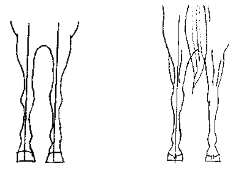
wide base
inside foot weight bearing

narrow base
outside foot weight bearing

toed out
(splay footed)
plaiting- in flight toes swing inward
results in abnormal wear on inside of foot
predisposes horse to medial splints
slight toe out may be desirable (decreased risk of carpal fracture)

toed in
(pigeon toed)
paddling- toes swing outward

out at the knee
bow legged
most affected horses are also toes in
predisposes to osteoarthritis of carpus/fetlock
predisposes to lateral suspensory branch desmitis & sesamoiditis

in at the knee
may predispose to splints
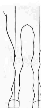
bench/offset knees
metacarpal 3 offset
medial aspect of joint bears abnormal weight
joint is no longer balanced

angular limb deformity (ALD)
deviation of the limb from its normal axis in the frontal plane
most common in the carpus but also seen in fetlock & tarsus
valgus or varus
usually some rotational deviation
valgus deformity
lateral deviation of limb distal to location of the deformity
carpal- distal portion of metacarpus deviates laterally & distal portion of radius deviates medially
usually toed out
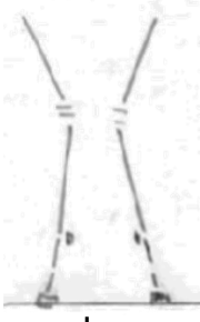
varus deformity
medial deviation of limb distal to location of the deformity
carpal- distal portion of metacarpus deviates medially & distal portion of radius deviates laterally
usually toed in

causes of ALD
acquired
asynchronous growth due to asymmetrical loading of growth plate in distal radius
trauma to growth plate (excessive exercise, contralateral limb lameness)
rapid growth from over-nutrition
mineral inbalances
congenital
in utero mal-positioning
laxity of collateral ligaments of carpus (surrounding soft tissue not strong enough to support carpus)
hypoplasia of cuboidal carpal bones (defective ossification)
ALD treatment
depends on age of foal, joint involved, severity of the angle
options:
confinement & altered nutrition
splints, braces, casts
surgical growth retardation
surgical growth stimulation/acceleration
timing for growth plate closures
distal metacarpus/tarsus= 3-6 months
rapid growth peak at 6 weeks
distal radius & tibia= 2 years
rapid growth peak at 3-4 months
rotational deformities
permanent rotation develops when a bone is abnormally loaded in both frontal & sagittal planes leading to rotation of the limb around the overloaded axis point
(temporary rotation of the limb from the elbow is not uncommon in narrow-chested foals & will self correct as the chest widens)

identifying deformities
radiography
clinical observation at rest
clinical observation at exercise
ideal forelimb conformation from the side
line bisecting elbow joint, carpus, & fetlock joint intersects ground 5cm behind heel
straight column from elbow joint to fetlock
fetlock angle 125º-135º

deviations from ideal forelimb conformation
back at the knee
over at the knee
back at the knee (calf-kneed)
knee inclines backward
behind a straight plumb line dropped from the middle of the forearm to the fetlock
carpal hyperextension overloads dorsal carpus
poor conformation leading to carpal fractures

over at the knees (bucked knees)
knee inclines forward
infront of a plumb line when viewed from the side
increased stress on flexor tendons/suspensory ligament

ideal hindlimb conformation from the side
line dropped from the tuber ischii should touch the point of the hock & plantar metatarsus and intersect the ground 7.5-10cm behind the heel

deviation from ideal hindlimb conformation
sickle-hocked
camped out behind
cause inefficiency in propulsion
post-legged (leg too straight)

hoof balance
even distribution of forces throughout the foot during weight bearing
imbalances caused by poor leg conformation
distortions caused by uneven loading of pressure from above
static hoof balance
horse standing still
examine all views of the feet and limbs
dorsal
palmar
lateral
solar

dorsopalmar hoof balance
lateral view
hoof pastern axis- angle of dorsal hoof wall should be same as line through long axis of pastern
53º-58º

broken back hoof pastern axis
long toe, under-run heel
common defect
often caused by poor foot care
result in increased strain on the deep digital flexor & delayed break-over
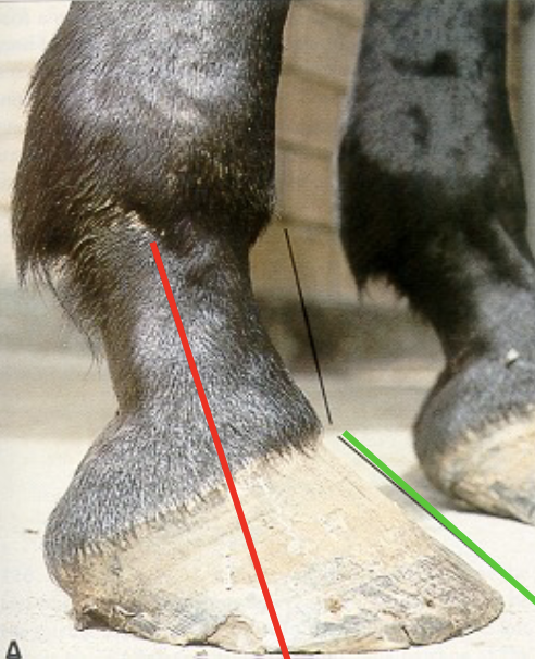
broken forward hoof pastern axis
club foot if angle >60º
caused by:
coffin joint flexion
increased strain on suspensory ligament
increased strain on superficial digital flexor tendon
increased pressure in the heel

mediolateral hoof balance
solar view
trimmed frog
frog should bisect hoof & shoe into 2 equal halves
frog width should be 50-66% of the frog length
length of the foot should approximate the width
both heels are equal length
line drawn across the weight bearing surface of the heels is perpendicular to axiss of the frog
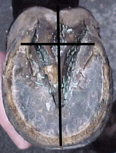
dynamic hoof balance
moving horse
gait from dorsal & lateral
foot landing- level to the ground (D) & level or just barely heel first (L)
weight bearing
foot flight
lameness
clinical sign/manifestation of signs of inflammation, pain, neurological disease, or mechanical defect that result in a gait abnormality characterized by limping
lameness distribution
60% front: 40% hind
70:30 with rider
95% of forelimb lameness problems occur distal to the carpus
stages of lameness examination
history
physical exam
evaluate gait
diagnostic anaesthesia
imaging
other tests
lameness exam equipment
head collar & lead rope (or bridle/chifney)
lunge rein
lunge whip
gloves
hoof testers
shoe removal equipment
pen to check back reflexes
level hard surface to trot on
lameness assessment form
lameness history
age- young= likely to be lame from DOD
specific use
used for particular type of performance are predisposed to particular causes of injury
onset- sudden or insidious
duration
progression- is horse improving
clinical signs- severe/mild, continuous/intermittent
past & current levels of training
previous injury & treatment
response to treatment for present lameness
lameness physical exam
limb conformation- conformational deficits predispose to musculoskeletal injury
posture of limbs- resting forelimbs is uncommon
muscular symmetry
atrophy
swelling
foot size- lame limb foot smaller if chronic
fetlock heights
joint/tendon sheath distension
increased soft-tissue thickness
manipulate joint to elicit signs of pain
palpate for increased digital pulses & heat in foot
gait
manner of walking/stepping
beat
number of foot strikes in a single stride cycle
step
distance between footprints of the 2 front/back feet