Nerve, Muscle, and Synapse Part 2
 Call Kai
 Learn
Learn Practice Test
 Spaced Repetition
 Match
Match Flashcards
 Knowt Play
1/71

There's no tags or description
Looks like no tags are added yet.
Name | Mastery | Learn | Test | Matching | Spaced | Call with Kai | Chat |
|---|
No analytics yet
Send a link to your students to track their progress
72 Terms
Electrical synapses have __synaptic delay. They are bidirectional (moving in __directions) and cells are connected internally and share signalling molecules through a shared cytoplasm (cytoplasmic communication).
no
2

In electrical synapses, there are ___junctions between the presynaptic and postsynaptic cell. electrical synapses work by ionic currents passing through ___, causing voltage changes in the neighbouring cell. these currents depolarize the postsynaptic cell and can trigger an action potential.
gap
gap junctions
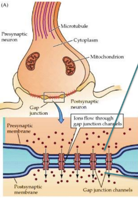
Presynaptic neurons release ___from their axon terminals and these bind to postsynaptic ___. axon terminals of presynaptic neurons have ___that contain neurotransmitter molecules.
neurotransmitters
receptors
vesicles


List what occurs at a presynaptic knob during an action potential (depolarization)
depolarization during the action potential opens voltage-gated Ca2+ channels in the plasma membrane of the presynaptic axon terminal.
Ca2+ flows into the neuron and activate processes that lead to the fusion of docked vesicles with the presynaptic terminal membrane.
vesicles interact with SYNAPTOTAGMIN and SNARE proteins.
neurotransmitters are released via exocytosis

The mechanism of electrical synapses has ions directly flowing through ___. Electrical synapse speed is very fast with __delay. it has ___identity. its plasticity (ability to adapt) is limited/extensive, and it can/cannot amplify signals
gap junction
no
intrinsic
limited
cannot
What does having no intrinsic identity mean
An electrical signal with no intrinsic identity means it lacks any permanent, predefined shape, size, or default form of its own. So, the receiving neuron acts as a fluid mirror that directly and proportionately copies the exact size, duration, and direction of the sender's voltage change. Its entire state is a blank canvas, dynamically and completely defined by whatever incoming data it receives.
The mechanism of chemical synapses has neurotransmitters flowing through the ___. chemical synapse speed is fast/slow. it converts the electrical signal into __then back into an electrical signal. it has a defined identity based on if the neurotransmitter and recptor are ___ or ___. its plasticity (ability to adapt) is limited/extensive, and it can/cannot amplify signals
synaptic cleft
slower (milliseconds delay)
electrical→chemical→electrical
excitatory or inhibitory
extensive
can amplify/modulate signals
What does it mean when a chemical synapse has intrinsic identity?
the signal gains a defined identity because its size, duration, and direction are determined by the specific neurotransmitter released and receptors it binds to (excitatory and inhibitory). the signal also evolves independently over time which depends on how fast the neurotransmitter binds, how fast it opens ion channels, and how fast it is cleared from the synapse (signaling is a separate process not a direct copy)
What are the two types of neurotransmitter receptors on the post synaptic membrane
ionotropic and metabotropic receptors
___receptors activates G protein first→indirect effects. the receptor and effector are different molecules. have a slower start but long lasting.
metabotropic receptors (aka G-protein coupled receptors)

___receptors are ion channels that open directly, are super fast and the receptor and effector are the same molecule.
ionotropic receptors (aka ligand-gated ion channels)

There are two types of postsynaptic potentials (PSPs)
excitatory postsynaptic potential EPSP
inhibitory postsynaptic potential IPSP
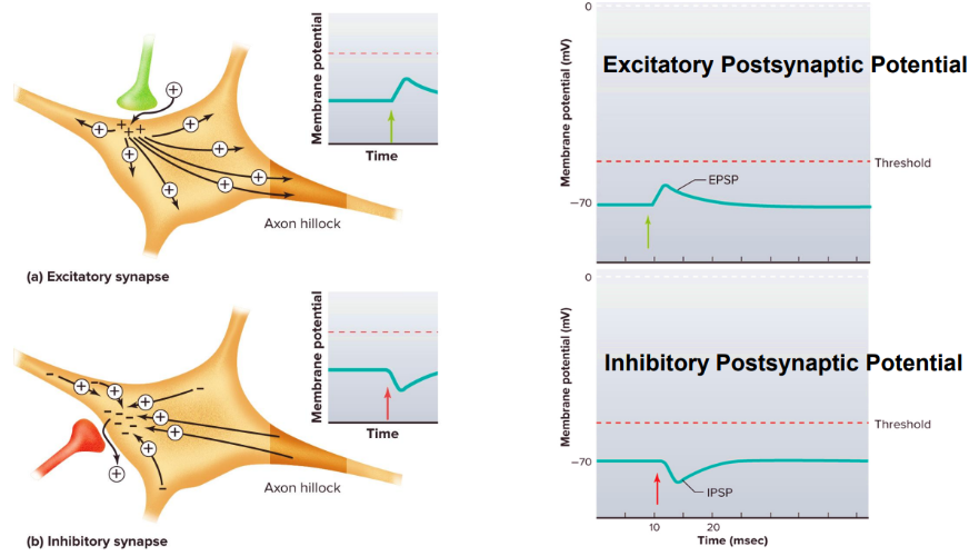
__PSPs slightly depolarize the postsynaptic cell. however, alone, they cannot generate an action potential (subthreshold)
excitatory
describe how an EPSP works
an excitatory neurotransmitter (glutamate) binds to a postsynaptic receptor and opens ligand-gated Na+ channels, causing them to open.
Na+ enters the postsynaptic cell and results in a small, subthreshold depolarization.
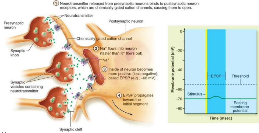
describe how an IPSP works
an inhibitory transmitter (ex. GABA, glycine) binds to the postsynaptic receptor and opens ligand-gated Cl- channels, causing them to open.
Cl- enters the postsynaptic cell and results in a small, hyperpolarization (more negative).
IPSPs reduce the likelihood that the postsynaptic cell will depolarize to the threshold potential and generate an action potential.

When multiple postsynaptic potentials from a single presynaptic axon occur close together in time, then add together. multiple small EPSPs can combine to reach a threshold and trigger an action potential
temporal summation
When postsynaptic potentails from multiple presynaptic neurons at differnt locations on the postsynaptic neuron add together. multiple small EPSPs combine to depolarize the membrane to threshold and trigger an action potential
spatial summation
both spatial and temporal summation occur at the same time in the __. for spatial summation to occur, postsynaptic potentials from different regions must also___. as a result, excitatory and inhibitory signals are combined into a single resopnse by the postsynaptic neuron.
brain
happen at the same time
the proces of summing together all the inputs into a pattern of action potential output in the postsynaptic cell
synaptic integration
How would you solve this question: A neuron sits at -70 mV and has a threshold of -50 mV. It then simultaneously receives 10 IPSPs of 0.5 mV each and 20 EPSPs of 1 mV each. Does the cell fire an AP?
first calculate the total voltage change of the IPSP using -mV values (bcs inhibitory). This is just simple multiplication.
then calculate the total voltage change of the EPSP using +mV values (bcs excitatory). this is also multiplication.
calculate the net voltage change: IPSP total + EPSP total
add that net voltage change to the RMP to see how hyperpolarized/depolarized it is and if that reaches the threshold.
Postsynaptic potentials are graded/not graded. they have/don’t have summation. they have/dont have a threshold. they have/dont have a refractory period. describe their conduction (passive/active?long/short distance?if they decay?). PSPs are very quick and mostly occur in __. they function to move voltage towards (_PSP) or away (_PSP) from the threshold.
graded
have summation (temporal+spatial)
no threshold
no refractory period
PSPs are passive, travel a short distance, and decays with distance.
occurs in dendrites and soma
towards=EPSP
away=IPSP
what does it mean for a signal to be graded
does the signal weaker over time and distance. varies depending on the stimulus
what happens when the PSP summation at the hillock reaches the threshold?
they trigger and action potential AP
action potentials are graded/not graded. they have/don’t have summation. they have/dont have a threshold. they have/dont have a refractory period. describe their conduction (passive/active?long/short distance?if they decay?). APs are very quick and mostly occur in__. they function as a reliable long-range signal that, once it reaches the synapse, causes___
not graded (all or none)
don’t have summation
usually have a threshold 15mV above RMP
do have a refractory period (absolute + relative)
their conduction is active, travels a long distance, and have no decay.
occur in axon hillock→synaptic terminal
reaches the synapse and causes neurotransmitter release which sets of new PSPs in the postsynaptic knob

the leakage of current through membrane leak channels (usually K+ leak channels) and cause a graded (postsynaptic) potential to lose height (amplitude) as they spread passively along the membrane
decremental conduction

__:a synapse in which one neuron’s axon terminal synapses onto the axon terminal of another neuron. one terminal controls how much neurotransmitter another terminal releases (but itself does not affect the postsynaptic neuron)
axo-axonic synapse
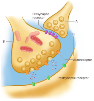
in axo-axonic synapse, a terminal can produce presynaptic inhibition or facilitation through decreasing or increasing __entry into another terminal and ultimately changing neurotransmitter release.
Ca2+ calcium ion
what are the three muscle types
skeletal
cardiac
smooth

cardiac muscles are located in__ and function to__. they are controlled involuntarily by ____. they can be striated (have stripes).
the walls of the heart
propel blood thorugh the circulatory system
autonomic nervous system, hormones, and other signals

smooth muscles are located in__and function to__. they are controlled involuntarily by__. they can regulate blood flow, move food, expel urin, and regulate airflow.
the walls of hollow organs and tubes (ex. arteries, GI tract, bladder, airways)
support internal movements by reducing the size of structures during contraction
autonomic nervous system, hormones, and other signals
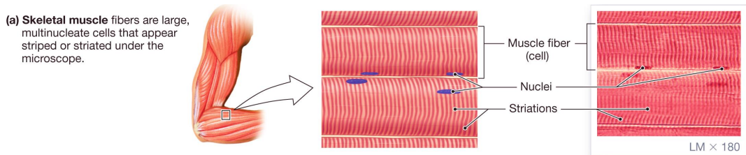
skeletal muscles are located in__and function to__. they are controlled voluntarily by__. they can be striated (have stripes).
attached to bones of the skeleton
supports and moves the skeleton
contractions which are initiated by action potentials in motor neurons
Skeletal muscle consist of myofibrils which are made of ___. the arrangement of thin and thick filaments along myofibrils gives skeletal and cardiac muscle fibers their striated (striped) appearance. thick filaments are made of __
myofibrils
myosin

Describe the structure of thick filaments and what they’re made of (4)
thick filaments are made of myosin which is made of 2 heavy chains twisted together to form a tail.
2 globular heads (cross-bridges) extend outward and pull on thin filaments during contraction.
each head has 2 light chains and binding sites for actin and ATP
the ATP-binding site acts as a myosin ATPase which breaks down ATP to provide energy

Describe the structure of thick filaments and what they’re made of (4)
thin filaments are made of actin, troponin, and tropomyosin. troponin and tropomyosin regulate muscle contraction.
actin is made of globular monomer proteins that join into 2 intertwined helical chains.
actin chains form the core of the thin filament, tropomyosin wraps around, and troponin stacks onto tropomyosin.
each actin molecule has a myosin-binding site

a type of endoplasmic reticulum specialized for the muscle cell
sarcoplasmic reticulum (SR)

enlarged SR regions that store Ca2+ calcium ions
terminal cisternae

lie between terminal cisternae and are continuous with the sarcolemma (aka plasma membrane)
transverse/t-tubules

action potentials travel through ___to carry signals into the muscle fiber
t-tubules
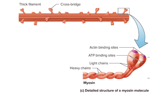
___:the activation of cross-bridges which are the force-generating sites within muscle fibers
contraction

CONTRACTION DOES NOT EQUAL SHORTENING
___: a motor neuron and all the skeletal muscle fibers it innervates (connects to to stimulate function)
motor unit

a single motor neuron can innervate many muscle fibers. however one musle fiber is controlled by a branch from __motor neuron(s). a whole muscle contains___ motor units.
only one
many

what happens during an action potential in a motor neuron
all the muscle fibers in its motor unit are stimulated to contract
a motor neuron originates in the __and extends large myelinated axons towards a muscle fiber.
the nerve’s axon terminal gets close to the muscle cell membrane (sarcolemma) which contains vesicles with the neurotransmitter ___.
the region of the muscle fiber plasma membrane that lies directly beneath the axon terminal is called the___
CNS (brainstem/spinal cord)
acetylcholine (ACh)
motor end plate


in a neuromuscular junction:
synaptic vesicles store and release acetylcholine (ACh) into the synaptic cleft.
Which binds to receptors on the motor end plate
Which triggers an action potential
the synaptic cleft between a motor neuron and the motor end plate is called a ___
neuromuscular junction (NMJ)

Describe how an action potential from a motor neuron is sent to a muscle fiber
Motor neuron action potential: an electrical impulse travels down the motor neuron axon to the axon terminal near the muscle fiber.
Ca2+ calcium ion enters: depolarization of the terminal opens voltage-gated calcium channels, causing Ca2+ to rush into the presynaptic terminal.
Acetylcholine release: Ca2+ triggers vesicles to fuse with the presynaptic membrane via SNARE proteins and synaptotagmin, releasing acetylcholine (ACh) into the neuromuscular junction.
ACh binding and Na+ influx: ACh diffuses across the cleft and binds to ligand-gated ion channels that function as receptors (ionotropic) on the motor end plate of the muscle fiber, opening them to allow Na+ to flow inside.
Depolarization and threshold: the influx of Na+ creates a local end-plate potential (graded) that depolarizes the adjacent muscle plasma membrane until it hits the threshold.
AP propagation: Once threshold is reached, an action potential propagants along the muscle plasma membrane.
Breakdown: Acetylcholinesterase is expelled by exocytosis breaks down ACh in the clef to end the signal.


Transmission normally happens in only one direction across the neuromuscular junction

__=local depolarization of the motor end plate
End-plate potential (EPP)
action potentials in the muscle fibers conduct their messages into the muscle fibers through ___
transverse (T) tubules

compare central (neuron-neuron) synapse and neuromuscular junction? how many potentials does each need to fire an AP?
central (neuron-neuron) synapses need the summation of many postsynaptic potentials to fire an action potential. one neuron recieves thousands of inputs.
vs NMJs, one presynaptic AP→triggers one muscle AP. each muscle fiber gets input from ONE axon
compare central (neuron-neuron) synapse and neuromuscular junction? do they experience excitatory or inhibitory signals?
central (neuron-neuron) synapse experiences excitatory AND inhibitory signals
skeletal NMJs ONLY experience EXCITATORY signals

E-C coupling describes the sequence of events where an AP in the motor end plate of a muscle fiber causes____and __of the muscle. describes how a surface electrical spike reaches deep into the muscle fiber to trigger it and how long it takes.
actin-myosin cross-bridge formation
contraction

List the steps of E-C coupling (3)
APs in muscle fibers conduct messages into muscle fibers through t-tubules
this initiates Ca2+ release from the lateral sacs of the sacroplasmic reticulum
Ca2+ ion channels are open for binding to TROPONIN and initiating the events of the SLIDING FILAMENT MECHANISM.
relaxation: Ca2+ is actively pumped back into the sacroplasmic reticulum which stops the contraction and lets the muscle relax.

What components are involved in the sliding filament mechanism?
thick filament (myosin with actin and ATP binding sites
thin filaments (actin with myosin binding sites and two regulatory proteins):
tropomyosin
troponin

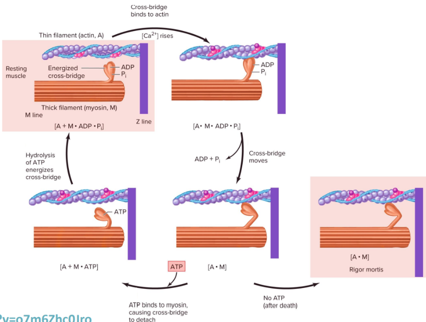
Describe how myosin binds and unbinds to ATP and actin, in reference to head conformations
Low-energy conformation: an ATP molecule binds to tits specific binding site on the myosin cross-bridge (head).
High-energy conformation: the myosin head hydrolyzes (breaks) the ATP molecule into ADP and an inorganic phosphate (Pi).
Actin binding: while still holding onto the ADP and Pi, the upright myosin head exposes its actin-binding site and forms a cross-bridge to pull the actin filament. this cause a muscle contraction.


Describe how Ca2+ ions regulate muscle contraction and relaxation
Tropomyosin covers the myosin-binding sites on actin subunits, preventing myosin heads from binding to actin and forming cross-bridges.
An action potential triggers Ca2+ ions to be released from the terminal cisternae of the sacroplasmic reticulum into the cytosol.
Ca2+ ions bind to troponin proteins positioned periodically along the tropomyosin strand.
Ca2+ binding changes troponin’s shape, pulling tropomyosin away from the myosin-binding sites and exposing them.
Energized myosin heads bind to the exposed binding sites on actin, forming cross-bridges.
Myosin heads bend in a power stroke, pulling the actin filaments and producing muscle contraction.
After contraction, Ca2+-ATPase (SERCA) pumps located in the scaroplasmic membrane use ATP to transport Ca2+ from the cytosol back into the SR.
As Ca2+ leaves the cytosol, Ca2+ detaches from troponin and the troponin/tropomyosin complex returns to its position covering the myosin-binding sites on actin. The muscle relaxes.

Describe ATP’s role in muscle contraction
An action potential triggers the release of Ca2+ ions from the terminal cisternae of the SR into the cytosol.
Ca2+ binds to troponin, causing a conformational change in the troponin-tropomyosin complex. Tropomyosin moves away from the myosin-binding sites on actin, exposing them.
When ATP is hydrolyzed into ADP and inorganic phosphate Pi, the myosin head is in an energized state. The energized myosin head binds to the exposed binding site on actin, forming a cross-bridge.
When the cross-bridge is formed, ADP and Pi are released, causing the myosin head to pull the thin filament toward the center of the sarcomere. This produces contraction (chemical energy→mechanical energy).
A new ATP molecule binds to the myosin head, causing it to detach from actin.
SERCA (Ca2+-ATPase) pumps in the SR membrane use ATP to pump Ca2+ from the cytosol back into the SR.
As Ca2+ conc. lessens, it dissociates from troponin and tropomyosin goes back to covering the myosin-binding sites on actin. The muscle relaxes.
In muscle contraction, the ____is whats being shortened. there is NO change in the lengths of the thick or thin filament, only in their amount of ___. this is known as the sliding-filament mechanism of muscle contraction.
sarcomere
overlap

Summarize E-C coupling including the sliding filament model
muscle action potential propagates along the sarcolemma and down the t-tubules.
receptors on the t-tubule sense the depolarization and change shape
this conformational change mechanically opens the receptor on the adjacent sarcoplasmic reticulum.
Ca2+ ions flood out of the SR into the cytosol.
Ca2+ binds troponin, dragging tropomyosin off the myosin-binding sites on actin.
cross-bridge cycling begins
the sarcomere shortens

____=a single contraction and relaxation cycle of a muscle fiber or motor unit in response to a single AP
muscle twitch
what are the three metabolic variations of muscle fiber types
slow-oxidative (type I)
fast-oxidative-glycolytic (type IIa)
fast glycolytic (type IIb or IIx)
___=a type of muscle fiber that fatigues quickly, used for fast powerful movements (ex. sprinting).
fast glycolytic (type 2b or 2x)
___=a type of muscle fiber that is fatigue-resistant, used for slow, sustained movements (ex. posture).
slow-oxidative (type 1)
___=a type of muscle fiber that is a combination of type I and type IIb.
fast-oxidative-glycolytic (type 2A)

most skeletal muscles include all 3 types of muscle fiber (Type 1, 2a, and 2b)
which muscle fiber has the lowest shortening velocity but maintains a steady maximum tension/force consistently over time.
slow-oxidative (type 1)
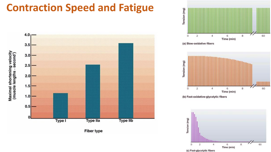
which muscle fiber has an intermediate shortening velocity (maintains high tension/force for a few minutes followed by a gradual decline with significantly lower but steady force (endurance))
fast-oxidative-glycolytic (type 2A)

which muscle fiber has the highest shortening velocity, contracting the fastest. produces very high initial tension/force but loses nearly all tension within 2-4 minutes.
fast glycolytic (type 2b or 2x)
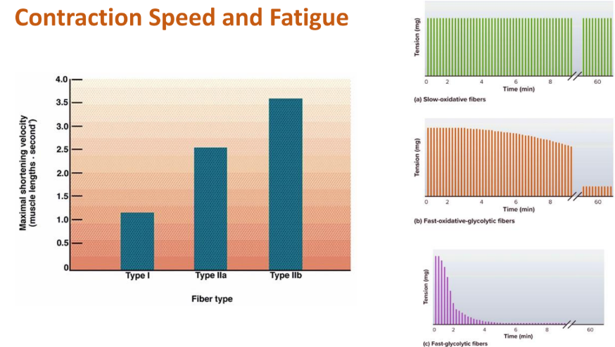
which muscle fiber has:
-gets most of their ATP through oxidative phosphorylation
-lots of mitochondria
-very red cells
-lower glycogen stores
-small in diameter
-endurance, fatigue resistant
slow-oxidative fibers (type 1)

which muscle fiber has:
-gets most of their ATP via anaerobic glycolysis
-few mitochondria
-few blood vessels and little myoglobin (white colour)
-lots of glycogen
-larger diameter (more myofibrils=stronger)
-powerful, fatigues more quickly
fast-glycolytic fibers (type 2a+2b)


Total muscle tension increases over time as additional motor units are recruited. They are all being used at the SAME TIME. the order goes type 1→type 2a→type 2b
training can improve the ability to recruit and activate 2b motor units easier, but it doesn’t skip 1 or 2a.