Chapter 9 - Musculoskeletal System: Part 1
 Call Kai
 Learn
 Practice Test
 Spaced Repetition
 Match
 Flashcards
 Knowt Play
1/64

There's no tags or description
Looks like no tags are added yet.
65 Terms
Axial Skeletion
Composed by the skull, vertebral column, sacrum, ribs, and sternum

Cervical Curvature

Thoracic Curvature
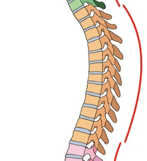
Lumbar Curvature

Pelvic Curvature
Made up by the sacrum and coccyx

Intervertebral disc
Connective tissue that separates the bodies of every vertebra and reduce friction as the vertebrae move. They also assist in absorbing shock

Atlas
The first of the cervical vertebrae. It articulates with the skull superiorly and the axis inferiorly. In Greek Mythology, the titan named ____ held the world. Simiarly, this vertebrae supports your skull!

Axis
The second cervical vertebra. The ___ allows you to shake your head back and forth. In other words, it allows your head to move along an ____.

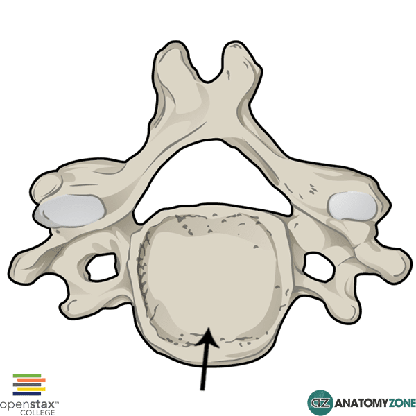
Body (of vertebrae)
Found on all vertebrae except atlas. The ___ transmits weight down the bodies of the vertebrae.
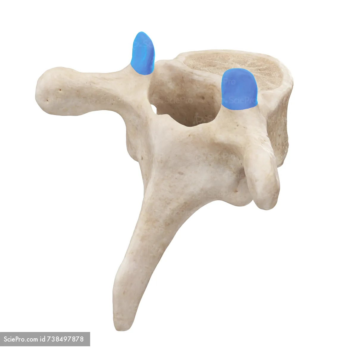
Superior Articular Process
These are the bony projections that point superiorly to articulate with the vertebra above.
Facets (for both superior and inferior articular processes)
The surfaces where the two bones physically articulate
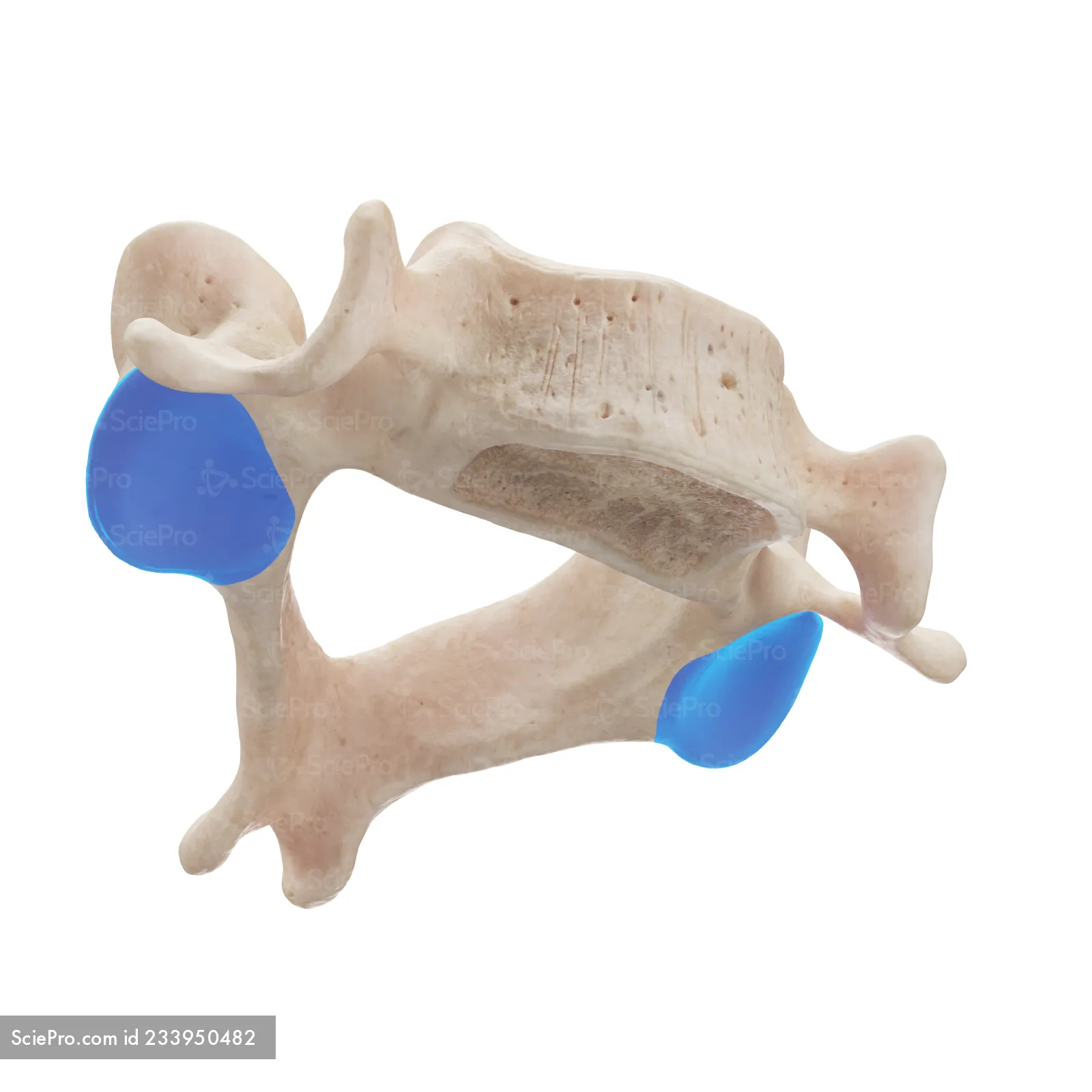
Inferior Articular Process
Very similar to the superior articular process; these are the bony projections that point inferiorly to articulate with the vertebra below.
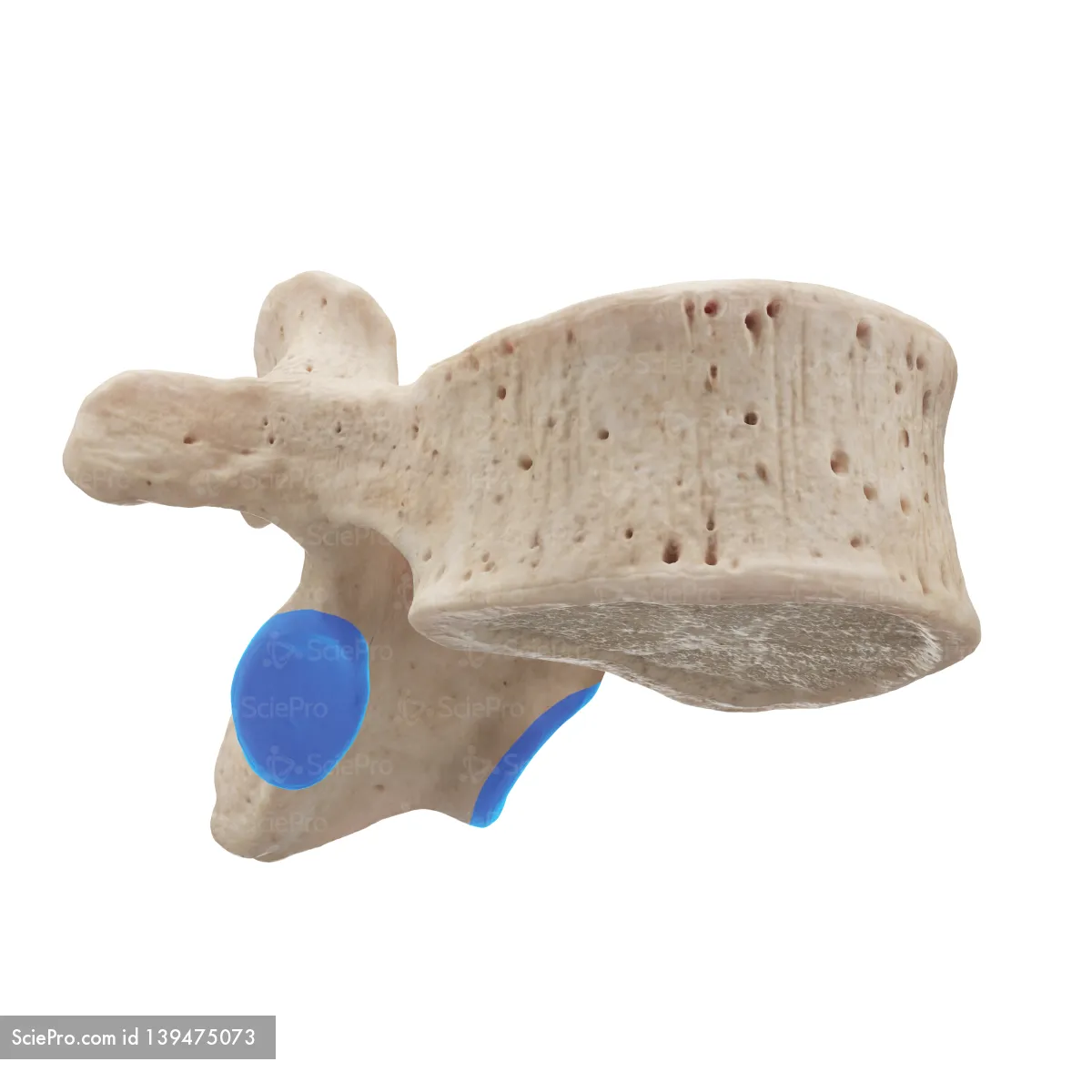
Transverse Process
This is the bony region that extends laterally from the body

Spinous Process
This is the somewhat sharp projection that extends posteriorly, opposite from the body of the vertebra.
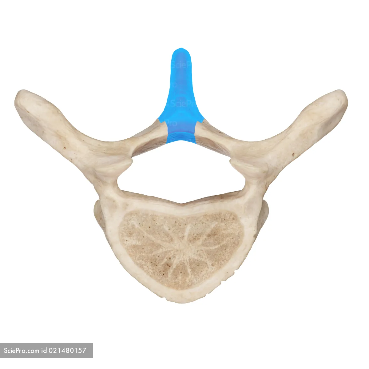
Vertebral Arch
The transverse and spinous processes arise from this bony arch.
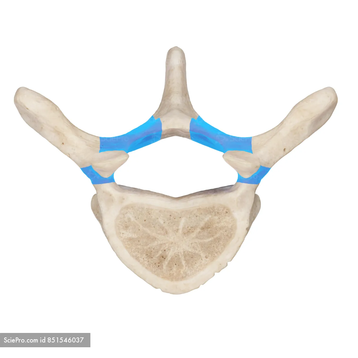
Vertebral Foramen
The opening formed by the vertebral body and arch

Vertebral Canal
All of the vertebral foramina will line up and together form this long tube. The nerves of the spinal cord run through this structure.

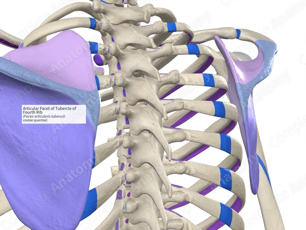
Rib Facets
On thoracic vertebrae, this structure (sometimes called costal facets) can be found on the body and on the transverse processes. Like other articular surfaces, they are covered with articular cartilage and appear smooth in real bone.
Body (of sacrum)
The main thick medial region of the sacrum.

Medial Sacral Crest
Formed from the spinous process of the fused vertebrae (feature of sacrum)

Auricular Surface
On the lateral faces of the sacrum, this forms the joint between the sacrum and ilium

Coccyx

True Ribs
Ribs 1 through 7; they articulate directly with the sternum via costal cartilage

False Ribs
Ribs 8 through 12; they either articulate with the sternum indirectly or not at all.

Floating Ribs
Ribs 11 and 12; do not articulate with the sternum directly at all.

Head
The articular facet on the ____ of each rib articulates medially with the rib facets of the thoracic vertebra.
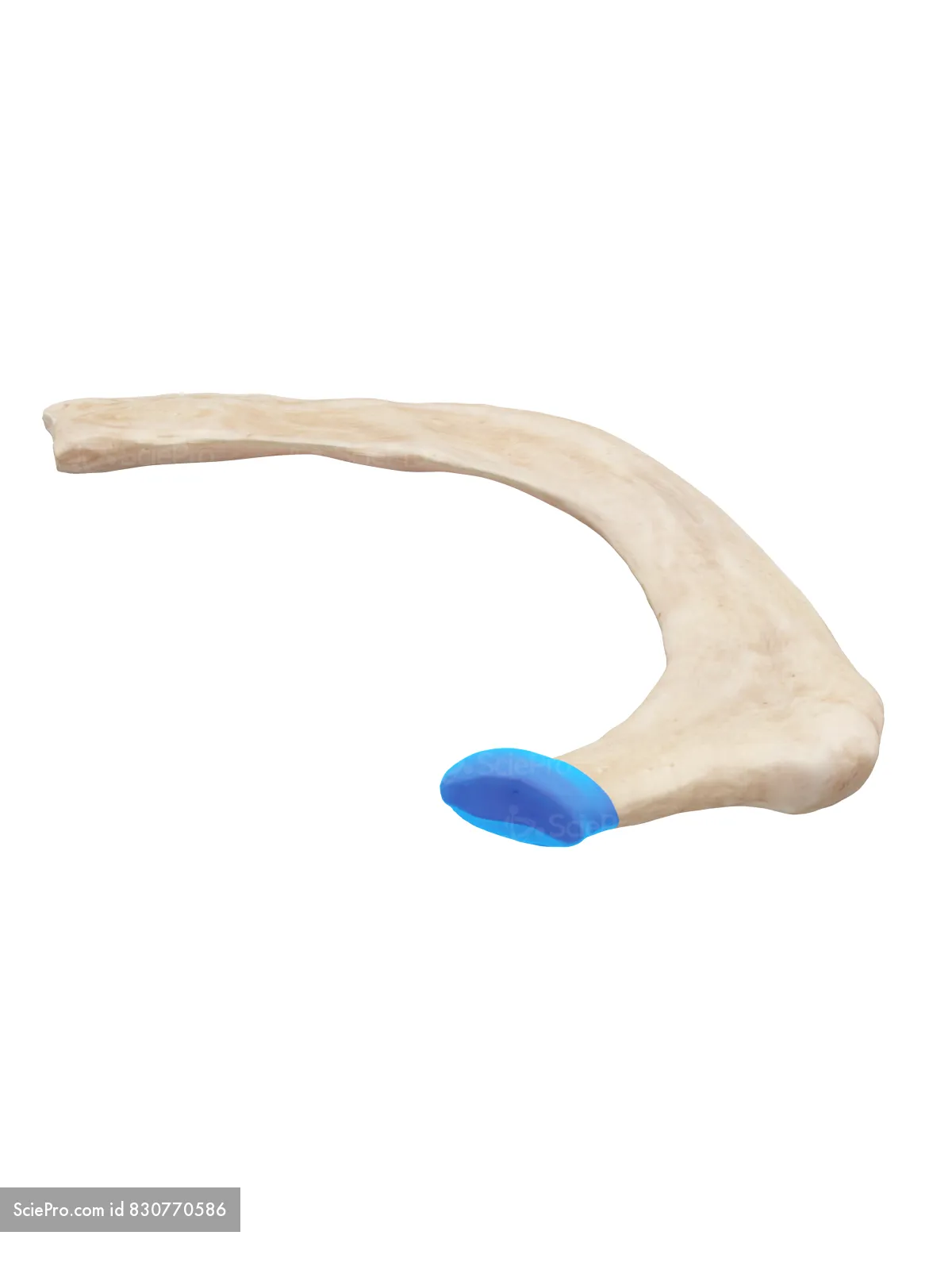
Tubercle
This small bumpy ridge is found just distal to the head.

Body (of rib)
This is the main part of the rib distal to the neck

Angle
Just distal to the tubercle, the rib makes a sharp bend, which is called the ____.

Body (of the sternum)
This is the large middle portion of the sternum

Facets (for the sternum)
This is where the costal cartilage associated with the ribs articulate

Manubrium
This is the upper part of the sternum

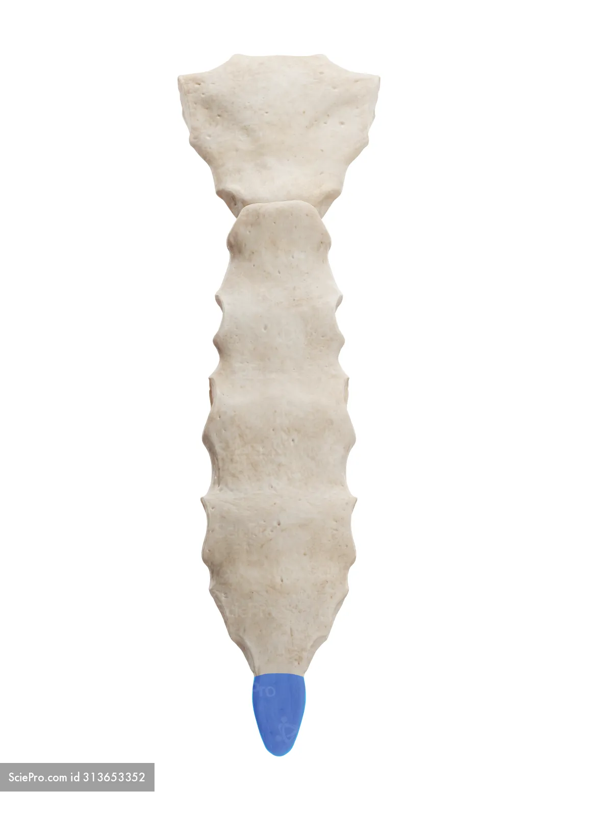
Xiphoid Process
This projection can be found inferior to the body of the sternum
Rectus Abdominis
There are two of this muscle, one on each side of the body midline. In individuals with low body fat, this pair of muscles forms a “six pack.”

Linea Alba
When viewed in cross section, the right and left rectus abdominis muscles are connected by a strong column of connective tissue called the ____ ____, which connects the xiphoid process to the pubic bone

External Oblique
Lateral to the rectus abdominis muscles, the ____ ____ muscles form the most superficial part of the abdominal wall. As you put your hands in your pockets, you sort of trace the V shape formed by the fibers of the right and left ____ ____ muscles.

Internal Oblique
Lateral to the rectus abdominis muscles, and hidden just underneath the external oblique muscles, are the ____ ____ muscles. These form the middle layer of much of the abdominal wall.

Transverse Abdominis
The deepest muscle layer of the lateral abdominal wall is the _____ _____ muscle. It lies below the internal oblique muscle. The fibers of this muscle run side to side, more like a belt around your abdomen.

External Intercostal Muscles
The fibers run on either side of your chest from superior-lateral to inferior-medial. This forms a V shape, mirroring the striations of the external oblique muscles in the abdomen. They may assist with inhalation by pulling the ribs upward.

Internal Intercostal Muscles
Fibers run on either side of your chest like an A shape, from superior-medial to inferior-lateral. They may assist with exhalation by pulling the ribs downward.

Innermost Intercostal Muscle
Fibers line the inside of your chest and run superior-inferior. This muscle may also assist with exhalation by pulling the ribs downward.

Diaphragm
The dome shaped muscle responsible for increasing the volume of your thoracic cavity to help you inhale. It forms the division between the thoracic cavity and the abdominal cavity.

Erector Spinae Muscle
Begins where it attaches to the lower back and hips. The muscle fibers run upward along the spine and attach to the skull.

Sternocleidomastoid
This muscle’s fibers extend from the mastoid process (the bony lump behind your ear) to the medial clavicle (your collar bone) and the manubrium of the sternum

Flexion
Decreases the angle between two bones or body parts at a joint
Extension
Increases the angle between two body parts, typically resulting in the straightening or opening of a joint
Hyperextension
A posterior chain strength exercise performed on a Roman chair, a 45-degree or 90-degree bench, or the floor to target the erector spinae, glutes, and hamstrings.
Lateral Flexion
Side-to-side bending movement of the spine or neck within the frontal plane
Rotation
The motion of a bone, limb, or body segment as it spins or turns around its own longitudinal or central axis.
Medial Rotation
A rotational movement where a bone or limb turns inward toward the midline of the body
Lateral Rotation
A twisting movement of a bone or limb around its long axis away from the midline of the body
Circumduction
A compound, circular anatomical movement that blends flexion, abduction, extension, and adduction in a smooth, continuous sequence. As you perform this motion, the stationary joint acts as the apex of a cone, while the distal end of the limb or body part traces a circle in space
Abduction
the anatomical movement of a limb or body part away from the midline of the body or from the centerline of a hand or foot
Adduction
Brings body parts back toward the midline
Supination
A rotational movement that turns the palm of the hand upward or rolls the foot outward so the weight rests on the outer edge
Pronation
A rotational or triplanar movement that turns the forearm and hand palm-down or rolls the foot inward during weight-bearing
Depression
A downward (inferior) movement of a body part, such as lowering the shoulders or opening the mouth (lowering the mandible)
Elevation
An upward (superior) movement of a body part in human anatomy, paired directly with depression as its downward counterpart
Protraction
A body part glides or moves forward (anteriorly) and laterally away from the midline
Retraction
Pulls a body part backward (posteriorly) and toward the midline of the body (medially)
Dorsiflexion
Upward bending movement of the foot and toes toward the shin
Plantar Flexion
Downward movement of the foot and ankle where the sole or toes point away from the shin and body
Eversion
the movement where the sole of the foot turns outward, away from the midline of the body in the frontal plane
Inversion
a movement where the sole of the foot tilts inward toward the midline of the body
Opposition of Thumb
the movement where the thumb rotates and flexes across the palm so that the pad of the thumb touches the pad or tip of another finger, especially the little finger