Wrist, Forearm & Elbow Anatomy + Positioning & Technical Factors
 Call Kai
 Learn
 Practice Test
 Spaced Repetition
 Match
 Flashcards
 Knowt Play
1/48

Earn XP
Description and Tags
https://quizlet.com/436526122/ap-elbow-diagram/
49 Terms

Label The Radius & Ulna (17)

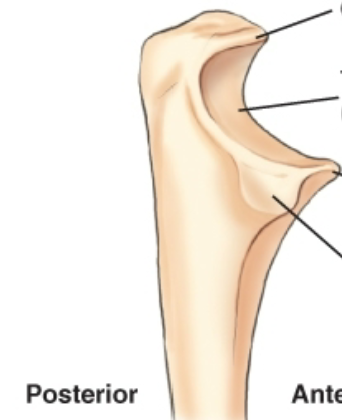
Label The Proximal Ulna (4)


Label the AP Elbow (11)


Label the Lateral Elbow (15)


What Are The Three Concentric Arcs?


Label the AP Elbow Image
A. medial epicondyle
B. trochlea
C. coronoid tubercle
D. radial head
E. capitulum
F. lateral epicondyle
H. olecranon process

Label the Lateral Elbow Image
G. superimposed epicondyles of the humerus
I. trochlear sulcus
J. trochlear notch
K. outer ridges of capitulum and trochlea
L. coronoid process
M. radial head
N. radial neck
Elbow Joints
synovial (diarthrodial)
elbow joint - ginglymus (hinge) type
proximal radioulnar joint - trochoidal (pivot) type
Wrist Movements
Ulnar Deviation
scaphoid
Radial Deviation
lunate, triquetrum, pisiform & hamate
Forearm Rotational Movements
Supinated (parallel) - AP Hand
Pronated (crossed) - PA HandZ

What Projections Is Which?
A. AP Elbow
B. AP Oblique Elbow Lateral Rotation??
C. AP Oblique Elbow Internal Rotation
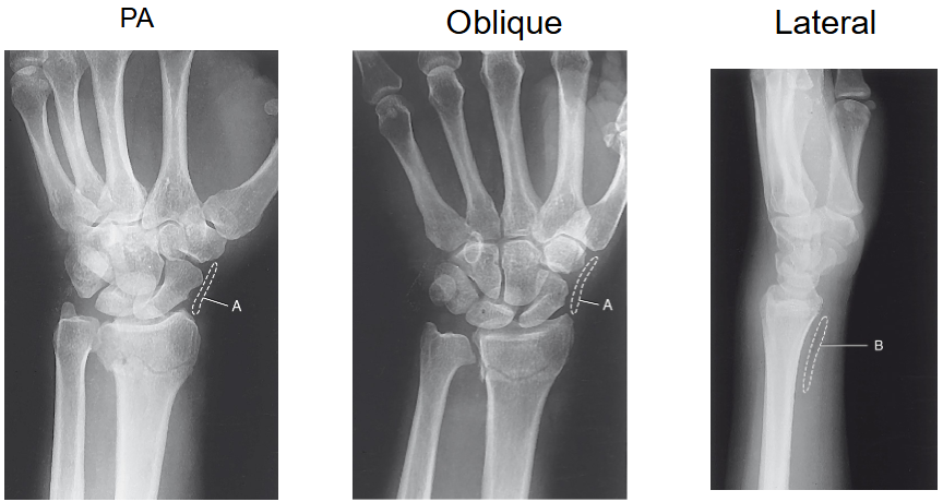
Label The Fat Pads & Stripes Pt. 1
A. Scaphoid Fat Stripe
B. Pronator Fat Stripe

Label The Fat Pads & Stripes Pt. 2
C. Anterior Fat Pad
D. Posterior Fat Pad
E. Supinator Fat Stripe
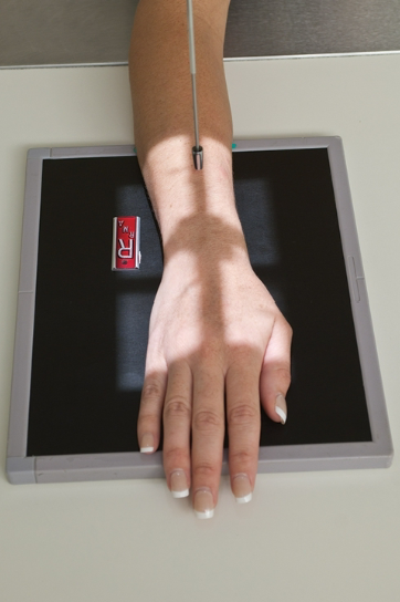
What Is This Projection?
PA Wrist Projection
PA Wrist Projection
patient is seated with arm extended
drop shoulder so it’s in the same plane as wrist
arch hand slightly so that carpals are in contact with IR
CR to midcarpal area
Evaluation Criteria
distal radius, ulna & carpals demonstrated
separation of distal radius & ulna
center of field at midcarpals
no rotation
exposure factors

What Is This Projection?

AP Wrist Projection
AP Wrist Projection
ALTERNATE TO PA WRIST
intercarpal spaces & wrist joint visualized better in AP position

What Is This Projection?
PA Oblique Wrist
PA Oblique Wrist
45° lateral rotation
may flex fingers to offer support if sponge is not available
CR to midcarpal area
Evaluation Criteria
distal radius, ulna & carpals demonstrated
head of ulna partially superimposed by distal radius
center of field at midcarpals
trapezium seen in its entirety
exposure factors


What Is This Projection?
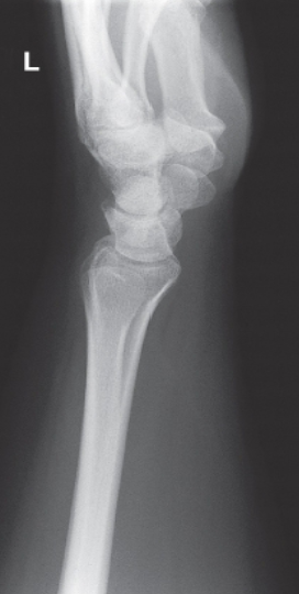
Lateral Wrist
Lateral Wrist
hand & wrist in true lateral
should in the same plane as wrist
CR to midcarpal area
Evaluation Criteria
distal radius, ulna & carpals demonstrated
center of field at midcarpals
no rotation
ulnar head superimposed over distal radius
metacarpals superimposed
exposure factors
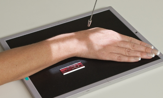
What Is This Projection?
PA Axial Scaphoid Ulnar Deviation
PA Axial Scaphoid Ulnar Deviation
evert hand toward ulnar side without moving forearm
CR angle 10°-15° toward elbow
angled proximally along long axis of forearm toward elbow
pointed at scaphoid
Evaluation Criteria
scaphoid clearly seen without superimposition
scaphoid not foreshortened
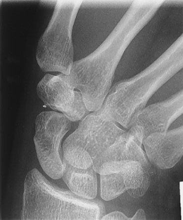
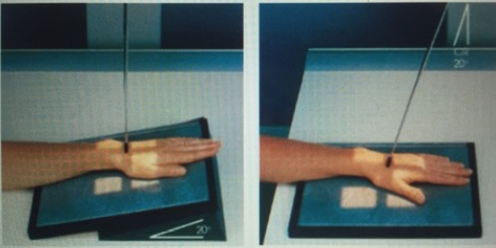
What Is This Projection?
PA Axial: Stecher Method
PA Axial: Stecher Method
CR to scaphoid
angle CR 20° toward elbow OR elevate image receptor 20°
MODIFIED STECHER: includes ulnar deviation
Evaluation Criteria
distal radius, ulna & carpals demonstrated
separation of distal radius & ulna
center of field at scaphoid
no rotation
exposure factors


What Is This Projection?
Radial Deviation
Radial Deviation
CR to midcarpals
invert hand toward radius


What Is This Projection?
Carpal Canal/Gaynor Hart Method/Tangential, Inferosuperior Projection
Carpal Canal/Gaynor Hart Method/Tangential, Inferosuperior Projection
ask patient to hyperextend (dorsiflex) wrist as much as possible (without lifting forearm/wrist)
rotate hand and wrist 10° internally (toward radius)
CR 25°-30° to hand
Evaluation Criteria
carpal sulcus demonstrated
pisiform & hamate separated
scaphoid in profile
exposure factors

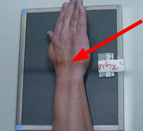
What Is This Projection?
Oblique Wrist with Medial Rotation
Oblique Wrist with Medial Rotation
hand rotated interally
Evaluation Criteria
triquetrum, hamate & pisiform well visualized

What Is This Positioning?
AP Forearm
AP Forearm
drop shoulder to be in same plane as forearm
epicondyles parallel to IR
patient to lean laterally to place forearm in true lateral position
CR to midforearm
include 1-1.5” beyond wrist & elbow joints on IR
Evaluation Criteria
carpals to distal humerus included
humeral epicondyles are in profile
slight superimposition of distal radioulnar joint
exposure factors

What Is This Positioning?
Lateral Forearm
Lateral Forearm
elbow flexed 90°
drop shoulder to same plane as forearm
wrist true lateral
CR to midforearm
for very muscular forearms, place support under hand & wrist as needed
Evaluation Criteria
carpals and distal humerus
included
elbow flexed 90°
head of ulna superimposed over radius
coronoid process superimposed by radial head
exposure factors


What Is This Positioning?
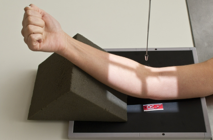
AP Elbow Fully Extended
AP Elbow Fully Extended
epicondyles parallel to IR
lean slightly laterally for true AP
interepicondylar plane parallel to IR
CR to mid-elbow joint
Evaluation Criteria
distal humerus, elbow joint space & proximal radius and ulna demonstrated
radial head, neck & tubercles separated or slightly superimposed by ulna
olecranon process seated in olecranon fossa

What Is This Positioning?
AP Elbow Partially Flexed (humerus parallel)


What Is This Positioning?
AP Elbow Partially Flexed (forearm parallel)


What Is This Positioning?
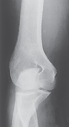
AP Elbow Medial Oblique
AP Elbow Medial Oblique
45° internal (medial) rotation, hand pronated
CR perpendicular to mid-elbow
Evaluation Criteria
coronoid process in profile
trochlea & medial epicondyle in profile
olecranon process seated in olecranon fossa

What Is This Positioning?
AP Elbow Lateral Oblique
AP Elbow Lateral Oblique
45° internal (medial) rotation, hand pronated
CR perpendicular to mid-elbow
Evaluation Criteria
radial head, neck & tuberosity in profile, free of superimposition by ulna
lateral epicondyle & capitulum in profile


What Is This Positioning?
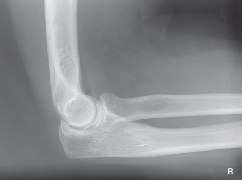
Lateral Elbow
Lateral Elbow
elbow flexed 90°
shoulder in same plane as elbow
wrist in true lateral position
provide support under wrist/hand for heavily muscled forearms
CR to mid-elbow joint
1 ½” medial to olecranon process
Evaluation Criteria
three concentric arcs visible
olecranon process in profile
½ of radial head superimposed by coronoid process
optimal exposure factors

What Is This Positioning?
Trauma Axial Lateromedial Coyle Method for Radial Head
Trauma Axial Lateromedial Coyle Method for Radial Head
CR to mid-elbow joint
CR 45° toward shoulder
90° flexion
for radial head
increase kVp
Evaluation Criteria
radial head & capitulum joint space open
radial head, neck & tuberosity in profile & free of superimposition

What Is This Positioning?
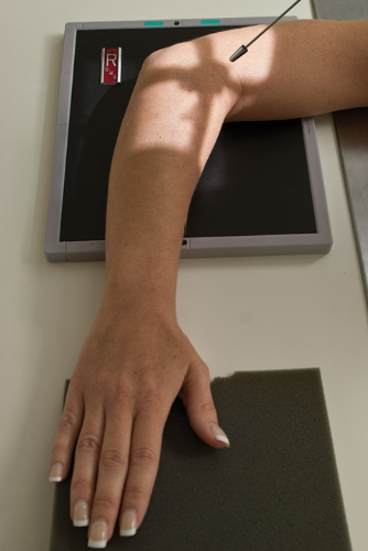
Trauma Axial Lateromedial Coyle Method for Coronoid Process
Trauma Axial Lateromedial Coyle Method for Coronoid Process
CR to mid-elbow joint
CR 45° toward shoulder
80° flexion
for coronoid process
Evaluation Criteria
coronoid process & trochlea joint space open
radial head & neck superimposed by ulna
optimal exposure factors
