M6 Roth- Extracellular Vesicles I + II: Discovery, Biogenesis, Transport and Function
 Call Kai
 Learn
 Practice Test
 Spaced Repetition
 Match
 Flashcards
 Knowt Play
1/15

There's no tags or description
Looks like no tags are added yet.
16 Terms
what are extracellular vesicles?
cells from all 3 domains produce EVs, by a non-canonical secretory pathway
these are vesicles involved in intercellular (not intracellular) cargo transport- for RNAs, proteins, metabolites and even DNA
they selectively load cell-specific cargo, protect it from extracellular degradation, and can target delivery to other cells, causing phenotypic changes
they are also found in all biofluids (blood, urine, milk, etc)
they are very small nanoparticles, about the same size as viruses
there are two major subtypes (but it is difficult to assign these to an EV unless its origin has been observed or we have EV-specific biomarkers):
small EVs/exosomes- derived from the endocytic pathway, by inward budding from the plasma membrane
large EVs/microvesicles- formed through outward budding of the PM, poorly understood but involves many of the same components (primarily tetraspanin webs)

how are small extracellular vesicles (endosomes) produced?
exosomes are derived from the endocytic pathway, by the receptor-mediated inward budding of the plasma membrane
inward budding produces an early endosome
these endosomes specifically take up cytosolic contents, including proteins, RNAs, DNA + metabolites, into intraluminal vesicles (ILV), to form a multivesicular body (MVB)
instead of fusing with lysosomes (for degradation at the vacuole, like the normal endocytic pathway), some MVBs fuse with the plasma membrane to release these extracellular vesicles out of the cell

how were extracellular vesicles discovered?
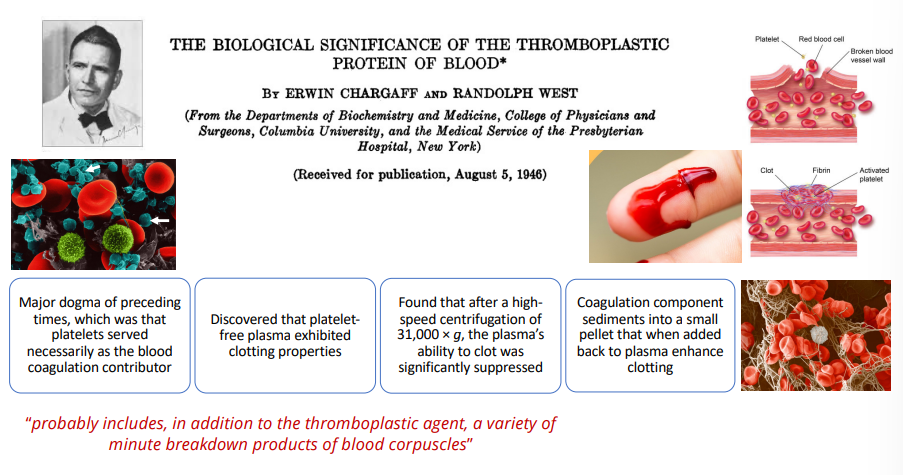
by coincidence, during a study on the clotting factors of blood
before this we believed that platelets were the primary blood coagulation factor, however the removal of platelets from plasma still resulted in clotting
centrifugation of the plasma (removing EVs), removed the clotting properties of blood- and adding the sediment back restored them
in fact, platelets release EVs that induce clotting (rich in phospholipids, ATP and contractile proteins)
EVs were finally identified using transmission electron microscopes, but were initially considered as artefacts, or cellular debris
how do we know EV packaging is selective and functional?
we know EV packaging is selective because mast cells EVs take up miRNA + mRNA, but not rRNA (not just generalised uptake of RNA)
this RNA is protected against RNAase degradation inside the vesicles
if EVs contain miRNAs, this means they could regulate phenotypic changes in a target cell by uptake of the exosomal contents, and miRNA binding to down-regulate target mRNA → cell-cell communication
this is exceptionally selective, since specific miRNA complements are exclusively packaged → with known functions in mast cell activity eg. tumorigenesis
mRNA cargo has been shown to be taken up by recipient cells, where they get translated- for example in glioblastoma (malignant tumour) cells, to increase angiogenesis (vascular tube formation) → can be used as a diagnostic marker

by what pathways is cargo specifically loaded into small extracellular vesicles (exosomes) during their biogenesis?
multiple pathways can be involved in small EV (exosome) production by intraluminal vesicle formation in multivesicular bodies (there is a lot of overlap between these pathways):
ESCRT-dependent pathway- canonical + default
syndecan-syntenin-ALIX pathway- non-canonical
tetraspanin (TET) pathway- non-canonical, conserved across all domains, this is the biomarker of EVs
ceramide pathway- non-canonical
how is cargo specifically loaded into small extracellular vesicles (exosomes) by the ESCRT-dependent pathway?
multiple pathways can be involved in small EV (exosome) production by intraluminal vesicle formation in multivesicular bodies (there is a lot of overlap between these pathways):
ESCRT-dependent pathway- canonical + default
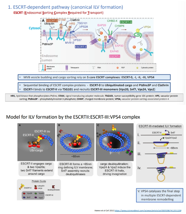
ESCRT → endosomal sorting complex required for transport
ESCRT-0 detects and binds ubiquitinated proteins targeted for vacuole degradation, as well as the EE membrane
this sequentially interacts with other ESCRTs (II, I, III) and clathrins to cluster cargo together
ESCRT-III defines the curvature and size of the ILV by forming a cargo-sequestering ring/spiral, and VPS4 cuts it off from the EE membrane
how is cargo specifically loaded into small extracellular vesicles (exosomes) by the syndecan-syntenin-ALIX pathway?
multiple pathways can be involved in small EV (exosome) production by intraluminal vesicle formation in multivesicular bodies (there is a lot of overlap between these pathways):
syndecan-syntenin-ALIX pathway- non-canonical
this pathway occurs independently from the canonical ESCRT biogenesis pathway involving ubiquitin and ESCRT-0, I and II
syndecan proteoglycans in the EE membrane act as coreceptors, binding to G-protein-coupled receptors (growth factor receptors) and to syntenins, via their cytosolic domain
these syntenins bind to each other and to tetraspanins to cluster, and recruit ESCRT-III components using the ALIX connecting protein
this triggers the formation of ILVs

how is cargo specifically loaded into small extracellular vesicles (exosomes) by the tetraspanin (TET) pathway?
multiple pathways can be involved in small EV (exosome) production by intraluminal vesicle formation in multivesicular bodies (there is a lot of overlap between these pathways):
tetraspanin pathway- non-canonical, conserved across all domains, this is the biomarker of EVs
this pathway occurs independently from the canonical ESCRT biogenesis pathway
tetraspanins contain 4 TM domains across the EE membrane and a large extracellular loop
these form tetraspanin webs/nanodomains at the membrane, binding to each other and to various partner proteins

how is cargo specifically loaded into small extracellular vesicles (exosomes) by the ceramide pathway?
multiple pathways can be involved in small EV (exosome) production by intraluminal vesicle formation in multivesicular bodies (there is a lot of overlap between these pathways):
ceramide pathway- non-canonical
ceramides are produced from sphingomyelin lipids in the membrane, by removing a large head group component
reducing the size of the head group causes structural negative curvature of the membrane

how are multivesicular bodies transported intracellularly to the plasma membrane?
instead of being transported to the lysosome + vacuole for degradation, some MVBs are transported to the plasma membrane to release their intraluminal vesicles as extracellular vesicles
MVBs are transported via microtubule motors (dynein + kinesin) to the plasma membrane, mediated by Rab GTPases
specific Rabs (7) are involved in direction to the lysosome, and others (27 + 35) are involved in direction + docking to the PM (alongside SNAREs etc), acting as a secretory vesicle

what are the mechanisms of extracellular vesicle uptake?
this process is quite poorly understood
proteins on the surface of EVs can either trigger uptake by membrane fusion and EV content release, or just direct changes in the target cell by target docking
endocytosis of EVs by recipient cells can be mediated by clathrin in the regular endocytic pathway, triggered by receptors on the EV surface (even inter-organismally)
phagocytosis and micropinocytosis can also be involved
how are extracellular vesicles involved in RBC iron uptake?
this was the experimental evidence for EVs being involved in secretion by the endocytic pathway
reticulocytes are immature red blood cells produced by the bone marrow
these have transferrin receptors (TfRs) in their plasma membrane that bind transferrin (Tf) bound to two iron
this triggers receptor-mediated endocytosis (using a clathrin coat) into the RBC to form an early endosome containing a membrane iron transporter and a proton pump
maturation of the early endosome into the late endosome, producing a more acidic lumen (influx of H+), causes iron to dissociate from Tf and be exported into the cytosol
Tf and TfR are recycled back to the plasma membrane and extracellular space
during reticulocyte maturation TfR are lost from the PM by internalisation into intraluminal vesicles and export as part of EVs, once the desired iron levels are achieved (30 minutes)

how are extracellular vesicles involved in reproductive physiology?
EVs are involved in sperm + egg development, implantation and maternal signalling, but we will focus on sperm motility and fertilisation
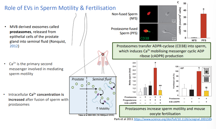
prostasomes (exosomes/small EVs) are released from the prostate gland into the seminal fluid
these increase sperm motility because they transfer a calcium uptake toolkit, increasing the intracellular calcium concentration and causing hyperactivation
this also increases the ability of sperm to fertilise the egg
how are extracellular vesicles involved in pathology and the immune response?
in certain COVID-19 patients, the EVs express an initiator of blood coagulation as a surface marker
these have biological activity as a clotting agent in the infection
this can also be used as a diagnostic marker for COVID-19 prognosis
EVs are also released by white blood cells to cause an immune response in T cells
this was the first experimental evidence for the biological function/activity of EVs → use in therapeutics

what are some examples of how extracellular vesicles are involved in non-mammalian systems?
a lot of skepticism initially because many didn’t believe EVs could permeate a fungal or plant cell wall
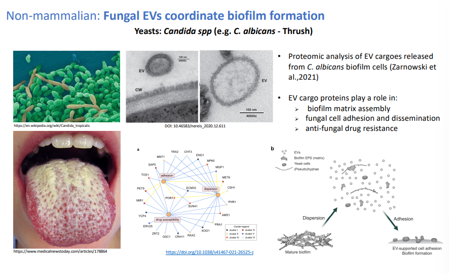
fungal EVs coordinate biofilm formation, fungal cell adhesion + antifungal drug resistance
EVs are specifically regulated + induced in the plant immune response- intercellularly, to transport defence-related proteins, and in plant-pathogen interactions, containing siRNAs to suppress virulence
at the same time, the pathogen produces EVs to suppress immunity in the plant host, in a continual arms race to suppress each other’s phenotypic immune responses
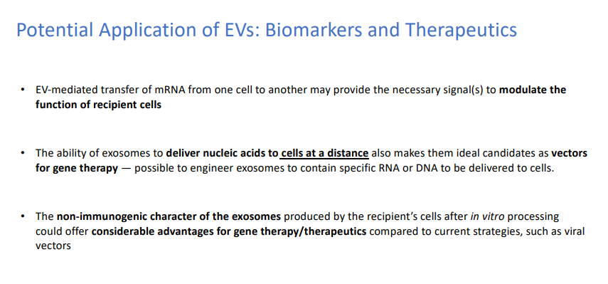
why can extracellular vesicles be used in therapeutics and diagnostics?
EVS are able to modulate the function of recipient cells, causing phenotypic changes → eg. released by white blood cells to induce an immune response in T cells
EVs can be used as diagnostic markers because the cargo they contain is specific and characteristic of the donor, eg. for tumour cells (since tumour-specific mRNA variants and miRNAs in EVs released by glioblastomas can be detected in serum), eg. for COVID-19 (blood clotting factor surface marker on EVs of infected patients)
Evs can be transported across large distances, so they are ideal candidates for vectors, eg in gene therapy for specific nucleic acid delivery
EVs produced by the patient’s cells are non-immunogenic