applied animal and health welfare - conduction system and output
 Call Kai
 Learn
 Practice Test
 Spaced Repetition
 Match
 Flashcards
 Knowt Play
1/26

There's no tags or description
Looks like no tags are added yet.
27 Terms
cardiac cycle
Contraction and relaxation of heart muscle
Systole –
contracting causing chamber to empty
Diastole –
relaxing and repolarising following contraction allowing chamber to fill with blood
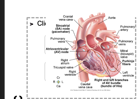
impulse for
heartbeat comes from sinoatrial (SA) node
located in wall of right atrium
pacemaker
charged atoms - cation
positively charged -
sodium (na+)
potassium (k+)
calcium (ca++)
anions
negatively charged
chloride (cl-)
polarisation
A SA node generates electrical current
-by movement of cations across the outer membranes of its cells
Cations are pumped out of the cell in a process called polarisation
results in outside of the cell having more positive charge than the inside
depolarisation- gates in
cell wall opened
cations fow into cell to equalise charge on either side of cell membrane
depolarisation generates an electrical current
-causes heart muscle to contract
systole
repolarisation
SA node automatically repolarises itself
then depolarises again
diastole
cardiac cycle - 1) pacemaker generates
wave of signals to contract

2 signals are delayed
at AV node

3)signals pass
to heart apex

4) signals spread
throughout ventricles
electrocardiogram (ECG/EKG)
Cardiac currents are the result of an electrical impulse
An instrument that detects electrical currents may be used to measure electrical activity of the heart
Electrocardiograph
Measures electrical current using metal electrodes attached to the skin
To produce an electrocardiogram

Correct lead placement vital
RED-
yellow-
green
black
N.B. In the UK, most commonly:
Red = Right forelimb
Yellow = Left forelimb
Green = Left hindlimb
Black = Right hindlimb

heart rate
Heart has to move adequate amount of blood to tissues in body
•To deliver oxygen and nutrients
•To remove waste products
heart must maintain sufficient
cardiac output to adequately perfuse tissues and meet these aims
heart rate and cardiac output
stroke volume
cardiac output
heart rate
calculation including
Cardiac Output (CO) = Stroke Volume (SV) x Heart Rate (HR)
larger animals have
larger stroke
slower
much greater tissue
larger hearts
volumes
-more blood pumped per beat
heart rates
mass to supply
heart rate based
in part on rate which SA node spontaneously depolarises
many factors can influence cardiac output and heart rate
stroke volume is a measure of
the volume of blood ejected from the left ventricle during one contraction or sytole
. If the cardiac output and stroke volume both decrease what has happened to the heart rate to maintain equilibrium?
it will increase
How could mitral valve stenosis affect the stroke volume?
Mitral valve stenosis impairs left ventricular filling, so there is a decrease in diastolic volume. This leads to a decrease in stroke volume.