Nursing 4 Exam II
 Call Kai
 Learn
Learn Practice Test
 Spaced Repetition
 Match
Match Flashcards
 Knowt Play
1/23

There's no tags or description
Looks like no tags are added yet.
Name | Mastery | Learn | Test | Matching | Spaced | Call with Kai | Chat |
|---|
No analytics yet
Send a link to your students to track their progress
24 Terms
determine if the following are associated with endocarditis or pericarditis
pericarditis
inflamation around the heart, is not always cuased by bacteria could be viral or fungal
signs: pain worsens with deep breathing, pain alleviated by leaning foward, pericardial friction rub, osler node, janeway lesion, roth spots
treated with colchincine, NSAIDS, and aspirin, pericardiocentesis
pericardial effusion- excess fluid in the pericardial sac, effects ventricle filling and ejection, cardiac tamponade=so much fluid its emergent to remove it (pericardiocentesis)
-pericardiocentesis alone often does not resolve acute tamponade. be prepared to provide adequate fluid volumes to increase cardiac output, and to prepare the patient for surgical intervention if tamponade recurs
-pulsus paradoxus-when sbp decreases by more than 10 during inspiration can see on a line

endocarditis
may occur after a strep throat infection—> rhumatic fever→ goes to endocardium
signs: presents with fever, petechiae, and joint pain
increased for iv drug users (this layer is connected to th blood stream)
testing: an echocardigram(transesophageal:back of throatd camera down to heart, transthoracic: camera outside of thoracic cavity). will shows structural abnormalities in the hearts structure
treated with antibiotics- long course and high dose
-monitor your wieghht daily and immediatley report a sudden increase in leg swelling or sob since at high risk for HF
The nurse is assessing a cardiac patient and notes the patient has a pericardial friction rub. How would the nurse describe the sound of a pericardial friction rub?
scratchy, high pitched sound
osler node for pericarditis

janeway lesion for pericarditis

roth spots for pericaritis
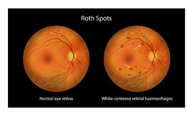
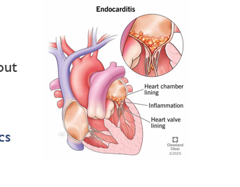
vegetation in endocarditis
damanged lining
-can become big, what out for clots
-eat away at valves—> regurgitation
-need blood cultures for antibiotics
-can lead to valvular disorders
-if bacteria get in through our mouth is sticky it cuases an increased risk( take antibiotics before dental procedures

the parts of the heart labeled
supeior an inferior vena cava
right atrium
tricuspid valve (opens during diaystole)
right ventricle
pulmonic valve (opens during systole)
pulmonary arteries (carries blood away from the heart)
lungs
pulmonary veins (back to the heart)
left atrium
mitral valve (during dyastole)
left ventricle
aortic valve (during systole)
aorta (to the rest of the body)
regurgitation vs. stenosis
regurgitation- cant fully close
stenosis- cant fully open

mitral valve stenosisis/reguritation
stenosis- causes atrial dilation bc the atrial muscle cannt compensate by becoming stronger like a ventricle..decreased afterload as it is not getting blood and decrease preload. SVR is unchanged or increased but think about what is does to the RV.. increases preload
-blood will go back to the lungs through the pulmonary veins, will shows signs of pulmonary congestion
-muscle aroudn the aorta will stretch
-could cuase a fib
afterload- decreased
preload-decreased
SVR- unchnaged/increased
reguritation-does NOT cause increased afterload as the LV becomes hypertrophied and dilated it is pushing blood through a working aortic valve and a low resistance mitral valve so normal/decreased afterload but increased preload and svr is normal to increased
-left atrium letting blood drop down and not holding what its suppossed to
-not alot of resistance (afetrlaod) the blood is flopping around
-muscle layer of left ventricle is going to dilate to accomidate for the excesisve blood which will make ti weaker
afterload- normal/decreased afterload
preload-increased(alot in the left ventricle)
SVR-normal/increased

aortic valve regurgitation and stenosis
stenosis- pressure overload, the heart muscle thickens—> ventricle space narrows—> at final stage of HF the muscle might end up strecthing like AR.. very icnreased afterload, svr unchnaged or increased
-pressure problem
-oxygenated blood stuck in the left ventricle
-blood backs uip and goes to the lungs
afterload- increased
preload- increased
SVR- unchanged/increased
regurgitation- volume overload—> heart muscle dilate and chamber gets bigger and weaker… still increased afterload becuase ventricle is tyring to push the extra blood through a hig pressure aorta. increased preload. icnreased SVR
-volume problem
afterlod- increased a little
preload-increased
SVR-increased

murmurs
systolic: MR.PASS
MR= mitral regurg
P= physiologic=benign murmurs
AS= aortic stenosis
S= systolic
Diastolic: ARMS
A= aortic
R= rgeuritation
M= mitral
S= stenosis
Mitral stenosis: apical/diastolic murmur(5)
Mitral reguritation: apical diastolic murmur(5)
Aortic stenosis: crescendo/decrescendo systolic murmur (1)
aoritc reguitation: diastolic high pitch/bloeing decrescendo (3)
tricuspid stenosis: diatsolic murmur (4)
tricuspid reguritation: blowing systolic murmur (4)

what should the aortic valve do during dyastole
aortic valve is suppsoed to close
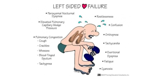
which valvular disorder can lead to left sided heart failure
cuased by MVS, MVR, AVS, AVR
expect : diuretics, ace, arb, blood thinner
goes back to lungs
which valvular disorder can lead to right sided heart failure
caused by TVS, TVR, PVS, PVR systemic congestion
meds: diuretics, would not use ace/dilators becuase it odesnt effect our veins/pumonary artery/lung vasculature like it does our arterial vasculature, it will just lower bp and not help

valve repair
MR:
-annuloplasty(makes valve ring smaller)
-transcatheter repair (clip)
MS:
-balloon valvuloplasty (no ewuipment left behind)

meds that effect preload, afterlaod, and stroke volume

heart valve replacment
mechanical heart valve-
-expetced ot last 20-30 years
-anticoagulants needed for like
bioprosthetic heart valve-
-low thrombogenicity
-must be replaced every 5-7 years
young patients getting mechanical valves, if it’s R sided the low pressure increased risk for clots so may opt for bioprosthetic even if younger pt
-try to repair first than replace
Henry is admitted with symptoms of systemic fluid volume overload. Which diagnostic test is the priority for the nurse to anticipate to confirm valvular disease?
transthoracic echcardiography
tricuspid valve stenosis/ regurgitation
stenosis-
afterload-decreased
preload- decreased
pulmonary vascular resistance- increased
-blood goes back into the peripheral system (cuasing peripheral edema)
regurgitation-
afterload- decreased
preload- increased
PVR- increased (BIG with IVDU bc the bacteria is introcuded here first)
patient would have labored breathing walking, jvd, distended abdomen, pitting edema, systolic murmur
indicated med order: furosemide, spironolactone
contriindicated: diltiazem, fluids, lisinopril

With repeat imaging we see that he still has severe tricuspid regurgitation and his right ventricle has become dilated.
The team decides that Henry is stable enough to consider a valve repair, but the leaflets are too damaged, and they decide to do a valve replacement. Which type of valve replacement would be considered for Henry?
a bioprosthetic valve becuase it carries a lowerr risk of valve thrombosis in a low pressure chamber
-left side of heart has alot of pressure, right side has lower pressure
He is ambulating well, his edema has resolved, and the nurse is preparing for discharge. Which statement by Henry indicates a need for further instruction?
A.“I need to weigh myself every morning before breakfast and call the clinic if I gain more than 3 pounds in a day.”
B.“I will make sure to take my blood thinner every day and get my blood drawn for monthly lab tests for the rest of my life.”
C.“I must inform my dentist about my valve replacement because I will need antibiotics certain dental procedures.”
D.“I will wash my chest incision gently with mild soap and water every day, pat it completely dry, and avoid putting any lotions or ointments on it.”
i will make sure to take my blood thinner every day and get my blood drawn for monthly lab tetss for the rest of my life
-dont need blood thinner