Circulatory System Week 1
 Call Kai
 Learn
 Practice Test
 Spaced Repetition
 Match
 Flashcards
 Knowt Play
1/78

There's no tags or description
Looks like no tags are added yet.
79 Terms
apex of the heart
portion: bottom part of the heart

base of the heart
portion: top part of the heart excluding vessels

right atrium of the heart
top right

right auricle of the heart
flap on right side slightly posterior

left atrium of the heart
top left

left auricle of the heart
flap on left side

right ventricle of the heart
bottom right

left ventricle of the heart
bottom left

anterior surface of the heart
region: anterior/front of the heart

diaphragmatic surface of the heart
region: the bottom

right pulmonary surface of the heart
region: right side touching lung

left pulmonary surface of the heart
region: left side touching lung

right border of the heart
(blue) right side

left border of the heart
(orange) left side

inferior border of the heart
(purple) below
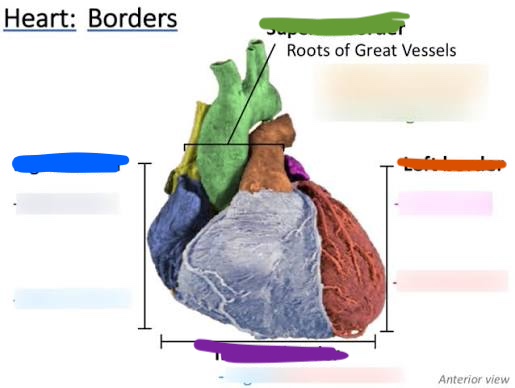
superior border of the heart
(above) green

coronary sulcus
depression: separates atrium + ventricles. wraps around entirely

anterior interventricular sulcus
divit on anterior side of heart

posterior interventricular sulcus
depression on posterior side of heart

aorta
the larger vessel

ascending aorta
the start/connects to atrium

arch of aorta
the arch

pulmonary trunk
on bottom

left pulmonary artery
shown on posterior side. regular left

right pulmonary artery
shown on posterior side. regular right

ligamentum arteriosum
in between aortic arch and pulmonary trunk. webbing. shown on body

superior vena cava
top part of vein (top of straw)

inferior vena cava
bottom part of vein (bottom of straw)

left superior pulmonary vein
(yellow) top left vein

left inferior pulmonary vein
(green) bottom left vein

right superior pulmonary vein
(purple) top right vein
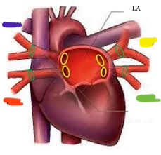
right inferior pulmonary veins
(orange) bottom right vein

right coronary artery
right above probe =probe in heart

right marginal artery
hanging artery on right side ( shaped

posterior interventricular artery
structure: inside posterior interventricular sulcus

left coronary artery
connects to pulmonary trunk, under probe

anterior interventricular artery
structure: inside anterior interventricular sulcus

circumflex artery
it follows down the anterior interventricular artery

1) right atrium 2) most of the right ventricle 3) part of the left ventricle 4) the SA and AV nodes
what does the right coronary artery supply
1) left atrium 2) part of the right ventricle 3) part of the right ventricle 4) interventricular septum
what does the left coronary artery supply
coronary sinus
internal structure: probe through thin skin

great cardiac vein
left lateral side shown from posterior side

middle cardiac vein
inferior to where coronary sinus is

fibrous pericardium
most external layer

parietal layer of the serous pericardium
(purple) outer layer

visceral layer of the serous pericardium
(green) directly on the heart; most internal

serous pericardium
(purple and green) collective

pericardial cavity
BQ ONLY: a potential space surrounding the heart between the parietal and visceral layers of serous pericardium
transverse pericardial sinus
BQ ONLY: behind the ascending aorta and pulmonary trunk- connects the left and right sides of the pericardial cavity
Endocardium
(green) inner most layer. traces inside

Myocardium
(purple) thick middle layer

Epicardium
(blue) outer most layer

interatrial septum
structure: pinching between two atriums

fossa ovalis
BQ: oval depression in the interatrial septum indicating the location of the fetal foramen ovale- where blood was shunted from right atrium to left atrium in the fetus
bean sitting in window. internal of right atrium

right atrium
chamber on right upper side

right auricle
ear like projection that lines artery

pectinate muscle
BQ: pectinate=comb shaped. the muscle has ridges like the teeth in a comb
the webbing/tree like muscle in right atrium

sinus venarum
shiny surface in right atrium

opening of the superior vena cava
space above probe/ probe through vena cavas

opening of the inferior vena cava
space below probe

opening of the coronary sinus
below fossa ovalis. probe inside

left atrium
chamber on left side

left auricle
elephant ear thing on left side

openings of the pulmonary veins
collective spaces: probes in back

interventricular septum
collective structure: thick muscle separating ventricles

muscular part of the interventricular septum
portion: lower

membranous part of the interventricular septum
portion: higher up

right ventricle
chamber on lower right side

right atrioventricular orifice
(doorway) space; opening from right atrium to right ventricle

tricupsid value
on right side (closed door)

papillary muscle
the carrot part

right tendinous cords
BQ: heart strings
leafy bit/ stringy hairs
NEED TO SPECIFY THE SIDE OF THE HEART

left tendinous cords
BQ: heart strings
leafy bit/ stringy hairs
NEED TO SPECIFY THE SIDE OF THE HEART
