Eukaryotic Module 6
 Call Kai
 Learn
Learn Practice Test
 Spaced Repetition
 Match
Match Flashcards
 Knowt Play
1/72

There's no tags or description
Looks like no tags are added yet.
Name | Mastery | Learn | Test | Matching | Spaced | Call with Kai | Chat |
|---|
No analytics yet
Send a link to your students to track their progress
73 Terms
chloroplast
located predominately in mesophyll cells of leaves
chloroplasts of higher plants are generally lens-shaped
approximately 2–4 μm wide and 5–10 μm long, and typically numbering 20–40 per cell
their dimensions make chloroplasts giants among organelles (as large as a mammalian blood cell)
chloroplasts arise by fission from preexisting chloroplasts or their nonpigmented precursors called proplastids
outer covering of a chloroplast consists of an envelope composed of two membranes separated by a narrow space
outer membrane of the envelope contains several different porins that exhibit selectivity toward solutes
inner membrane of the envelope is highly impermeable, substances moving through this membrane do so only with aid of variety of transporters
thylakoid membrane: internal membrane of the chloroplast organized into flattened membranous sacs
much of the photosynthetic machinery is part of the internal membrane: light-absorbing pigments, complex chain of electron carriers, ATP synthesizing apparatus
arranged in orderly stacks called grana
space inside a thylakoid sac is the lumen
space outside the thylakoid and within the chloroplast envelope is the stroma, which contain enzymes responsible for carbohydrate synthesis
contains chlorophyll molecules and protein complexes that compromise the energy-transducing machinery of the chloroplast
high protein content and unusual in having relatively little phospholipid
high percentage of galactose-containing glycolipids
both fatty acids of these lipids contain several double bonds, making the lipid bilayer of the membrane highly fluid
fluidity of the bilayer facilitates lateral diffusion of protein complexes through the membrane during photosynthesis
stroma: contains small double stranded circularf DNA molecules and prokaryotic like ribosomes
DNA of a chloroplast is a relic of the genoome of an ancient bacteiral endosymbiont
mitochondria vs chloroplast
outer membrane of mitochondrion and outer membrane of chloroplast contain several different porins
matrix of mitochondria and stroma of chloroplast contain small double stranded circularf DNA molecules and prokaryotic like ribosomes
both have a double membrane
both generate ATP using electron transport chain and proton gradient
mitochondria is found is almost all eukaryotic cells while chloroplasts only in plants and algae
inner membrane of mitochondrion is folded inward to form cristae while chloroplast inner membrane is smooth and encloses a third internal thylakoid membrnae system
evolution of earth’s life forms
the earliest life forms were heterotrophs that depended on an external source of organic compounds, which were formed abiotically.
the appearance of autotrophs, which manufactured their own organic nutrients from the simplest inorganic molecules, such as carbon dioxide and hydrogen sulfide, tremendously boosted the evolution of life
two types of autotrophs
utilize the energy stored in inorganic compounds such as ammonia, hydrogen sulfide, or nitrites.
utilize the radiant energy fromt he sun
needs more energy since the oxygen atom has much stronger affinity for its electrons
cyanobacterium
cyanobacteria switched from using hydrogen sulfide to using water as their source of electrons, which required evolving a very strong oxidizing agent
this process produced molecular oxygen as a waste product, changing Earth's atmosphere and life.
symbiotic evolution of chloroplasts
An ancient O2-producing cyanobacterium took up residence inside a primitive eukaryotic host cell and gradually transformed into the chloroplast organelle
Most genes from the cyanobacterium were transferred to the plant cell's nucleus; therefore, polypeptides in modern chloroplasts are encoded by both the nuclear and chloroplast genomes, and all modern chloroplasts descend from a single, ancient symbiotic relationship.
photosynthetic plants
provides the most of the organic nutrients for the lives on the planet
leaf anatomy

chloroplasts of higher plants
generally lens-shaped
approximately 2 to 4 μm wide and 5 to 10 μm long
numbering 20-40 per cell
membranes of chloroplast
outer membrane
inner membrane
internal membrane

outer membrane
similar to the outer membrane of an mitochondrian
the outer membrane of a chloroplast envelope contains porins
porins are integral proteins that have a relatively large channel surrounded by a barrel of b strands.
exhibit selective permeability towards various solutes

inner membrane
highly impermeable
substances moving through the inner membrane needs the aid of a variety of transporters
internal membrane
arranged in stacks of disc-like thylakoids that are physically separate from the outside double membrane that forms the envelope.
contains much of the photosynthetic machinery
light-absorbing pigments
complex chain of electron carriers
ATP-synthesizing apparatus

thylakoids
flattened membranous sacs
space inside a thylakoid sack is the lumen
space outside the thylakoid and within the chloroplast envelope is the stroma
thylakoids arranged in orderly stacks are called grana thylakoids

chloroplast structure

membranes + spaces in chloroplasts

thylakoid membranes
high protein content, relatively little phospholipid
contain high percentage of galactose-containing glycolipids
both fatty acids of these lipids contain several double bonds, making the bilayer highly fluid

chloroplast genome
after taking residence in a proeukaryotic cell, most chloroplast genes were lost or transferred into the nuclear genome
only small, double-stranded, circular DNA molecules are present in the chloroplast stroma
depending on the organism, chloroplasts contain between 60 to 200 genes involved in either gene expression or photosynthesis
vast majority of chloroplast proteins (200-3500) are encoded by nuclear DNA synthesized in the cytosol and imported into the chloroplast by a specialized transport machinery
chloroplasts are reproduced by fission from preexisting chloroplasts or their nonpigmented precursors- proplastids
photosynthetic metabolism
in 1931, van Niel proposed that oxygen released during photosynthesis of green plants comes from splitting water (H2O) molecules, not from carbon dioxide
van Niel studied sulfur bacteria and green plants
van Niel’s general equation for photosynthesis
CO2 + 2H2A ——> (CH2O) + A2
arrow is light
H2A is oxidized (loses hydrogen)
CO2 is reduced (gains hydrogen to form carbohydrates)
photosynthetic equation for sulfur bacteria and its byproducts
CO2 + H2S ———> (CH2O) + S2
arrow is light
byproduct is elemental sulfur (instead of oxygen like photosynthetic green plants)
H2S is electron/hydrogen donor
photosynthetic equation for green plants
CO2 + H2O —————> (CH2O) + O2
arrow is light
water (H2O) replaces H2S as the hydrogen donor
confirmation of van Niel’s hypothesis
1941- Samuel Ruben and Martin Kamen experiemented on suspensions of green algae using 18O replacing the common isotope 16O
labeled C[18O2] and unlabeled H2O
unlabeled CO2 and labeled H2[18O]
the O2 released from the experiment was labeled, which confirmed van Niel’s hypothesis that light energy is used to split H2O to form O2.
photosynthesis in chloroplast vs aerobic respiration in mitochondrion
photosynthesis in chloroplast is essentially the reverse of mitochondrial respiration

two series of reactions in photosynthesis
light-dependent reactions: energy from sunlight is absorbed and stored as chemical energy in two biological molecules (ATP and NADPH)
light-independent reactions: carbohydrates are synthesized from CO2 using the energy stored in ATP and NADPH molecules produced in light-dependent reactions
absorption of light
photons: packets of energy light travels in
energy cotent of a photon: E=hc/λ
h: Planck’s constant
c: speed of light in a vacuum
step 1 (ground state): electron is at rest in its stable, lowest-energy orbital
step 2 (excitation): when a photon of light hits the molecule, the electron absorbs the photon’s energy, jumping to a higher outer energy shell
step 3 (relaxation): the high energy state is unstable and lasts for a fraction of a second (10^-9 s). the electron rids itself of the excess energy to return to the ground state
once excited, the energy takes one of two paths to return to ground state
fluorescence pathway: chlorophyll will release this energy as heat and a lower-energy photon of light
electron acceptor pathway: the energy released is captured by primary electron acceptors in chloroplasts, which passes the energy in the ETC to photosynthesis

photosynthetic pigments
organisms containing chlorophyll are primary light-absorbing photosynthetic plants
secondary pigments: secondary light collectors that draw excess energy away from excited chlorophyll and dissipate it as heat
chlorophyll molecules are primary light-absorbing pigments while carotenoids are secondary/accessory pigments that support chlorophyll
when a plant absorbs too much sunlight, carotenoids deaw the excess energy away from excited chlorophyll molecules and dissipate it as heat, preventing damage to the cell

structure of chlorophyll
porphyrin ring with conjugated double bonds that function in light absorption
hydrophobic phytol chain that keeps chlorophyll embedded in photosynthetic membrane

structure of B-carotene
a linear system of conjugated double bonds

absorption spectrum for chlorophyll a, b and b-carotene
chlorophylls absorb the strongest in the violet, blue, and red regions
carotenoids absorb into the green region

action spectrum and absorption spectrum of chlorophyll a,b and b-carotene
the red line (action spectrum) indicates the relative efficiency with which light of various wavelengths is able to promote photosynthesis in the leaves of a plant
an action spectrum can be generated by measuring the O2 produced by leaves following exposure to various wavelengths
green line is the combined absorption spectrum of all pigments

light-dependent reactions
light absorbing reactions of photosynthesis occur in large pigment protein complexes called photosystems

chlorophyll molecules in a chloroplast
not all chlorophyll molecules in a chloroplast are directly involved in conversion of light energy into chemical energy
in 1932, Robert Emerson and William Arnold used green algae Chlorella and flashing light of extremely short duration at saturating intensity
one molecule of oxygen was released during a brief flash of light for every 2500 molecules of chlorophyll present
a minimum of 8 photons must be absorbed to produce 1 molecule of O2.
the experiment shows that chlorophyll molecules work together rather than individually
300 molecules of chlorophyll is needed
photosynthetic unit
hundreds of chlorophyll molecules act together as one photosynthetic unit
reaction-center chlorophyll: transfers electrons to electron receptors
the only member of the unit that can do this
the bulk of pigment molecules do not directly participate in the conversion of light energy to chemical energy, but are responsible for light absorption
light-harvesting antenna
pigment molecules form the light light harvesting antenna
absorbs photons of varying wavelength
very rapidly transfers the energy (excitation energy) to the pigment molecule at the reaction center
light-harvesting antenna and reaction-center
transfer of excitation energy from one pigment molecule to another is sensitive to the distance between molecules
chlorophyll molecules of the antenna are kept in close proximity and proper orientation by noncovalent linkage to integral membrane polypeptides
less than 1.5 nm separation
“rule” that operates among antenna pigments is that energy can only be passed to a pigment molecule that absorbs light of equal or longer wavelength/lower energy than that absorbed by the donor pigment
the energy transferred to a chlorophyll of the reaction center absorbs light of longer wavelength than its neighbors
once the energy is received by the reaction center, the electron excited by light absorption can be transferred to its acceptor

oxygen formation
evolution of organisms capable of using H2O as an electron source was accompanied by major changes in the photosynthetic machinery
energetics of oxygenic photosynthesis is the reason for these changes
O2-H2O couple standard redox potential: +0.82 V
NADP+-NADPH couple standard redox potential: -0.32 V
the difference between the redox potentials of these two couples provides measure of minimal energy that the system must absorb to remove an electron from H2O and pass it to NADP+ under standard conditions (1.14 V)
it is estimated that during actual operations in the chloroplast, more than 2 V of energy are untilized to carry out the reaction
one mole of photons of 680 nm wavelength (red light) is equivalent to a change in redox potential of 1.8 V
it is theoretically possible for one photon red light to boost an electron to an energy level required to reduce NADP+ under standard conditions
the process is accomplished, however, through the combined action of two sequestial light-absorbing reactions
photosystems
large pigment protein complexes where light-absorbing reactions of photosynthesis occur
two types of photosystems are required to catalyze the two light-absorbing reactions in oxygenic photosynthesis
photosystem II (PSII): boosts electrons from an energy level below that of water to a midway point
photosystem I (PSI): raises electrons from a midway point to an energy level well above that of NADP+
PSII and PSI act in series
although have different photochemical reactions, they are similar in protein composition and architecture in photosynthetic plants and bacteria
shared properties suggest the photosynthetic reaction centers have evolved from a common ancestral structure

reaction center of the two photosystems
PSII reaction center: a chlorophyll dimer referred to as P680
p for pigment, 68p for the wavelength of light absorbed most strongly by this pair of chlorophyll
PSI reaction center: chlorophyll dimer referred to as P700
when sunlight strikes a thylakoid membrane, energy is absorbed by antenna pigments of both PSII and PSI and passed to the reaction centers of both photosystems
electrons of both reaction-center pigments boosted to an outer orbital, and each electron is transferred to a primary electron acceptor
after losing their electrons, the reaction centers become positively charged and the electron acceptors turn negatively charged
this separation of charge within the photosystems is the light reaction (conversion of light to energy) because positively charged centers act as attractants for electrons, and negatively charged acceptors act as electron donors
separation of charge within each photosystem sets the stage for the flow of electrons along chain of specific carriers
structure of PSII
consists of >20 different peptides most of which are embedded in the thylakoid membrane
two proteins, D1 and D2, together bind the P680 reaction centerchlorophyll dimer and cofactors involved in electron transport
separate pigment-protein complex light-harvesting complex II (LHC II) is situated outside the core of the photosystem
binds both chlorophylls and carotenoids
uses absorbed light energy to generate proton gradient across the thylakid membrane
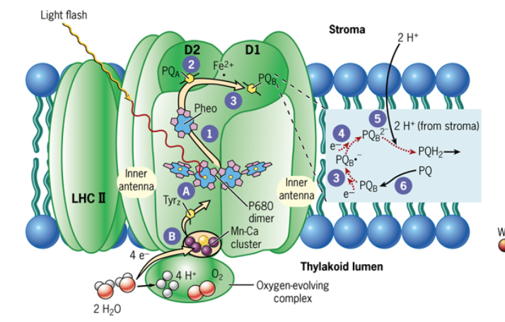
first step: flow of electrons from PSII to plasto-quinone
first step in PSII activation is the absorption of light by an antenna pigment
light energy from antenna pigments to PSII reaction center
the excited reaction center P680* transfers a single photo-excited electron to a closely associated chlorophyll-like pheophytin molecule
Pheo is the first electron transfer and it generates a separation of charge in PSII (P680 and Pheo)
P680+ and Pheo- are generated
step 2: pheo and plastoquinone
pheo- transfers its electrons to a molecule of plastoquinone (PQA)
PQA is bound near the outer (stromal) side of the membrane
PQA (plastoquinone)
lipid soluble molecule similar in structure ot ubiquinone
step 3: PQA
PQA transfers its electron to a second plastoquinone (PQB) to produced a semireduced form of the molecule, plastoquinol (PQB-) that remain firmly bound to the D1 protein
with each of these transfers, the electron moves closer to the stromal side of the membrane
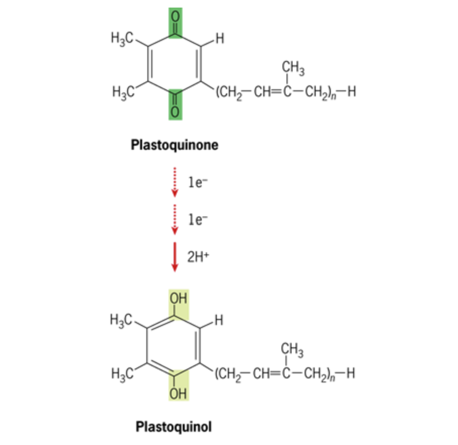
step 4: PQB2-
a second photon sends a second energized electron along the path from P680 to PQA to (PQB-), forming PQB2-
step 5: PQH2
PQB2- combines with two protons to form PQH2
The reduced PQH2 molecule dissociates from D1 protein and diffuses into the lipid bilayer
step 6: PQ
The displaced PQH2 is replaced by a fully oxidized PQ molecule derived from a small “pool” of plastoquinone in the bilayer

flow of electrons from water to PSII
all of these electrons come water and are released when water is split into protons and molecular oxygen
the redox potential of the oxidized form of P680 (1.17 V) is sufficiently strong enough to pull tightly held (low-energy) electrons from water (+ 0.82)- thus splitting the water molecule
photolysis: splitting of water during photosynthesis
formation of one molecule of oxygen during photolysis is thought to require the simultaneous loss of four electrons from two molecules of water
yet, one PSII reaction can only generate one positive charge or oxidizing equivalent at a time
solution to the problem: S-state hypothesis by Pierre Joliot and Bessel Kok, allowing the photosystem to accumulate the four oxidizing equivalents needed to oxidize water
there is a cluster of five metal atoms associated with the D1 protein of PSII at its luminal surface: four Mn atoms and one Ca atom
cluster is stabilized and protected by an oxygen-evolving complex that accumulates 4 oxidizing equivalents by transferring 4 electrons, one at a time, to P680+
a tyrosine residue on the D1 protein (Tyrz) mediates the electron transfer between Mn and P680+
intermediate electron carrier
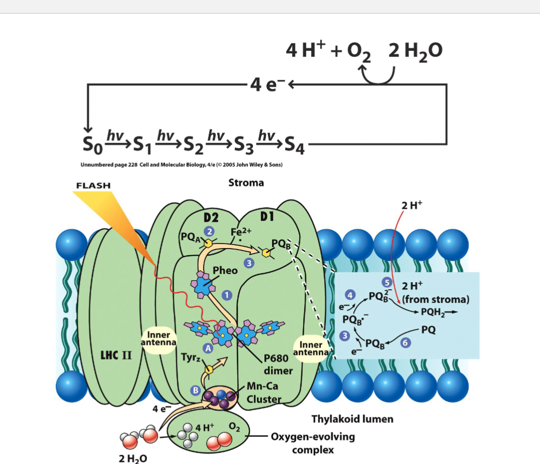
4 step cycle (S states)
S0 to S4
every flash of light pushes the system up by one state
Once it hits S4 it immediately releases oxygen and resets back to S0
kinetics of O2 release
because you need 4 steps/flashes to release oxygen, the peaks in the graph happen every 4 flashes
the first peak occurs after 3 flashes because dark-adapted chloroplasts primarily rest in the S1 (already possessing 1 oxidizing equivalent) rather than S0
Mn clusters are in the S1 state
aqs the number of flashes increases, the oscillations damp out due to desynchronization.

flow of electrons from PSII to PSI
step one: two molecules of PQH2 are formed by accepting 4 electrons from PSII and combining with 4 protons from the stroma
step two: PQH2 diffuses through the lipid bilayer and binds to a large multiprotein complex, cytochrome b6f
step 3: electrons are transferred from cytochrome b6f to a mobile, water-soluble, copper-containing, peripheral protein, plastocyanin (PC)
step 4: PC transfers electrons to P700+
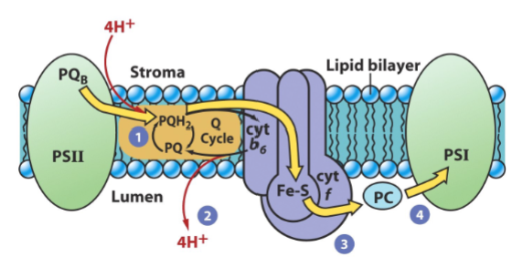
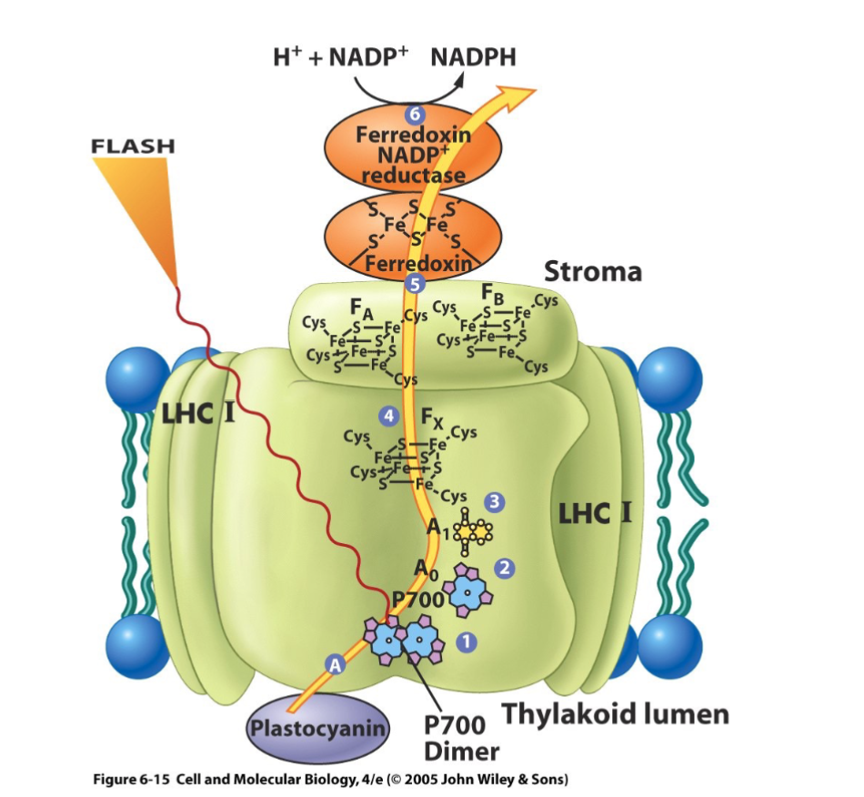
flow of electrons from PSI to NADP+
step 1: following energy absorption, P700* transfers an electron to a separate monomeric chlorophyll a molecule (A0)
step 2: from A0- to a phylloquinone molecule (A1)
step 3: from A1- to an iron-sulfur cluster (Fx)
step 4: from Fx- to two iron-sulfur clusters (FB and FA)
step 5: from FB and FA to a small water-soluble iron-sulfur protein, ferredoxin
step 6: reduction of NADP+ by ferredoxin-NADP+ reductase
overview of photosynthetic electron transport
2H2O + 2NADP+ ——→ 1O2 + 2NADPH
PSII handles water splitting and oxygen release
generates a strong oxidizing agent capable of producing O2 from water
the ETC moves the electrons and builds proton gradient
protons are pumped from the stroma into the thylakoid lumen
ATP synthase uses that proton gradient to make ATP
PSI finishes the job by making NADPH
generates a strong reducing agent capable of producing NADPH from NADP+
light reactions uses 8 photons to produce 3 things needed for the Calvin Cycle (O2, ATP, NADPH)

mitochondria vs chloroplast proton-motive force
in mitochondria: proton motive force is the electrochemical potential
in chloroplast: the proton motive force is largely due to a gradient of pH
gradient is across the thylakoid membrane
photophosphorylation
making ATP using light
two ways- noncyclic and cyclic
ATP is synthesized in the thylakoid membrane
NADPH is synthesized in the stroma of thylakoid membrane
noncyclic photophosphorylation
linear electron flow
PSII to PSI to NADP+
as electrons move down the chain, they pump protons to build a proton motive force, which drives ATP synthase to make ATP alongside making NADPH and oxygen
electrons do not cycle back
cyclic photophosphorylation
sometimes the plant needs extra ATP without making extra NADPH so electrons from PSI loop back into electron transport chain to pump more protons and make ATP, but they do not end up at NADP+
carried out by PSI independent of PSII
after electron is passed to ferredoxin, it is passed back to the electron-deficient PSI to complete the cycle
energy released from the flow of the electron is utilized to translocate protons by the cytochome b6f complex and proton graident is generate for ATP synthesis
cyclic phosphorylation provides additional ATP for carbohydrate synthesis and other ATP-requiring activities in the chloroplast.
summary of light-dependent reactions

discovery of light-independent reactions
“dark reactions”/ calvin cycle
after world war II, melvin calvin and colleagues andrew benson and james bassham began to study enzymatic reactions by whch carbon dioxide was assimilated into the organic molecules of the cell
used long-lived radioactive isotope of carbon (14C) and two dimensional paper chromatography to study green algae chlorella cell suspension
algal suspension was drained into container of hot alcohol (stops enzymatic activity and extracts soluable molecules)
extracts of cells were placed as a spot on chromatographic paper and subjected to 2D chromatography
photographs were developed by pressing against the chromatogram and kept in the dark to locate the radioactive compounds at the end of the procedure
after photographic development, identification of the radiolabeled compounds on the autoradiogram was made by comparison to known standards
carbohydrate synthesis in C3 plants
if the incubation period after adding 14C was very short, one radioactive spot on the chromatogram predominated,
compound that formed the spot was PGA (3-phosphoglycerate)
an intermediate of glycolysis
because the first intermediate was three-carbon molecule, plants using this pathway to fix CO2 are referred to as C3 plants

light-indepedent reaction
the initial acceptor is a five-carbon compound: ribulose 1,5-biphosphate (RuBP)
RuBP condenses with CO2 to produce a six-carbon molecule
the 6-carbon molecule rapidly fragments into 2 molecules of PGA
both reactions are carried out in the stroma by a large multisubunit enzyme, ribulose bisphosphate carboxylase (Rubisco)
rubisco is a very low efficient enzyme (3 CO2/sec) therefore is present in large amount (1/2 of leaf proteins)
calvin cycle
three main parts:
carboxylation of RuBP to form PGA
reduction of PGA to glyceraldehyde 3-phosphate (G3P or GAP)
regeneration of RuBP
summary of calvin cycle
3CO2 + 9ATP + 6NADPH ——> glyceraldehyde-3-P + 9 ADP + 8 Pi + 6 NADP+
first step: RuBisCO attaches a CO2 molecule to a RuBP
regulation of calvin cycle
regulation by light: light activated electron transfer is linked to pumping H+ into thylakoid disks
pH in the stroma increases to about 8
alkaline (basic) pH activates stromal calvin cycle enzymes: RuBP carboxylase, fructose-1,6-bisphosphatase & sedoheptulose bisphosphatase
the light-activated H+ shift is countered by Mg2+ release from thylakoids to stroma
RuBP carboxylase (in stroma) requires Mg2+ binding to carbamate at the active site
reduced thioredoxin (the form more prevelant in the light) activates several calvin cycle enzymes, including fructose-1,6-bisphosphatase, sedoheptulose-1,7- bisphosphatase, and RuBP carboxylase activase, by reducing specific disulfides in those enzymes to thiols
redox control of the calvin cycle
in light, ferredoxin is reduced, and a fraction of these electrons are transferred to the small protein thioredoxin, which reduces the disulfide groups of certain calvin cycle enzymes, maintaining them in an active state
in dark, electron flow to thioredoxin ceases, the sulfhydryl groups of the regulated enzymes become oxidized to the disulfide state, and the enzymes are inactivated.

redox control of the calvin cycle
picture for the dark

photorespiration (C2 Cycle)
light-dependent metabolic process in plants where enzyme rubisco fixes oxygen instead of carbon dioxide
which consumes energy and releases previously fixed carbon
wasteful pathway: instead of making two molecules of 3-PGA, it makes one molecule of 3-PGA and one toxic, 2-carbon molecule called phosphoglycolate. The plant has to spend energy (ATP and reducing power) to recycle this byproduct, releasing previously fixed CO2 in the process, reducing photosynthetic efficiency
rubisco catalyzes two distinct reactions:
rubisco binds RuBP, which adopts the enediol form. The enediol form of RuBP can be attacked by either CO2 or O2. The predominant direction of the rubisco-catalyzed reaction is determined by the ratio of CO2/O2 available.
rate of the reaction is determined by O2/CO2 ratio, temperature, and stomatal closure
temperature rises, RuBisCO’s affinity for CO2 drops
stomatal closure: when a plant is stressed by heat or lack of water, it closes its stomata (pores) to prevent water loss. This traps oxygen produced by the light reactions inside while blocking new CO2 from entering, causing the internal CO2/O2 ratio to drop and photorespiration to increase
occurs in the chloroplast but needs cooperation from chloroplast, peroxisome, and mitochondria
step one: RuBisCO binds O2 to RuBP, producing 2-phosphoglycolate, which is quickly converted to glycolate
step two: glycolate travels to the peroxisome, where it is oxidized into glyoxylate and then converted into the amino acid glycine.
step three: two molecules of glycine are brought into the mitochondria, and are combined to form serine, which releases a molecule of CO2 and ammonia
step 4: serine travels back to the peroxisome to be converted to glycerate and glycerate enters the chloroplast, where an ATP molecule phosphorylates it into 3-PGA, allowing it to finally rejoin the normal calvin cycle

C4 plants
In 1965, using [14C]O2, Hugo Kortschak found that in sugarcane the first compound appeared contained 4-carbon skeletons (predominately oxaloacetate and malate). These 4-carbon compounds resulted from combination of CO2 with phosphoenolpyruvate (PEP), which is the first of the C4 pathway
C4 plants, such as sugarcane, corn, and sorghum, live in a dry and hot environment.
they are adapted to close their stomata to prevent water loss, which also decreases the CO2 concentration in the leaves
the enzyme responsible for linkage of CO2 and PEP (PEP carboxylase) can fix CO2 even when the leaf stomata are almost entirely closed and the CO2 level in the cell is very low
C4 metabolism
CO2 enters the mesophyll cell and is bonded to phosphoenolpyruvate (3C) to form oxaloacetate (4C) by enzyme PEP carboxylase
the oxaloacetate is converted into malate (4C) and shuttled into the chloroplast of the bundle sheath cell through plasmodesmata.
the malate is decarboxylated to form pyruvate (3C) and CO2
the CO2 can be used in the Calvin cycle in the bundle sheath cell.
the pyruvate is cycled back to the mesophyll cell, where it can be used to regenerate phophoenolpyruvate
C4 plants are able to generate high CO2/O2 ratios in the local Rubisco environment, thereby favoring the process of CO2 fixation over that of photorespiration

comparison of C3 and C4 plants structures

comparison of C3 and C4 metabolism
first carbon product:
C3: 3 carbon compound 3-phosphoglycerate
C4: 4 carbon compound oxaloacetate
enzyme for initial CO2 fixation:
C3: rubisco
C4: PEP carboxylase
anatomy/cell structure:
C3: no specialized bundle-sheath cell separation; standard chloroplast arrangement
C4: bundle-sheath cells surround leaf veins
separation of reactions:
C3: carbon fixation and the Calvin cycle happen in the same cells
initial fixation in mesophyll cells; Calvin cycle in bundle-sheath cells
photorespiration
C3: high (Rubisco binds O2 in hot/dry conditions, wasting energy)
C4: Minimized/Suppressed (PEP carboxylase does not bind O2 and CO2 is concentrated around Rubisco)
water/thermal efficiency:
C3: lower efficiency in high heat; loses more water through open stomata
C4: high efficiency; saves water and thrives in warm, bright conditions
best environment:
C3: cool, wet, or moderate climates
C4: hot, sunny, and arid/tropical regions
CAM plants
many desert plants, such as cacti, have another biochemical adaptation that allows then to survive in very hot, dry habitats.
these are also C4 plants but instead of segregating the C4 and C3 pathways in different parts of the leaf, they separate them in time instead.
CAM stands for Crassulacean Acid Metabolism because it was first studied in members of plant family Crassulaceae
night vs morning processes in CAM plants
night:
CAM plants take in CO2 through their open stomata (they tend to have reduced numbers of them).
the CO2 joins with PEP to form the 4-carbon oxaloacetic acid.
this is converted to 4-carbon malic acid that accumulates during the night in the central vacuole of the cells.
morning:
the stomata close (thus conserving moisture as well as reducing the inward diffusion of oxygen).
the accumulated malic acid leaves the vacuole and is broken down to release CO2.
3. the CO2 is taken up into the calvin (C3) cycle