RBC
 Call Kai
 Learn
Learn Practice Test
 Spaced Repetition
 Match
Match Flashcards
 Knowt Play
1/65

There's no tags or description
Looks like no tags are added yet.
Name | Mastery | Learn | Test | Matching | Spaced | Call with Kai | Chat |
|---|
No analytics yet
Send a link to your students to track their progress
66 Terms
What type of tissue is blood? What is it made of?
Blood = Connective tissue with fluid (plasma) and cellular part
Plasma (46 - 63%):
Water (92%)
Plasma proteins (7%)
Regulatory proteins (eg. hormones and enzymes)
Other solutes (eg. nutrients, gases and wastes)
Cells (37 - 53%):
Leukocytes
Granulocytes (neutrophils, eosinophils, basophils)
Agranulocytes (lymphocytes, monocytes)
Erythrocytes
Platelets

Describe normal body fluid compartments
% of TBWT that blood makes up (cat vs. dog)
% TBWT made of water (+ compartments)
Movement of fluid between compartments
Blood: 5 - 10% BWT
Cat = 55mL blood/kg BWT
Dog = 90mL blood/kg BWT
Water: 60 - 70% BWT is water
67% intracellular fluid (ICF)
33% extracellular fluid (ECF)
Plasma in blood (IVF)
Interstitial fluid (ISF)
Movement: Membrane are permeable to water → free exchange until equilibrium reached
Extramedullary haematopoiesis (EMH)
Liver and spleen produce blood cells when the bone marrow does not work
Describe formation of blood cells and what is required
Formation: Derived from primitive (pluripotential) stem cells in the bone marrow
ALL blood cells have a limited lifespan and are incapable of self-renewal
Bone marrow produces steady supply of replacements to replace old cells
Requirements: Cytokines stimulate differentiation of the stem cells into committed (unipotential) cell lines forming each blood cell type eg.
Erythropoietin (EPO) → RBC production
Thrombopoietin
Granulocyte and macrophage colony stimulating factors (CSF)
Describe EIGHT organs involved in haematopoiesis (+ functions of each organ)
Bone marrow
Main source of blood cells (RBC, WBC and PLT)
Stores iron for heme synthesis
Source of stem cells for T and B lymphocytes to be produced elsewhere
Liver = Major haematopoietic organ during 1st ½ of embryonic life (bone marrow takes over BUT retained EMH potential as adult)
Spleen
Produces T/B lymphocytes and plasma cells from bone marrow stem cells
Stores RBCs and iron
Removes old RBCs from circulation
Retained haematopoietic potential (EMH)
Lymph nodes = Produce T/B lymphocytes and plasma cells from bone marrow stem cells to participate in Ab production and CMI
Thymus = Controls differentiation of bone marrow stem cells into T lymphocytes
Macrophages of monocyte phagocytic system (MPS) = Macrophages lining the sinusoids of the spleen, liver, lymph nodes and bone marrow complete intracellular haemolysis
Kidney = Hypoxia stimulates renal tissue to produce EPO for erythropoiesis
Stomach and intestinal mucosa
Describe the differentiation of multipotential haematopoietic stem cell (haemocytoblast)

Erythropoiesis
Location
Duration
Process
3 Pools of RBCs in the body
Location: BONE MARROW (also liver and spleen EMH)
Duration: ~7 days from initial stimulation of stem cells by EPO to reticulocyte release
Process:
Pluripotential stem cells → Erythroid stem cell
Erythroid stem cells → Rubriblasts in response to EPO which begins to produce Hb and other proteins
Rubricytes produced from each rubriblast
Rubricyte → Metarubricyte (final nucleated stage)
Metarubricyte → Reticulocyte with no nucleus (triggered when RBCs reach critical Hb content)
Reticulocytes remain in bone marrow for 2 - 3 days
Reticulocytes released into circulation for final maturation as a erythrocyte

Pools of RBCs:
Erythrocyte precursors in bone marrow
Mature erythrocytes in blood
Mature erythrocytes in spleen
Regulation of Erythropoiesis
Stimulus
4 Functions of EPO
Species-specific responses
4 Other requirements
Stimulus: Accelerated RBC production if demand for O2 is great (eg. anaemia) OR hypoxia detected by renal tissue → Stimulates production of EPO in the kidney
Functions of EPO:
Stimulates stem cells to differentiate into rubriblasts (instead of other cell lines)
Promotes Hb synthesis
Reduces maturation time of RBC precursors
Stimulate early release of immature reticulocytes
Species Variation: Dogs > Cats > Cows response to EPO
Horses do NOT release reticulocytes (do not respond to EPO) → Requires bone marrow aspirate to determine regenerative response
Other Factors: Deficiency → Anaemia
Essential vitamins (B12, B6, folic acid)
Essential minerals (iron, copper, cobalt)
Hormones (GH, ILGF, testosterone, thyroid)
IL and colony-stimulating factors
Name the following species each RBC belong to (+ description)
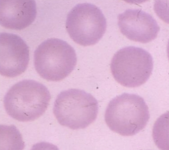 |  |  |
 |  |  |
DOG Biconcave with obvious central pallor | CAT
| GOAT
|
DEER Sickle-shaped | CAMELID Elliptical | BIRD, REPTILE, FISH Nucleated and elliptical |
Describe the structure of erythrocytes
Hb content%
Hb structure
Heme structure
Organelles and energy source
Shape and 2 functions
Content: RBCs = 95% Hb
Structure:
Hb = 4 globin subunits which are linked and folded about a central heme molecule
Heme = Protoporphyrin ring with an atom of iron for O2
Energy: No mitochondria or ribosomes → Energy source from glucose in plasma ONLY
Shape: Biconcave →
Increase SA to allow more Hb to attach to RBC membrane for gas exchange
Assists RBC deformation through small capillaries
Lifespan of 3 different blood cells
Erythrocytes (months)
Larger the animal → Longer the lifespan
Cat = 70d, dog = 120d, horse = 145d, cow = 160d
Platelets (days)
WBCs (hours and replaced 2x daily)
Extravascular Haemolysis
Normal function
Normal MoA of extravascular haemolysis
Outcomes of the components of RBC breakdown
Function: Old/damaged RBCs that have reached the end of their lifespan are removed from circulation for membrane degradation
Allows their components to further broken down and recycled (spleen #1)
aka. intracellular haemolysis (within macrophages of the spleen, liver and bone marrow OUTSIDE blood vessels)
Physiological process which becomes pathological if occurring at a rapid rate
MoA:
Blood flows through the spleen
Old/worn out cells have different structure and contain proteins that the splenic macrophage recognises
RBCs enter the macrophage's cytoplasm to undergo haemolysis by digestive enzymes
Haemoglobin (Hb) is released from lysed RBCs and broken down into heme, globin and iron
Components of RBC Breakdown:
Heme = Further broken down into bilirubin and carried by albumin to liver for processing and elimination as bile in faeces
Bilirubin = Yellow/orange
Globin = Broken down into AA for recycling
Iron = Incorporated into haemosiderin (or ferritin) for storage in macrophages
Haemosiderin = Yellow/brown

Extravascular vs. Intravascular Haemolysis
Definition
Severity
Signs
5 Mechanisms (+ DDx)
EXTRAVASCULAR HAEMOLYSIS (90%)
Definition: Haemolysis that occurs by splenic macrophages NOT within the blood vessels
Physiological OR pathological
Severity: More chronic than intravascular haemolysis (NO haemoglobinaemia or haemoglobinuria → Hb recycled and NOT lost in urine)
Signs:
No haemoglobulinaemia or haemoglobulinuria
Hyperbilirubinaemia (overwhelming of liver)
Abnormal RBC morphology (eg. erythrocytic parasites, spherocytes, schistocytes)
Positive Coomb’s test
Splenomegaly
DDx:
Reduced RBC deformability due to membrane alteration (eg. blood parasites, immune-mediated anaemias, microangiopathies)
Impaired glycolysis or low ATP content in RBCs (eg. pyruvate kinase deficiency)
Immunoglobulins/C3 opsonisation of RBC membranes (eg. IMHA)
INTRAVASCULAR HAEMOLYSIS (10%)
Definition: Lysis of RBCs within the lumen of the blood vessels (fragile) resulting in Hb released directly into the plasma
Usually pathological (small amounts occur normally but bound to haptoglobin)
Severity: More severe than extravascular haemolysis due to urine loss of Hb
Signs:
Haemoglobinaemia and haemoglobinuria
Decreased PCV, decreased RBC and [Hb]
Increased artifactual MCHC (agglutination)
Hyperbilirubinaemia (jaundice/icterus) with exceeded liver capacity to remove and conjugate bilirubin + bilirubinuria
Altered RBC morphology (eg. Heinz bodies, schistocytes and parasites)
Positive Coomb’s test
Decreased serum haptoglobin concentration (protein binding free Hb and complex removed by kidney)
DDx:
Complement-mediated lysis (eg. immune-mediated anaemia)
Oxidative injury → Denaturation of Hb to Heinz bodies and lysis of RBCs (eg. chemical and plant toxins)
Osmotic lysis due to RBC membrane alterations, infusion of hypotonic fluids or drinking lots of water
Erythroparasites
Leptospirosis and clostridium

Pathology of haemoglobinuria due to intravascular haemolysis
What occurs normally to free Hb?
What occurs when in excess?
Some free Hb dissociates into globin dimers which binds haptoglobin in plasma to form the haptoglobin-haemoglobin dimer complex
Hpt-Hb complex prevents filtration of Hb through the glomerulus for renal excretion, and is instead taken to the liver to be broken down into Fe and bilirubin
When excess Hb in plasma is present, the Hpt is saturated and unbound Hb is filtered by the kidneys
Most unbound Hb is reabsorbed by tubular epithelial cells to be converted to haemosiderin
When the capacity for tubular resorption of Hb is exceeded, free Hb is present in the urine
Iron Metabolism
3 Storage sites
Normal metabolism
Effects of anaemia on iron metabolism
Transferrin
Definition
2 Causes of decreased transferrin concentration
2 Causes of increased transferrin concentration
Storage:
Within RBCs (50 - 70%)
Within macrophages of bone marrow, spleen and liver (25 - 40%)
Small amounts in other molecules (myoglobin and enzymes)
Metabolism: Very little iron is lost
Iron released from RBC breakdown is almost enough for normal erythropoiesis + small contribution from absorption from gut
Anaemia: Increases iron demand due to accelerated erythropoiesis
Increased efficiency of intestinal Fe absorption
Release iron from tissue macrophages within haemosiderin or ferritin
Transferrin:
Definition: Apotransferrin that transports iron in the blood → Used to assess iron levels in blood
Decreased [transferrin]
Iron deficiency
Acute/chronic inflammation
Increased [transferrin]
Haemolytic anaemia
Hyperadrenocorticism
TWO Methods of CBC Analysis
Advantages
Disadvantages
Information gathered
ONE: Automated analyser (commercial or in-clinic labs)
+ve: Good at counting
-ve: Unreliable at characterising
Information
Erythrogram: Count, Hb, HCT, MCH, MCHC, retics
Leukogram: Count and differential BUT not reliable
Thrombogram: Count
TWO: Blood smear exam
+ve: ESSENTIAL part of haematological exam
Necessary for characterising morphology (eg. inclusions, change in shape etc.)
Check if the automated analyser was accurate
Area: Monolayer = Transition between the body and feathered edge of the blood smear (blood cells JUST separated)
Information:
RBC morphology: Size, colour, shape, other (structure and arrangement)
Check the automated analyser counted correctly
WBC morphology: Differential and stages
PLT number (clumping)
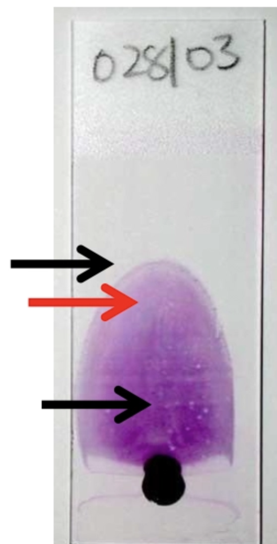
Describe EIGHT assessments of erythrocytes in the CBC
Haematocrit (HCT) = Volume of erythrocytes that make up a L volume of blood
Calculated value (HCT = MCV x RBC)
Unit: L/L (proportion between 0 and 1)
Packed cell volume (PCV) = % of blood composed of erythrocytes
Manually measured estimated of HCT by centrifuge
Unit: %
Red cell count (RBCs) = Number of erythrocytes per unit volume of blood
Unit: # erythrocytes x 10^12/L
Necessary for MCV and MCH calculations
Haemoglobin concentration (Hb) = Amount of Hb per unit volume of blood (lyse RBCs in sample to calculate)
Unit: g/L
Most direct indication of O2 carrying capacity of blood
Absolute reticulocytes (absolute retics) = Number of reticulocytes per unit volume of blood
Absolute superior: In anaemic animals, % does NOT account for relative increase in reticulocytes due to the reduced numbers of mature erythrocytes
Mean corpuscular volume (MCV) = Volume of an average RBC (size)
Unit: fL (femolitre)
Helps to classify anaemias
Mean corpuscular haemoglobin concentration (MCHC) = Amount of Hb in a litre of RBC (vs. Hb concentration = amount of Hb in whole blood including plasma)
Unit: g/L
Influences the colour and pallor of the RBC MCHC = Hb/HCT = MHC/MCV
Mean corpuscular haemoglobin (MCH) = Amount of Hb per average RBC
Unit: Picogram (pg)
Little information provided
MCH = Hb/RBC
Mean Corpuscular Volume (MCV)
4 DDx for macrocytosis
3 DDx for microcytosis
Macrocytosis:
Regenerative anaemia = Reticulocytosis
IMHA (artifactual increase in MCV and decreased RBC count as agglutinated cells are counted as one)
Poodles
FeLV
Microcytosis:
Iron deficiency anaemia (less Fe to synthesise Hb = smaller cell as RBC adjusts to Hb content)
PSS
Japanese Akitas and Shibas
Structure of a reticulocyte
Anucleated
Slightly larger than mature erythrocyte
Still with some cytoplasmic organelles (mitochondria and ribosomes) = slightly basophilic
Hypochromic (pallor) due to fewer Hb
Mean Corpuscular Hb Concentration (MCHC)
2 DDx for hypochromasia
DDx for hyperchromasia (+ 6 examples)
Hypochromasia: Dogs = Bigger central pallor and narrow rim of Hb
Iron deficiency (lack of Fe = lack of Hb)
Regenerative anaemia = Reticulocytosis (immature RBCs have fewer Hb)
Hyperchromasia: Artifactual (each RBC has finite Hb carrying capacity)
Haemolysis (extracellular Hb)
Heinz bodies
Marked spherocytosis
Lipaemia
RBC shrinkage due to EDTA
Crenation
Advia scattergram
Graph produced by the in-lab CBC machines where each dot represents an RBC
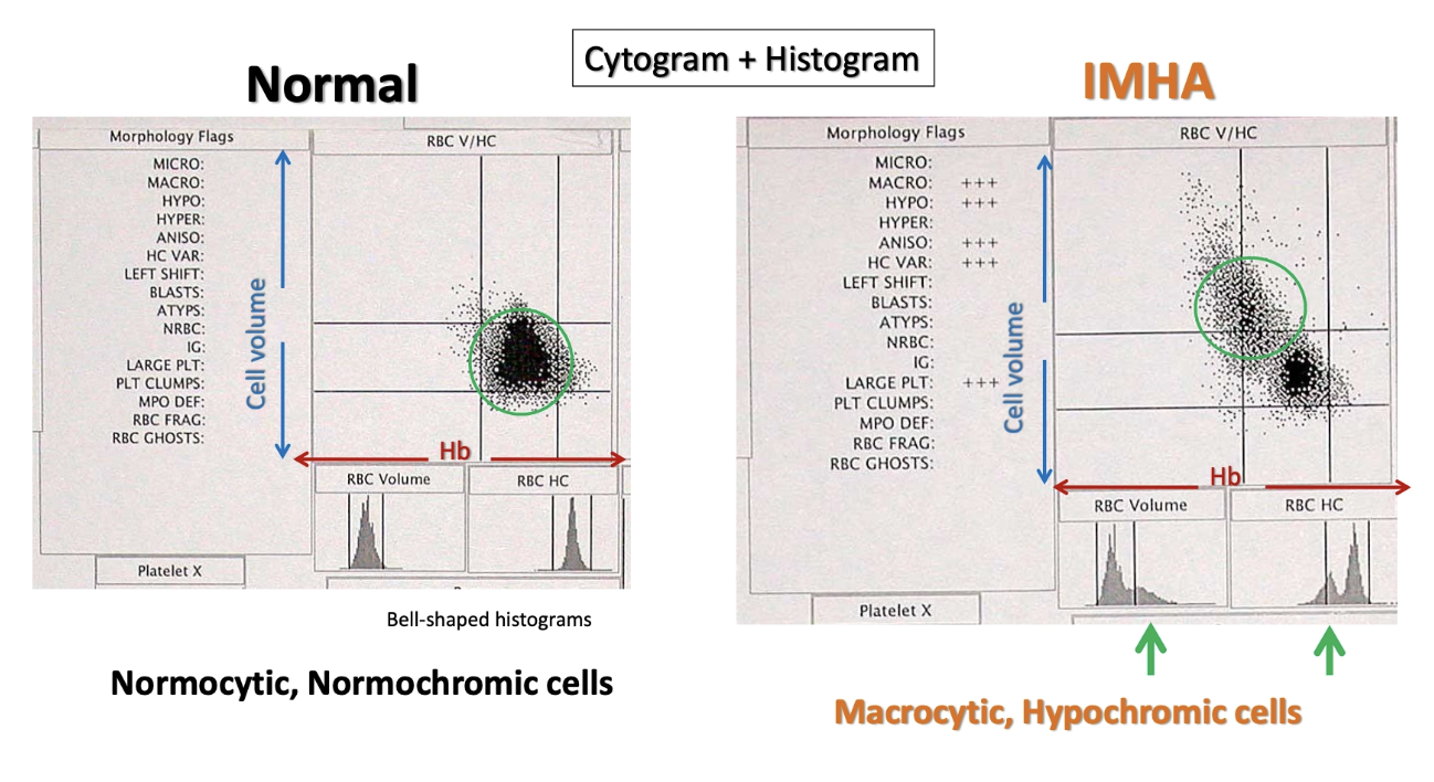
Describe SIX features to assess with RBC on blood smear (+ examples)
ONE: NUMBER
Severe anaemia = More gaps between RBCs
Cannot accurately estimate number from blood smear alone
TWO: SIZE
Anisocytosis = Variation in cell size (large/small/both)
Indicative of a regenerative anaemia (larger reticulocytes) BUT mild anisocytosis is normal in cats

THREE: COLOUR
Polychromasia = Variation in cell colour and staining affinity due to:
[Hb] which affects redness and pallor (increases with maturity)
RNA produces blue (ribosomes and mitochondria decrease with maturity)

FOUR: SHAPE
Poikilocytosis = Non-specific abnormally shaped erythrocyte
Acanthocyte
Echinocyte
Schistocyte
Keratocyte
Spherocyte
Eccentrocyte
Codocyte/leptocyte
FIVE: OTHER STRUCTURES
Basophilic stippling
Heinz bodies
Howell-Jolly bodies
Metarubricytes
Siderocytes
SIX: ARRANGEMENT IN MONOLAYER
Agglutination
Rouleaux formation
Hallmarks of regenerative anaemia
3 Hallmarks on CBC
3 Hallmarks on blood smear
CBC:
Increased reticulocyte count
High MCV = Macrocytosis
Low MCHC = Hypochromasia
Blood Smear:
Anisocytosis
Polychromasia
± Basophilic stippling in large animals
TWO methods of reticulocyte staining
Appearance
Which can ID punctated reticulocytes?
Diff-Quick routine blood staining
Polychromatophil = Reticulocyte that is larger and more basophilic due to more ribosomes in the cytoplasm
New methylene blue stain (NMB)
Stains RNA of immature erythrocytes blue → Blue dots on reticulocytes (can also identify punctated reticulocytes)
Aggregated = More organelles in cytoplasm and larger cells that can be identified on Diff-Quick stain (and NMB)
Punctated = More mature erythrocytes which have fewer smaller organelles and can ONLY be identified on NMB stain (indicate more chronic regenerative anaemia)

Name the cell
Definition/appearance
2 DDx
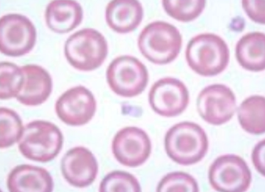
Codocyte/Leptocyte
Definition: Excessive membrane resulting in membrane folding and a ring of pallor
DDx: Little significance unless seen in almost every cell
99% artifactual due to increased EDTA
Hepatic or lipid disorders
Name the cell
Definition/appearance
DDx

Eccentrocyte
Definition: Clear area to the side of the cell and lack of central pallor as Hb shifted to one side
DDx: Oxidative damage of Hb (commonly seen with Heinz bodies)
Name the cell
Definition/appearance
DDx
Location

Spherocyte
Definition: Small, darkly staining, spherical erythrocytes which lack central pallor caused by partial phagocytosis my macrophages in spleen and liver
Membrane of the RBC close the phagocytosed gap and it becomes smaller
DDx: IMHA
Easier to detect in dogs as they have a normal centre of pallor
Location: ID in monolayer
Name the cell
Definition/appearance
2 DDx

Keratocyte
Definition: Blister-like vesicle which can rupture → 2 long horns on one side of the cell
aka. Blister cell, helmet cell, apple stem cell
DDx:
Iron deficiency
Intravascular trauma (DIC, non-laminar flow or vascular neoplasia)
Name the cell
Definition/appearance
4 DDx

Schistocyte
Definition: Erythrocyte fragments formed from intravascular trauma → Shearing of RBCs
DDx:
DIC (fibrin clots shear RBCs)
Vascular neoplasia (eg. HSA)
Non-laminar flow (eg. valvular disease)
Iron deficiency (fragmentation of weaker cells exposed to NORMAL forces)
Name the cell
Definition/appearance
7 DDx

Echinocyte
Definition: Multiple, short, evenly-spaced, uniform projections
aka. Burr cell
DDx:
Artifact #1 (slow drying of blood smear, underfilling EDTA tubes, improper smearing, crenation DDx)
Common in cats
Renal failure
Lymphoma
Chemotherapy
Rattle snake bites
Post-exercise in horse
Name the cell
Definition/appearance
3 DDx

Acanthocyte
Definition: Cell with few, unevenly-spaced, irregular projections due to change in cholesterol/phospholipid concentrations in RBC membrane
aka. Spur cell
DDx:
Dog with splenic haemangiosarcoma
Dog with iron deficiency
Cat with hepatic lipidosis
What is this?
Definition
2 DDx
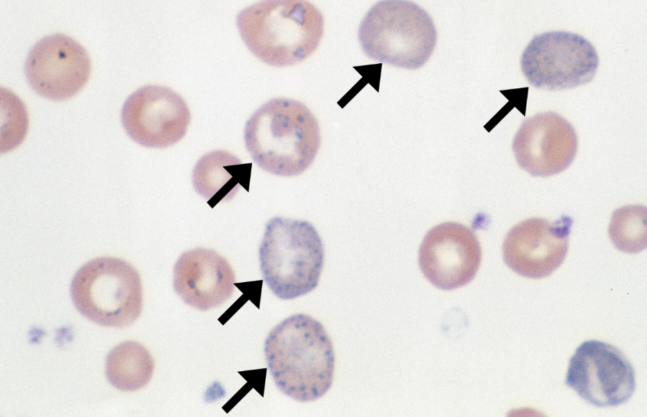
Basophilic Stippling
Definition: RBCs with blue dots (ribosome aggregations) in cytoplasm
DDx:
Regenerative anaemia (reticulocytosis)
Lead poisoning
What is this?
Definition
3 DDx
Alternative stain

Heinz Bodies
Definition: Unstained blobs at the edge of RBCs on normal stain (blue on NMB stain)
DDx: Oxidative damage → Hb denaturation on the internal RBC surface
Oxidative chemicals (brassicas, paracetamol and onions)
Most normal cats have <10% RBCs with Heinz bodies
>10% = diabetes mellitus, hyperthyroidism and lymphoma
Stain: NMB stain = Blue Heinz bodies

What is this?
Definition
3 DDx

Howell Jolly Bodies
Definition: Small (1µm), purple, spherical inclusion within erythrocytes = Retained nuclear remnants
DDx:
Regenerative anaemia
Chronic steroid therapy in dogs
Reduced splenic function
What is this?
Definition
5 DDx
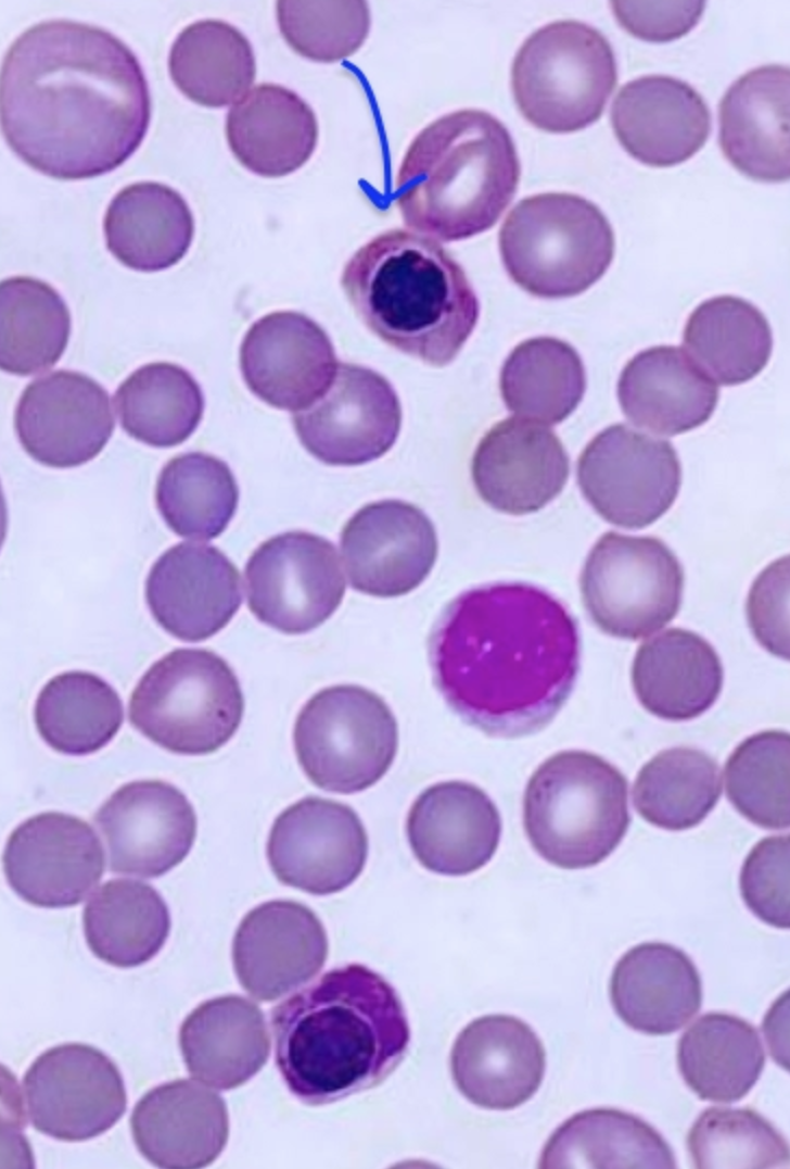
Metarubricytes
Definition: Last nucleated stage of erythrocyte
DDx:
Severe regenerative anaemia
Lead toxicity
Non-functioning spleen
Haemangiosarcoma
Increased corticosteroids
What is this?
Appearance
2 DDx
Alternative stain

Siderocytes
Definition: Mature erythrocytes with iron-containing (siderotic) cytoplasmic inclusions
DDx:
Taranaki anaemia (decreased Mg in cattle)
Lead toxicity
Stain: Iron granules stain positive with Prussian blue
TWO Abnormal Arrangements of RBCs in the Monolayer
Appearance
DDx
How to differentiate
AGGLUTINATION
Appearance: RBCs grouped in irregular clumps (grapes)
DDx: Ag-Ab complexes (eg. IMHA)
MUST ID in monolayer (thicker layer causes cells to appear clumped)

ROULEAUX FORMATION
Appearance: Linear stacking of cells
DDx: Increased plasma protein content
Hyperfibrinogenaemia (acute inflammation)
Hyperglobulinaemia (plasma cell myeloma)
NORMAL in horses

Differentiate: Saline dispersion test (rouleaux disperse)
Bone marrow examination
Function
Always send what?
6 Indications
Function: Confirm or exclude DDx based on haematology (when no definitive dx is reached on haematology)
Send: Always sent peripheral blood smear for CBC at the same time
Indications:
Non-regenerative anaemia
Persistent leukopenia
Persistent thrombocytopenia (with no obvious cause)
Atypical cells suggestive of leukaemia in peripheral blood
Monoclonal gammopathies
Determine regenerative anaemia in horses
Erythrocytosis
Definition
4 Causes
Definition
Clues
DDx
Definition: Increased erythrocyte concentration in blood (PCV, RBC and [Hb] above RR)
Causes:
Greyhound
Relative Erythrocytosis = Erythrocyte mass is SAME, but plasma volume is reduced
Clues: Increased TP and dehydration (skin tent and enophthalmos)
DDx:
Dehydration #1 (eg. vomiting/diarrhoea, diuresis, reduced fluid intake)
Endotoxaemia (net movement of water out of vessels via widespread vasodilation)
Physiological Erythrocytosis = Transient (<2hr) increased erythrocyte mass due to increased adrenaline (SNS) → Smooth muscle contraction of the spleen → RBCs released into circulation
NO pathological significance
Clues: Fear, pain, excitement (NOT stress) eg. young horse #1
NORMAL TP
± Concurrent physiological neutrophilia
Absolute Erythrocytosis = True increase in erythrocyte mass (longer term change)
Primary Erythrocytosis = Increased erythrocyte produced independent of EPO (normal PO2)
Rare condition due to myeloproliferative neoplasia (bone marrow) resulting in uncontrolled division of RBCs without EPO via clonal expansion = Polycythaemia vera
Secondary Erythrocytosis = Increased erythrocyte production in response to increased EPO levels
Appropriate = Increased EPO associated with tissue hypoxia which is detected by renal interstitial cells and stimulates produced of EPO
High altitude
Chronic respiratory disease causing poor oxygenation of blood
Cardiovascular shunts
Inappropriate = Increased EPO WITHOUT hypoxia (normal PO2)
DDx: EPO-secreting renal tumours or lesions (renal cysts, hydronephrosis, infarcts)

Overview of Anaemia
Definition
5 Clinical signs
3 Mechanisms
Definition: Reduction in RBC mass resulting in reduced RBC count, PCV/HCT and usually [Hb]
Clinical Signs:
Pale MM
Lethargy
Reduced exercise tolerance
Increased RR
Increased HR ± murmurs due to turbulence
Mechanisms:
Decreased RBC production
Increased RBC destruction (haemolysis)
Increased RBC loss (haemorrhage) COMBINATIONS of them too
Fill out the following table
Classification of Anaemia | DDx |
Normocytic, normochromic | |
Macrocytic, hypochromic | |
Microcytic, hypochromic | |
Microcytic, normochromic |
Classification of Anaemia | DDx |
Normocytic, normochromic | Non-regenerative anaemia |
Macrocytic, hypochromic | Regenerative anaemia (polychromatophil) |
Microcytic, hypochromic | Iron deficiency anaemia (lack of Hb) |
Microcytic, normochromic | Early iron deficiency anaemia |
List 7 other tests to help diagnose the cause of anaemia
Estimation of plasma protein = Indicates regenerative anaemia due to blood loss (low) OR haemolysis (normal)
Coombs test for suspected immune-mediated haemolytic anaemia
Microcytic anaemia → Check faeces for blood (chronic blood loss from GIT disease causing iron deficiency anaemia)
Serum iron concentrations
Ehrlichiosis check for dogs with thrombocytopenia and hyperglobulinaemia (not in NZ)
FeLV and FIV test for anaemic cats
EIV test for anaemic horses
Describe the THREE types of anaemia
Definition
Timing
Causes
ONE: REGENERATIVE ANAEMIA
Definition: Bone marrow response to anaemia by increasing synthesis of EPO which accelerating erythropoiesis and release of immature RBCs into circulation
Shorter lifespan of metarubricytes and reticulocytes and poorer O2 carrying capacity
Timing: First visible 3 - 5 days loss and peak 7 days
Causes:
Haemorrhage (RBC lost as whole blood)
Acute vs. chronic blood loss
Internal vs. external blood loss
Haemolysis (RBC ONLY destruction)
Intravascular
Extravascular
TWO: PRE-REGENERATIVE ANAEMIA
Definition: Not enough time elapsed for evidence of bone marrow regenerative response to be seen in the peripheral blood smear
Timing: Sample taken within 3 - 5 days post-loss
Diagnosis: Differentiate by repeating the CBC 3 - 5 days later to observe for change (i.e. reticulocytes present)
NON-REGNERATIVE ANAEMIA
Definition: Lack of expected bone marrow response (bone marrow cannot produce enough RBCs to replace those lost normally)
Timing: Diagnose with at least 2 CBCs to assess change in reticulocyte count (stays low = non-regenerative, increases = pre-regenerative → regenerative)
Causes:
Deficiency of essential erythropoietic factor
Toxins inhibiting bone marrow function
Infection
Neoplasia
Defective maturation of RBCs
Deficiency in stem cells
Timeframe of acute blood loss
Time Post-Haemorrhage | RBC Parameters | Other Signs |
<4hr | ||
4 - 12hr | ||
3 - 5d | ||
7d | ||
10 - 14d |
Time Post-Haemorrhage | RBC Parameters | Other Signs |
<4hr | NORMAL
| ± PLT increase and mild neutrophilia by 3hr |
4 - 12hr | Reduced PCV
| Reduced TP |
3 - 5d | Reticulocytosis | |
7d | Peak reticulocytosis | Normal TP |
10 - 14d | Normal | Normal |
Chronic Haemorrhage
Definition
Clinical signs
RBC changes
3 DDx
Definition: Anaemia develops slowly without hypovolaemic shock and provides the body time to adapt
Clinical Signs: May develop AFTER drop in PCV
Regenerative response is evident initially with low TP, but it becomes progressively less regenerative overtime due to iron deficiency
RBC: Iron deficiency anaemia = Microcytosis and hypochromasia
DDx:
Internal parasitism (eg. haemonchosis, hookworms, coccidiosis)
External parasitism (eg. fleas, ticks, lice)
GIT ulcers
Internal Haemorrhage vs. External Haemorrhage
Definition
Process and regenerative response
Causes
Internal Haemorrhage
Definition: Haemorrhage into tissues or a body cavity
Process: Most RBCs resorbed by lymphatics and recirculate within 24 - 72hr
Remaining RBCs are phagocytosed and lysed with the iron and plasma proteins reutilised → Greater regenerative response
Difficult to differentiate from haemolysis due to normal TP (rapid resorption via lymphatics)
Causes:
Bleeding in lungs, liver, within body cavities
Bleeding disorders (eg. haemophilia, thrombocytopenia)
Bleeding tumours
External Haemorrhage
Definition: Whole blood loss from body (RBC, plasma protein, iron)
Process: Decreased TP with reduced regenerative response (plasma proteins and iron lost)
Causes: Haemorrhage from
GIT
Respiratory tract
Skin
Urinary tract
Overview of Haemolytic Anaemia
Definition
2 Signs/clues
2 Types
Definition: Accelerated RBC destruction which cannot be balanced by increased erythropoiesis
Signs:
Normal TP
Larger regenerative anaemia and more reticulocytes due to recycling of iron (retained in plasma)
Types:
Intravascular haemolysis
Extravascular haemolysis
9 DDx for regenerative anaemia and 5 DDx for non-regenerative anaemia
Regenerative Anaemia:
IMHA (5)
Neonatal isoerythrolysis (3)
Iron deficiency anaemia (4)
Sporidesmin toxicity (3)
Leptospirosis (3)
Chronic copper deficiency (3)
Blood parasites (3)
Brassica redwater (2)
Bovine post-parturient haemoglobinuria (2)
Non-Regenerative Anaemia:
Anaemia of chronic inflammatory disease
Anaemia of chronic renal failure
Iron deficiency anaemia (4)
Lead toxicity (4)
“Taranaki” Anaemia (2)
Immune-Mediated Haemolytic Anaemia (IMHA) (5)
Primary vs. secondary
Pathogenesis
Species variation
Dog (aetiology and signalment)
Cat (aetiology and signalment)
Horse (aetiology and signalment)
6 Clinical features
6 Laboratory findings
Diagnosis (+ 3 -ve)
Primary vs. Secondary:
Primary IMHA = Autoimmune in which the body attacks own RBC Ag
Secondary IMHA = Antigenic alteration in RBC due to infectious agents, inflammatory agents, neoplasia or drugs
Pathogenesis:
Ab developed against Ag on the RBC surface with complement fixation
Recognised by macrophages in the spleen and bone marrow
Extravascular haemolysis which develops to intravascular (specific diseases may directly be intravascular)
Species Variation: Dogs > Cats » Horses
Dogs = Primary #1
Females > Males
Cocker spaniel, standard poodle, rough coated collie, old English sheepdog
Cats = Secondary #1 (Mycoplasma haemofelis, FeLV or neoplasia)
Worse prognosis than dogs (cannot be treated with corticosteroids)
Horses = Secondary and RARE
Clinical Features: Regenerative extravascular ± intravascular haemolytic anaemia
General anaemic signs
Haemoglobinaemia
Icterus
Splenomegaly (more splenic macrophages)
± Hepatomegaly (icterus-related)
± Fever (infectious agent or neoplasia)
Laboratory Findings:
Moderate - severe anaemia (low HCT, Hb, RBC)
Marked regenerative response (anisocytosis, polychromasia and reticulocytes ± nRBC)
SPHEROCYTES and ghost cells (no Hb)
Agglutination which is grossly visible if severe
± Leukocytosis with neutrophilia and left shift
± Thrombocytopenia
Diagnosis: Positive Coomb’s test = Detect anti-RBC Ab (reagent to species-specific anti-Ig and/or C3b)
-ve:
Helps confirm IMHA but has poor Sn and Sp
NOT necessary if gross agglutination is observed on smear
Does NOT work for other suspected immune-mediated diseases

Neonatal Isoerythrolysis (3)
Species signalment
Pathogenesis
Diagnosis
Treatment
Species: Foals #1
Pathogenesis: Regenerative extravascular haemolytic anaemia
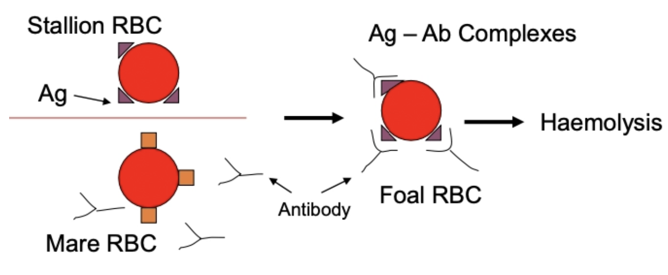
Foal inherits RBC Ag from stallion which are incompatible with the mare
Small amounts of the foetal RBC Ag enter the maternal circulation across the placenta due to haemorrhage
Maternal Ab raised to foal RBC Ag and concentrate in mare's colostrum
Foal is normal at birth, but develops clinical signs within 24 - 48hr of colostrum intake
RARE in maiden mares as the first foal is the exposure (insufficient time for adaptive immune response)
Diagnosis: Test mare for anti-RBC Ab against stallion's RBCs 2 - 3 weeks prior to foaling Coomb's OR haemolytic tests
Treatment: Withhold mares colostrum in first 24 - 36hr and feed substitute
After delay, foal can feed safely as not enough Ab absorbed to cause haemolysis
Iron Deficiency Anaemia (4)
3 DDx
Features on blood smear
Diagnosis
DDx: Fairly common
Chronic blood loss (esp. GIT) #1
Suckling animals (piglets in commercial operations) which have no soil and milk is low in Fe
Puppies and kittens heavily infected with fleas and hookworms
Blood Smear: Regenerative → Non-regenerative as iron in tissues is depleted
Microcytic ± hypochromic
RBC fragility = Schistocytes keratocytes, blister cells and apple stem cells
Thrombocytosis in 50% of cases
Diagnosis:
Tests for transferrin saturation (low indicates iron deficiency anaemia)
Blood Parasites (3)
2 MoA causing haemolysis
Risk factor
Agents (cat, sheep, cow, pig, dog)
2 Methods of diagnosis
MoA:
Immune-mediated extravascular haemolysis (parasites bind outside of RBC and Ab bind to increase macrophage phagocytosis)
Intravascular haemolysis (direct eg. Babesia)
Risk: ONLY causes disease in immunosuppressed individuals or those with concurrent disease (incidental findings in NZ)
Agents:
Cat = 3 types of mycoplasma (M. haemofelis most pathogenic)
Small purple dots on outside of RBC surface
Take blood smear IMMEDIATELY after sampling as parasites fall off
Sheep = Mycoplasma ovis in NZ but no clinical significance
Cow = Mycoplasma wenyoni and Theileria orientalis
Pig = Mycoplasma haemosuis
Dog = Babesia spp. and Anaplasma spp. (EXOTIC)
Tick-borne parasites (competent vectors in NZ)
Diagnosis:
Blood smear
PCR assay which have higher specificity and sensitivity

Brassica Redwater (2)
Signalment
Aetiology
Clinical signs
Bovine Post-Parturient Haemoglobinuria (2)
Signalment
Aetiology
Clinical signs
Brassica Redwater
Signalment: Ruminants
Aetiology: Brassica crops (kale/rape and Field Mustard)
Unpredictable and transient toxicity causing oxidative damage
Clinical Signs: Regenerative intravascular haemolytic anaemia
Heinz bodies
Bovine Post-Parturient Haemoglobinuria
Signalment: Sporadic disease of multiparous, high-producing dairy cows (within 4 weeks of calving)
Aetiology: Phosphorus deficiency or Northland cattle
Clinical Signs: Regenerative intravascular haemolytic anaemia
10 Toxic causes of oxidant-induced haemolytic anaemia
Onion (dog and cat)
Brassica (contain SMCO)
Wilted red maple leaves (camelid and horse)
Pistacia leaves (horse)
Nitrates (ruminants)
Zinc (dog)
Copper
Moth balls (dog)
Skunk musk (dog)
Paracetamol (cat)
Non-Regenerative Anaemia
Definition
Classification
Definition: Associated with bone marrow that cannot produce enough RBCs ± other cell lines involved
Classification: Whether or not other cell lines are affected too
Pancytopenia due to aplastic anaemia
RBC line only
Pure red cell aplasia
Red cell hypoplasia
Aplastic Anaemia
Definition
2 Causes
Definition: Decrease in ALL cell lines (red, white and platelets) = Pancytopenia
Causes:
Damage to stem cells (drugs, hormones or toxins)
eg. Chemotherapy or lead toxicity
Myelophthisic anaemia (replacement of functional haematopoietic tissue with another tissue eg. neoplasia, fibrous tissue, bone = osteopetrosis)
Pure Red Cell Aplasia
Definition
Incidence
Example
Definition: Increased destruction of RBC line
Prevalence: RARE intrinsic bone marrow problem
Example: IMHA of erythroid precursors in dogs
Red Cell Hypoplasia
Definition
4 Examples
MoA
DDx
Definition: Externally-originating bone marrow condition resulting in decreased erythropoiesis
Examples:
Anaemia of chronic disease #1
MoA:
Inflammatory cytokines suppress erythropoiesis (IL-1, TNF-⍺, IFN-ɣ)
Iron sequestration
Reduced RBC lifespan
DDx: Chronic disease due to inflammation, infection, trauma or neoplasia
Anaemia of chronic renal disease
MoA: Decreased EPO (also other causes like chronic inflammatory disease mechanisms)
DDx: Chronic renal failure
Anaemia of endocrine disease
DDx: Hyperthyroidism and hypoadrenocorticism
Anaemia of nutritional deficiency
DDx: Iron deficiency (chronic loss NOT inadequate intake), copper, vitB6, cobalamin, cobalt deficiency
Lead Toxicity (4)
Signalment
Pathogenesis
2 Features on blood smear
Signalment: Young dogs
Pathogenesis: Lead inhibits breakdown of ribosomal RNA and heme synthesis
Blood Smear: Mild non-regenerative (or poorly regenerative) anaemia
Inappropriately high metarubricytes and polychromatic cells for mild anaemia
Basophilic stippling of non-cattle RBCs esp. in metarubricytes
Taranaki Anaemia (2)
Species signalment
Aetiology
2 Clinical signs
2 Features on blood smear
Signalment: Rare disease in Taranaki dairy cows ± chronic udder oedema
Aetiology: Long-term magnesium deficiency
Clinical Signs:
Poor BCS with low milk yield
Mild non-regenerative anaemia
Blood Smear:
Moderate - severe metarubricytes
Variable siderocytes
EXAMPLE: Greyhound with poor performance

Question: Provide description and interpretation for the case
Description
Mild erythrocytosis (high Hb, PCV, RBC)
TP - unremarkable
Interpretation
Given that the total protein did not reveal any significant findings (eg. high TP indicative of dehydration), and Greyhounds normally have a high PCV, this animal is normal. The reference ranges provided are not specific for greyhounds.
EXAMPLE: 8-month-old Friesian calf with marked exercise intolerance and rapid RR. MM cyanotic and heart sound abnormal.

Question: Provide an interpretation and diagnosis for the case
Interpretation: Marked erythrocytosis (high Hb and PCV) likely due to inadequate oxygenation of blood (indicated by the exercise intolerance, rapid RR and cyanosis) caused by a cardiovascular shunt (consistent with the heart murmur). More indicative of absolute erythrocytosis due to marked increase, and there is normal protein (less likely to be relative). Physiological is less likely as it is a cow. The cardiovascular shunt would prevent Hb from picking up O2 in the lungs, resulting in more deoxygenate Hb in the blood which would cause the cyanotic membranes. The kidneys detect hypoxia and secrete EPO in response to increase erythropoiesis in the bone marrow to release more RBCs necessary to increase O2 carry capacity of blood.
Mild leukocytosis (increased total WBCs) characterised by moderate mature neutrophilia and monocytosis. Indicative of inflammatory disease that is chronic in ruminants (also consistent with normal fibrinogen which means it is less likely to be acute), likely due to the hypoxia of the tissues. Also indicative of a stress leukogram and a result of chronic disease causing increased production in corticosteroids.
Diagnosis: Cardiovascular shunt (heart disease)
Classification: Absolute secondary appropriate erythrocytosis
EXAMPLE: Cat HBC and immediately brought into vet. MM pale and owner reports significant blood loss

Question: Provide an interpretation and diagnosis
Interpretation: All parameters within normal reference ranges as cat was brought into clinic immediate after haemorrhage occurred. Not enough time has elapsed to observe any changes in blood as this was haemorrhage and blood was lost in equal proportions of blood cells to plasma.
Diagnosis: External haemorrhage <4hr ago (cat will become anaemic but is not apparent yet from CBC)
EXAMPLE: 3-year-old Friesian been off milk for 4 days and depressed

Question: Provide and interpretation and diagnosis
Interpretation: Moderate anisocytotic, hyperchromic (high MCHC) regenerative (high reticulocytes, basophilic stippling and nRBC present) anaemia (low Hb and PCV). Hyperchromasia likely artifactual due to EDTA artefact or haemolysis within the tube (RBC cannot be more hyperchromic as finite room for Hb).
This regenerative anaemia may be due to blood loss or destruction. I think we are leading towards a blood loss due to the marked panhypoproteinaemia (low TP) characterised by moderate hypoalbuminaemia and hypoglobulinaemia (low Albumin and Globulin) as RBC destruction does NOT change hypoproteinaemia. This low protein also is indicative of external haemorrhage as is is a marked decrease (internal haemorrhage: protein can be resorbed into circulation).
Mild mature neutrophilia (increased segmented neutrophils) likely due to corticosteroid response due to being unwell. It is unlikely to be physiologic as it is an adult cow. It could be mild inflammation, but the normal fibrinogen means this is less likely. Monocytopenia is unlikely clinically relevant.
Diagnosis: Regenerative anaemia due to external blood loss (abomasal ulceration is a common cause)
Classification: Moderate regenerative anaemia likely due to external haemorrhage
EXAMPLE: 2-year-old Labrador presented for vomiting and seizures

Question: Provide an interpretation and diagnosis
Erythrocyte changes: Moderate, normocytic (normal MCV), normochromic (normal MCHC), pre-regenerative or non-regenerative (normal absolute retics.) anaemia (low Hb, PCV, RBC) with high nucleated RBC and moderate basophilic stippling.
Leukocyte changes: None
Diagnosis: Lead toxicity. Basophilic stippling in non-ruminant species is highly indicative of lead toxicity as is the inappropriately high number of metarubricytes for mild - moderate anaemia observed.
Classification: Mild to moderate, normocytic, normochromic, non-regenerative anaemia