BIO 137 LAB Exam 2
 Call Kai
 Learn
 Practice Test
 Spaced Repetition
 Match
 Flashcards
 Knowt Play
1/85

There's no tags or description
Looks like no tags are added yet.
86 Terms
________ is a tough, but flexible tissue in the body. It contains COLLAGEN and ELASTIC fibers. The cells, called chondrocytes, are located in depressions, called lacunae, in the matrix.
Cartilage
Name the three types of cartilage.
1. Hyaline-Provides structure, cushions joints
2. Elastic-Function is Flexibility
3. Fibrocartilage- resists compressive forces
_______ cartilage appears red and white on the lab slide. Features to know are chondrocytes in the lacuna. It's function is to provide structural support and cushion joints. Its location is in the nose, larynx, trachea, between the ribs and sternum and at the ends of long bones.
Hyaline
________ cartilage appears red, black with white spaces on the lab slide. The features on the slide to know are the elastic fibers. Its function is flexibility and it is found in the earlobe and the epiglottis.
Elastic. Remember those E's!
_______ has nearly parallel collagen fibers. It appears blue and white on the lab slide. Its function is to resist compressive forces. It is found in the intervertebral discs, pubic symphysis, and the meniscus of the knee.
Fibrocartilage
_____ tissue is a living tissue consisting of three types of cells.
Bone
Mature bone cells are called ___________.
Osteocytes
In bone tissue, the osteocytes are located within shallow depressions, called ______, in the hard bony matrix.
lacunae
The basic structural unit of COMPACT bone is called the _____________.
Haversian system or osteon.
The osteon consists of a central opening called the __________.
Haversian or central canal.
In bone tissue, the concentric rings surrounding the haversian or central canal are called _________.
Lamellae
In bone tissue, very small canals run perpendicular to the lamellae. These tiny canals are called _______.
Canaliculi
______ tissue looks like tree rings on the lab slide. Its function is support, protection, mineral storage, and attachment sites for muscles. Its location is bones.
Bone
______ tissue consists of a liquid matrix, called plasma, and formed elements which include erythrocytes, leukocytes, and thrombocytes.
Blood
The formed elements of blood include what?
1. Erythrocytes (red blood cells)
2. Leukocytes (white blood cells)
3. Thrombocytes (platelets)
In blood tissue, ________ are small and pink. They lack a nucleus. The center is depressed and usually appears lighter than the rest of the cell.
Erythrocytes
In blood tissue, ________ are usually stained purple. They are fewer and larger than the erythrocytes. Most either have a bi-lobed or multi lobed nucleus.
Leukocytes
In blood tissue, ________ are small cell fragments that are stained blue or purple. They are smaller than the erythrocytes and are scattered between the cells in the tissue.
Thrombocytes
The function of _____ tissue is the transport of nutrients, gases, wastes, etc.
Blood
The ________ system includes the skin, hair and nails.
integumentary
Name the three main layers of the skin.
1.Epidermis (outermost layer)
2. Dermis (Main Middle Layer)
3. Hypodermis (innermost layer)
The _______ is stratified squamous epithelial tissue of 4 to 5 distinct layers.
Epidermis
Name the 4 layers of the epidermis.
1. Stratum corneum - outmost layer
2. Stratum granulosem- second layer
3. Stratum spinosum- thickest layer of the epidermis
4. Stratum basale- bottom most layer.
The ______ is dense irregular connective tissue. It consists of two main layers. The uppermost layer is the papillary layer and the thicker reticular layer.
Dermis
The thin uppermost layer of the dermis called the _____ layer, is folded into peg like projections called dermal papillae.
papillary layer
The thick ______ layer of the dermis consists of several appendages, including hair follicles, sebaceous glands, arrector pili muscles, sweat glands and Meissner's and Pacinian corpuscles.
reticular
Most _____ glands are formed from the hair follicle and are located beside the hair follicle.
sebaceous (oil) gland
The ___________ muscle is attached to the hair follicle. When it contracts, it pulls the hair follicle up and forms a "goose bump".
Arrector Pili
The _____ glands are located throughout the dermis and have a duct that leads outside the skin.
Sweat
__________ corpuscles are small, encapsulated sensory nerve endings located near the top of the dermis that are the gentle touch receptors.
Meissner's
________ corpuscles are large, plate like sensory nerve endings located near the bottom of the dermis and are the deep pressure receptors.
Pacinian
The ________ consists of adipose tissue and is not considered part of the skin. It is below the skin.
Hypodermis
The _______ system consists of bones and ligaments.
Skeletal
Bones can be classified on the basis of shape. What are the 4 common classifications of bone?
1. Long bones
2. Short Bones
3. Irregular bones
4. Flat Bones
_____ bones are longer than they are wide. They include all bones of the limbs, EXCEPT the patella, wrist bones and ankle bones.
Long
_____ bones are roughly cube shaped. They include the wrist bones and ankle bones.
Short
A ______ bone is a type of short bone that is shaped like a sesame seed and forms in a tendon. The patella is an example of one.
Sesamoid
_____ bones are flattened bones, They include the sternum, scapulae, ribs and most skull bones.
Flat
_______ bones are irregularly shaped bones. They include the vertebrae and hip bones.
Irregular
The shaft of a long bone is called the _______.
Diaphysis
Each knob like end of a long bone os called an ______.
Epiphysis
The canal that runs through the diaphysis of a long bone is called the _________.
Medullary cavity
The medullary cavity of long bone is surrounded by a thin layer of porous spongy bone, which is surrounded by a thicker layer of _________.
Compact bone.
The epiphyses of a long bone contain _________.
Spongy bone
The first seven pairs of ribs are true ribs because they attach directly to the sternum via the __________.
costal cartilage
The last five pairs of ribs are false ribs. Why are they called this?
Because they either attach indirectly to the sternum or not at all.
The last two pairs of ribs are ______________ ribs because they do not attach to the sternum at all.
Floating
The ______ ends of the ribs attach to the costal cartilage. In lab, the anterior end on most of the model ribs are broken.
anterior
The ______ is the breast bone. It is composed of three bones fused together, the manubrium, the body and the xiphoid process.
Sternum
The top bone of the sternum is the ________. It has three notches. One jugular notch and two clavicular notches.
Manubrium
The bottom bone of the sternum is the ________. It is used as a point of reference for the Heimlich maneuver.
xiphoid process
Each rib has three parts we must know. Name them.
1. ANTERIOR END-articulates with the costal cartilage.
2. The rib HEAD articulates with the thoracic vertebrae
3. The TUBERCLE is a small bump on the posterior end of the rib that articulates with the vertebrae below the vertebrae that the rib head articulates with.
The ______ bone is found between the chin and the thyroid gland on the anterior neck. It serves as an attachment site for several muscles that help elevate the larynx during swallowing. It also supports the tongue.
Hyoid. It is the only bone in the body that does not articulate with another bone. A fractured hyoid during an autopsy is an indicator of strangulation.
There are 7 cervical vertebrae, 12 thoracic vertebrae and 5 lumbar vertebrae. There is 1 sacrum and 1 coccyx.
The sacrum is five fused vertebrae, the coccyx consists of 4 or 5 fused vertebrae below the sternum.
The ______ is also known as the tailbone.
Coccyx
The _____, the first cervical vertebrae (c1), articulates with the occipital condyles of the skull. It is the only vertebrae WITHOUT A BODY. It also has no SPINOUS PROCESS.
Atlas
The second cervical vertebrae, also known as the _____, has a prominent tooth like process called the DENS that projects superiorly from the body of the vertebrae and fits into the anterior portion of the vertebral foramen of the atlas.
Axis
All cervical vertebrae has THREE holes in them. The transverse foramina are two additional hole openings in the transverse process.
1. The transverse processes
2. The transverse foramina
The _____ vertebrae are a bit larger than cervical vertebrae in order to support more body weight. When viewed from the side they resemble an elephant.
Thoracic
The ______ vertebrae have large, thick bodies to support much body weight. The vertebral foramen is triangular. From the side resembles a moose.
Lumbar
All 7 cervical vertebrae have a vertebral foramen that is larger than the body. They also have two transverse foramina, which are two additional openings in the transverse process. True or false.
True. Cervical vertebrae are the only vertebrae with three holes.
The _____ is a large, triangular bone that articulates with the fifth lumbar vertebrae. It consists of FIVE fused vertebrae.
Sacrum.
There are four pairs of sacral foramina that allow for the passage of spinal nerves. At the top of the dorsal side is the superior sacral canal, which forms a passageway for the spinal cord. At the inferior end is the sacral hiatus, which is the opening for the exit of inferior spinal nerves. True or false.
True.
Name the markings on the sternum.
1. Manubrium
2. body of sternum
3. xiphoid process
4. the jugular notch
5. the clavicular notch
Name the four sutures of the skull.
1. Coronal Suture
2. Sagittal Suture
3. Lambdoid Suture
4. Squamous Suture
The _______ skeleton includes the shoulder girdle, upper limbs, pelvic girdle, and lower limbs.
Appendicular
The ___________ includes the scapula (shoulder blade) and the clavicle (collar bone).
Shoulder or Pectoral girdle
What are the markings on the scapula we are supposed to know?
Lacrimal process, coracoid process, glenoid fossa, lateral border, medial border and spine.
In lab, the white scapulas are all right scapulas. The only left scapula looks older and more brittle, is yellowing and has 2 large holes.
The proximal limbs are the _______ and _________ .
Femur and Humerus
Both the femur and humerus are large and have a distinct rounded head, allowing for a great range of movement, What is the easiest way to tell them apart?
The FEMUR has a distinct NECK separating the head from the rest of the bone, while the humerus lacks such a neck.
On the femur, the head points towards the middle of the body and is on the same side as the medial condyle.
The patellar surface (the notch between the lateral and medial condyles) faces forward or anteriorly.
The ______ limbs are the tibia, fibula, ulna and radius.
distal.
On the tibia, the medial condyle points towards the center of the body and is on the same side as the medial malleolus.
The ______ is a large, heavy bone that has potential to be confused with the femur or humerus. But note that the tibia has a superior end that is rather flat topped and lacks any sort of rounded head.
Tibia
What are the parts of the tibia we are supposed to label?
Lateral condyle, medial condyle, tibial tuberosity, anterior crest (shin), medial malleolus.
The _______ is distinguished by being longer and proportionally more slender than any other bone. Also note that it is enlarged and POINTED at each end.
fibula
The ____ is best identified by the distinctive U shaped notch made by the olecranon and coracoid processes.
Ulna.
Think U for Ulna!!
The radius' most distinctive feature is the nearly perfectly round, flat topped head. It looks like a horse hoof.
In lab, the male pelvis is yellow and the female pelvis is white. and more u-shaped.
Name the three fused bones that make up the pelvis.
1. Ilium
2. Ischium
3. Pubis
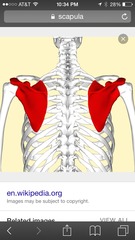
Orientation of the scapula
Orientation of the tibia

Orientation of the femur

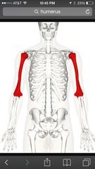
Orientation of the humerus
Orientation of the clavicle
Acromial is lateral
Sternal is medial
