UNC SPHS.582 Midterm 2
 Call Kai
 Learn
 Practice Test
 Spaced Repetition
 Match
 Flashcards
 Knowt Play
1/192

There's no tags or description
Looks like no tags are added yet.
193 Terms
deaf/hard of hearing/person with hearing loss
An impairment characterized by deficits in language and speech development because of a diminished or non-existent auditory response to sound. (Hearing impaired is discouraged)
genetic hearing loss
Hearing loss caused by the presence of an abnormal gene within one or more chromosomes (50-60%)
non-genetic hearing loss
caused by inflammatory disease, toxicity, noise, or injury that inflicts damage on any part of the auditory system (25-30%)
congenital hearing loss
hearing loss present at birth
acquired hearing loss
hearing loss that develops after birth
progressive hearing loss
hearing loss that increases over time
non-progressive hearing loss
hearing loss that is stable over time
syndromic hearing loss
hearing loss associated with a more pervasive syndrome such as Pendred syndrome
nearly 400 syndromic forms of deafness have been described
non-syndromic hearing loss
hearing loss that has no other associated findings
familial hearing loss
hearing loss that runs in the family
sporadic (de novo) hearing loss
hearing loss occurs when a child or individual has hearing loss caused by a brand-new genetic mutation that did not come from either biological parent
risk factors for permanent hearing loss in children
family history, in utero infection, craniofacial anomalies, low birth weight (
newborn hearing screening
newborn infants through six months require physiologic measures (ABRs and OAEs)
hearing screening is now a standard of care in all 50 states (required by law in 43)
EHDI guidelines
newborn hearing screening by 1 month - newborn nursey
diagnosis of hearing loss by 3 months - audiologist/ENT
intervention by 6 months - audiology / early intervention specialists ongoing management/monitoring/management
importance of early diagnosis and intervention
Educational benefits (neuro-typical children who are identified early and receive appropriate intervention are performing at age level. for those with co-occurring conditions, impact can be significantly reduced)
Relative to the past, children with congenital hearing loss are able to access spoken languge via hearing technologies (HAs/CIs)
D/HH+: co-occurring conditions
ADHD, autism, developmental delays, fine or gross motor impairments, learning disabilities, genetic conditions, language disabilities, genetic conditions, language impairments, vision impairments
expected skills for children who are D/HH
use of nonverbal communication, such as eye contact, gesture use, direction of facial expressions
shared enjoyment and joint attention
social initiations, vocalizations/signs directed toward others
functional and imaginative play skills
behavioral measures
subjective measures that require patient participation in some way (ex. audiogram)
physiologic measures
objective measures that the patient does not need to actively participate (ex. tympanometry)
cross-check principle
no single test result obtained during an assessment should be considered valid until you have obtained results across multiple measures to determine weather they agree and to make a diagnosis
acoustic immittance
measures how energy flows through the system from outer to middle ear
acoustic impedance
opposition to the flow of energy
acoustic compliance
ease of the flow of energy
tympanometry
an objective measure of middle ear function that evaluates how the tympanic membrane and middle ear system respond to changes in air pressure
middle ear pressure
def: tells us about the pressure within the middle ear space, identified near the location of the peak of the tympanogram
normative range: children/adults = -150 to +50
tympanic membrane compliance
def: also called admittance, tells us about how easily the tympanic membrane and middle ear system moves
normative range: children = 0.3-0.9, adults = 0.3-1.4
ear canal volume
def: tells us the volume or size of the ear canal between the probe and tympanic membrane
normative range: children = 0.4-1.0, adults = 0.6-1.5
tympanogram
the graph of plotted results from a 226 Hz tone delivered into the ear canal while the pressure is changed within the sealed canal, resulting in a measurement taken at the probe that plots the flexibility of the tympanic membrane and the ossicles
Type A tympanogram
normal middle ear pressure, normal compliance, normal ear canal volume
clinical example: healthy outer/middle ear
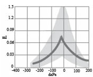
Type As tympanogram (shallow)
normal middle ear pressure, reduced compliance, normal ear canal volume
clinical example: fixation of ossicles, eardrum abnormalities

Type Ad tympanogram (deep)
normal middle ear pressure, abnormally high compliance, normal ear canal volume
clinical example: disarticulation of the ossicles

Type B tympanogram (normal volume)
"flat," no compliance of pressure peak indicated, normal ear canal volume
clinical example: middle ear effusion

Type B tympanogram (large volume)
"flat," no compliance of pressure peak indicated, increased ear canal volume
clinical example: perforation of the TM, patent PE tube

Type B tympanogram (small volume)
"flat," no compliance of pressure peak indicated, reduced ear canal volume
clinical example: occluding cerumen, probe hitting against the ear canal wall

Type C tympanogram
excessive negative middle ear pressure, normal or reduced compliance, normal ear canal
clinical exmple: eustachian tube dysfunction, may be associated with a cold, allergy, or URI

high frequency tympanometry
For infants younger than ~6 months, a 1000 Hz probe tone is used
typical tympanometry probe tone frequency
226 Hz
acoustic reflex physiology
an involuntary/reflexive muscle contraction of the middle ear in
response to very loud sound via stapedius & tensor tympani
muscles
ipsilateral
presenting sound and measuring in same ear
contralateral
presenting sound in one ear, measuring in the opposite ear
acoustic reflect threshold (ART)
o Present and normal (for adults 70 - 90 dB HL)
o Present and elevated (for adults 95 - 105 dB HL)
o Absent (for adults no response at or above 105 dB HL)
right ipsilateral acoustic reflex
sound is presented to the right ear, measuring response with probe in the right ear

left ipsilateral acoustic reflex
sound is presented to the left ear, measuring response with probe in the left ear

right contralateral acoustic reflex
sound is presented to the right ear, measuring response with probe in the left ear

left contralateral acoustic reflex
sound is presented to the left ear, measuring response with probe in the right ear

otoacoustic emissions (OAEs)
an objective measure of the integrity and function of the outer hair cells of the cochlea
OAEs setup: probe
measured using a small probe in the ear canal, quiet environment, can be measured in babies and adults
OAEs interpretation: present
There is minimal or no conductive hearing loss affecting outer/middle ear
The frequencies tested show normal or no worse than mild hearing loss in the cochlea
Outer hair cell function intact, but pathology could still be retrocochlea
OAEs interpretation: present but reduced
the outer hair cells in your cochlea (inner ear) are still functioning and producing echoes, but the amplitude or signal strength is lower than what is expected for a completely healthy, normal-hearing ear
OAEs interpretation: absent
There could be significant conductive hearing loss affecting outer/middle ear
There could be damage or pathology to outer hair cells from sensorineural hearing loss
OAEs physiology
because OAEs require sound to travel through the middle ear in
both directions, outer or middle ear problems can affect the results
basilar membrane
a structure that runs the length of the cochlea in the inner ear and holds the auditory receptors, called hair cells.
outer hair cells/prestin
a specialized motor protein in the OHCs that converts changes in membrane voltage directly into rapid cell contraction and elongation, which acts as a mechanical amplifier to boost hearing sensitivity and frequency selectivity
frequency tuning
frequency lowering (FL) technology and frequency response fine-tuning in hearing aids
distortion product OAEs (DPOAEs)
Evoked by 2 tones (Frequency 1 & Frequency 2), test frequencies 1000-8000 Hz
spontaneous OAEs (SOAEs)
Responses generated without auditory stimulus presented
About 60% of normal hearing humans have SOAE
transient-evoked OAEs (TEOAEs)
Responses generated to brief clicks or tone bursts
ototoxicity monitoring with OAEs
OAEs can be used to monitor cochlear function in patients receiving ototoxic medication
Changes in OAEs may occur before a patient notices a change in hearing
auditory evoked potentials
objective tests that measure electrical responses from the brain or brainstem pathways in response to sound
can provide information on if a hearing loss is present, degree of hearing, and differential diagnosis
auditory brainstem response (ABR)
Objective measure of auditory nerve and brainstem activity in response to sound
Electrical activity is recorded using electrodes placed on the scalp
The resulting waveform provides information about neural activity along the auditory pathway
Commonly used to assess hearing in infants and patients who cannot reliably complete behavioral testing
Can provide frequency specific information and be used to estimate hearing thresholds
ABR setup
To start, perform otoscopy to confirm clear ear canals
Prepare the skin and place electrodes on the forehead and near ears
Place insert earphones in the ears
Present brief, repeated sounds such as clicks or tone bursts
Electrodes record the electrical response of the auditory nerve and brainstem structures
Responses to many presentations are averaged together to identify the ABR waveform
Repeat recordings to confirm response reliability
Test each ear separately
ABR interpretation: abnormal results
may be associated with: Sensorineural hearing loss (mild - profound), Conductive hearing loss can affect ABR response, Retrocochlear pathology (e.g., tumor along central system), Auditory neuropathy spectrum disorder (ANSD)
ABR generators
Wave I: portion of the auditory nerve
Wave II: portion of the auditory nerve
Wave III: superior olivary complex
Wave IV: Pons, lateral lemniscus
Wave V: Midbrain, lateral lemniscus, and inferior colliculus
Wave VI/VII: Undetermined

ABR testing in infants/children
ABR is very sensitive to movement and muscle activity
The patient needs to be quiet and still (ideally asleep)
Young infants can often complete ABR during natural sleep
Older infants and children may require sedation or anesthesia when natural sleep testing is unsuccessful
When appropriate, ABR may also be coordinated with another sedated procedure or surgery
purpose of audiologic case history
understand the patient's primary concern, identify symptoms and risk factors for hearing loss, help determine which tests may be needed, provide context for interpreting audiologic test results, identify findings that may require medical referral
case history questions: adults vs. children
adults: ask patient directly about their hearing, symptoms, family history, and risk factors
children: heavily relying on caregiver, ask about primary concern, pregnancy/birth/prenatal history, medical history, family history, developmental milestones, and speech and language development
otitis
infection of the ear
otalgia
ear pain
otorrhea
drainage from the ear
aural fullness
pressure sensation in the ear
tinnitus
noises present in the ear (buzzing, ringing, etc)
vertigo
spinning sensation
basal cell carcinoma
outer ear, most common and least severe type of skin cancer, it is slow-growing and painless, not hearing loss associated, occurs from excessive UV exposure
exostoses
outer ear, bony growth in the ear canal, sometimes referred to as "surfer's ear," occurs most commonly in males, typically does not require any type of treatment, not expected to affect audiometric results unless growths close up ear canal
otitis externa
outer ear infection; symptoms include itchiness, aural fullness, redness, swelling, otalgia, and otorrhea; treatment by ENT or PCP includes antibacterial ear drops or topical steroids to improve inflammation of the ear canal
microtia
outer ear, occurs when the pinna is underdeveloped or malformed and may occur unilaterally or bilaterally, middle/inner ear structures may or may not be affected, can be surgically corrected by creating a pinna from cartilage from other parts of the body, may also use hearing aids that bypass outer ear and vibrate the inner ear directly
atresia
outer ear, an absence or closure of the external auditory meatus and it may co-occur with microtia, middle/inner ear structures may or may not be affected, can be surgically corrected by creating a pinna from cartilage from other parts of the body, may also use hearing aids that bypass outer ear and vibrate the inner ear directly
perforated tympanic membrane
outer ear; occurs when there is a hole in the eardrum (can be partial or full rupture); likely to report otalgia, otorrhea, or hearing changes; may occur from sudden pressure changes (barotrauma), traumatic noise, or self ear cleaning; HL configuration will depend on the size/shape/location of the perforation, treatment includes myringoplasty or tympanoplasty
tympanosclerosis
outer ear; occurs when there is calcification or scarring of the tympanic mebrane; likely to occur after recurrent ear infections or trauma to the eardrum; often asymptomatic, but may co-occur with other middle ear disorders; if extensive, may stiffen the tympanic membrane and cause conductive HL
Treacher Collins syndrome
outer ear, microtia and aural atresia, middle ear ossicular chain abnormalities also common, often associated with conductive hearing loss, inner ear function is often preserved
CHARGE syndrome
outer ear, genetic syndrome affecting multiple body systems , associated with small or shorter ears with unusual configuration (atresia may be present), likely to have inner ear issures (semicircular canal hypoplasia) and other cochlear abnormalities, associated with conductive, sensorineural, or mixed hearing loss
eustachian tube dysfunction
middle ear; occurs when there is an issue with the eustachian tube regulating pressure within the middle ear space, resulting in negative pressure buildup in the middle ear; commonly associated with allergies, congestion, and URI, often occurs with a retracted tympanic membrane and aural fullness; treatment includes the Valsalva procedure or Toynbee procedure; can be treated surgically in severe cases with balloon dilation
otitis media
middle ear infection; higher incidence in children relative to adults; viruses and/or bacteria can reach the middle ear from the nasopharynx via the eustachian tube; may be acute, recurrent, or associated with persistent effusion; symptoms include otalgia, redness, inflammation, fever, and decreased hearing
dormant otitis media
middle ear; patient starts treatment for otitis media and notices improvement and subsequently stops taking antibiotics; the unresolved otitis media lays "dormant" for a few weeks/months until overt symptoms of otitis media return; will often be more difficult to resolve because it may be a stronger strain and less susceptible to antibiotics
patulous eustachian tube
middle ear; occurs when the eustachian tube is chronically open; allows sound and pressure to travel between the nasopharynx and middle ear; autophony is a common symptom; chewing noise and breathing may be annoying; hearing is often normal and tymps likely to appear normal; diagnosed with a special continuous tympanogram
autophony
symptom of patulous eustachian tube where patient feels the "head in a barrel" sensation and may perceive own voice as overwhelmingly loud
pressure equalization (pe) tubes
middle ear; treatment option for patients with chronic or unresolved otitis media to moderate pressure between the middle ear and outer ear
patent PE tube
middle ear; when the tube is open and functioning properly and can be observed on tympanometry
cholesteatoma
middle ear; a benign mass in the middle ear space; commonly observed in patients with a history of recurrent ear infections; symptoms include foul-smelling otorrhea, otalgia, aural fullness, possible tinnitus, and difficulty hearing; generally unilateral and progressive; requires medical management and mastoidectomy and tympanoplasty may be recommended as treatment
mastoidectomy
middle ear; surgical removal of diseased or infected mastoid cells from mastoid bone
tympanoplasty
middle ear; surgical repair of the tympanic membrane
otosclerosis
middle ear; presents with abnormal bone growths on the middle ear bones and usually starts on the stapedial footplate and may spread across the ossicular chain; the bony growths can cause fixation of some or all of the ossicles, resulting in limited sound energy transmitted to the cochlea; slow growing and progressive; most common in women and strong genetic component; generally both ears are affected; symptoms include gradual hearing loss, tinnitus, and possible dizziness, Schwartze sign, & Lombard voice reflex; treatment includes stapedectomy
schwartze sign
red glow seen through the tympanic membrane and produced by increased vascularity of the promontory in some cases of otosclerosis
lombard voice reflex
the involuntary tendency of speakers to raise their voice volume, pitch, and duration when background noise increases above roughly 40 to 50 dB
stapedectomy
the surgical removal of the top portion of the stapes bone and the insertion of a small prosthetic device known as a piston that conducts sound vibrations to the inner ear
ossicular chain disarticulation
middle ear; occurs when there is a separation of the middle ear bones; may be caused from head trauma, fracture to the skull, chronic otitis media, or cholesteatoma; 50% of temporal bone fractures result in ossicular chain injury; symptoms include tinnitus, hearing loss, otalgia, possible vertigo; diagnosis can be made with CT scan; treatment includes ossicular chain reconstruction
common symptoms of inner ear disorders
hearing loss, poor word recognition, tinnitus, vertigo, dizziness, facial weakness
noise-induced hearing loss (NIHL)
inner ear; a sensorineural hearing loss caused by excessive noise exposure; may result from chronic or acoustic trauma; damage is typically permanent; symptoms include hearing loss and tinnitus
chronic trauma
damage to hearing from exposure to noise over time
acoustic trauma
damage to hearing from a single intense sound