Week 8 ,, Cestodes
 Call Kai
 Learn
 Practice Test
 Spaced Repetition
 Match
 Flashcards
 Knowt Play
1/64

There's no tags or description
Looks like no tags are added yet.
65 Terms
Proglottids
Segments that form from the body ,, New segments are continuously formed in the neck region just behind the tapeworm's head (scolex), pushing older segments toward the rear. As they move back, they mature and eventually become "gravid" (packed with fertilized eggs)
Which tape worm doesn’t require an intermediate host?
Hymenolepiasis
Hymenolepiasis Definitive Host
Humans, rats, or mice
Hymenolepiasis Intermediate Host
Insects (grain beetle)
Why is autoinfection possible with Hymenolepiasis?
The eggs passed in the feces are immediately infective and can be ingested by the same host
Which Hymenolepiasis species is most commonly found in humans?
H. nana
H. diminuta (“Rat Tape Worm”)
Rats are the definitive host and humans are an incidental host
Hymenolepis nana Infective Stage
Ingestion of cysticercoid-infected arthropods and embryonated eggs in feces ,, Autoinfection can occur if eggs remain inside the intestines
Hymenolepis nana Diagnostic Stage
Embryonated eggs in feces
Hymenolepiasis Clinical Features
Generally asymptomatic ,, Prolonged infection can lead to diarrhea, weight loss, weakness, vomiting, and seizures
Hymenolepiasis Geographical Location
Occurs worldwide but more frequent in warm, dry regions ,, Crowded environments with poor sanitation
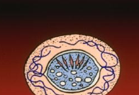
Which species does this ovum belong to?
H. nana

Which species does this ovum belong to?
H. diminuta
H. diminuta Microscopic Features
Scolex (head) has four suckers with an unarmed rostellum
H. nana Microscopic Feature
Scolex (head) has four suckers and a short rostellum with hooks (armed rostellum)
What is Taenia saginata also called?
Beef tapeworm
What is Taenia solium also called?
Pig tapeworm
Taenia saginata Geographic Distribution
Worldwide ,, Commonly found in regions where people have a tendency of eating raw beef (Africa, Latin America, Eastern Europe, and Asia)
Taenia saginata Infective Stage
Cysticercus in the tissue of the intermediate host
Taenia saginata Diagnostic Stage
Eggs and gavid proglottids in feces and are passed to the environment
T. saginata Clinical Features
Generally asymptomatic ,, Migrating proglottids can cause inflammation of the appendix and bile duct
Taeniasis
Adult tapeworm in intestine usually asymptomatic or abdominal discomfort
Cysticercosis
Larvae move out of the intestine into the host's tissue
Neurocysticercosis
Damage of the CNS by larval cysts ,, Symptoms include a wide range of neurological manifestations, including seizures, headache, intracranial hypertension, and psychiatric disturbances
T. solium Symptoms
Cysticercosis, neurocysticercosis, and cysticercosis
When should the stool specimen for T. solium be collected
On three different days

Which species does this egg belong to?
Taenia spp.
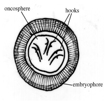
Which species does this egg belong to?
Taenia spp.
Scolex of T. solium
Contains 4 suckers with rostellum containing two rows of hooks
Scolex of T. saginata
Contains 4 large suckers without rostellum or rostellar hooks
Proglottids of T. saginata
12-30 lateral uterine branches
Proglottids of T. solium
7-13 lateral uterine branches

Identify the Species
T. saginata

Identify the Species
T. solium
Taenia solium Laboratory Diagnosis
Examination of feces for eggs or proglottids ,, Biopsy of infected tissue ,, ELISA
What is Diphyllobothriasis also known as?
Fish tapeworm
Which is the largest human tapeworm?
Fish tapeworm

Identify the Parasite
Diphyllobothrium latum

Identify the Parasite
Diphyllobothrium latum
Diphyllobothrium latum Infective Stage
Infected crustacean is ingested by small freshwater fish ,, The procercoid larva is released and develops into a plerocercoid larva
Diphyllobothrium latum Diagnostic Stage
Unembryonated eggs in feces
Diphyllobothriasis Geographic Distribution
Usually seen in the Northern Hemisphere ,, Infection can occur worldwide by exportation of freshwater fish
Diphyllobothriasis Clinical Features
Can be asymptomatic and last for decades ,, May cause diarrhea, vomiting, weight loss, and abdominal pains ,, Intestinal obstruction ,, Cholecystitis
Dipylidium caninum Definitive Hosts
Dogs and cats
Dipylidium caninum Intermediate Hosts
Fleas
Dipylidium caninum Geographical Distribution
Worldwide distribution ,, Infections have been reported in Europe, the Philippines, China, Japan, Argentina, and the United States
Dipylidium caninum Diagnostic Stage
Proglottids containing egg packets
Dipylidium caninum Infective Stage
Cysticercoid larva that develops inside the intermediate host

Identify the Species
Dipylidium caninum
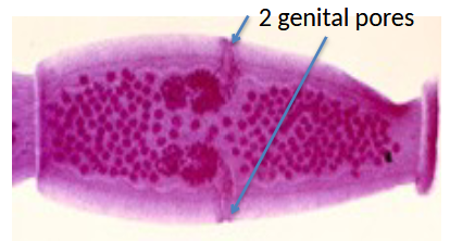
Identify the Species
Dipylidium caninum
Dipylidium caninum Clinical Features
Generally asymptomatic ,, Can be chronic and include mild diarrhea, stomach upset, and itching around the rectum
Diphyllobothriasis Laboratory Diagnosis
Microscopic identification of the eggs in stool ,, Examination of proglottids in stool ,, Rosette-shaped ovaries ,, Colonoscopy
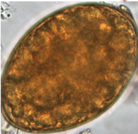
Identify the Species
Diphyllobothriasis

Identify the Species
Diphyllobothriasis
What is Echinococcus granulosus also known as?
Dog tapeworm or hydatid tapeworm
Echinococcus granulosus Geographical Distribution
Occurs worldwide in sheep-raising areas
Echinococcus granulosus Infective Stage
Ingestion of eggs due to close contact with infected dogs
Echinococcus granulosus Diagnostic Stage
Hydatid cyst located in liver, lung, or other sites
Echinococcus granulosus Intermediate Host
Sheeps
Echinococcus granulosus Accidental Host
Humans
Echinococcus granulosus Symptoms
Expanding cysts cause pressure and pain, which can lead to destruction of the tissue around the area where the cyst is located
Echinococcus granulosus in the Liver
Most common site ,, If cysts gets large enough then jaundice and portal hypertension develop ,, Rupture of cysts can cause death or anaphylactic shock
Echinococcus granulosus in the Lungs
If the cyst becomes large enough, coughing, shortness of breath, and chest pains can develop
Echinococcus granulosus Laboratory Diagnosis
Biopsy of liver, lung, or other organ that contains scoleces, brood capsules, hydatid sand, or daughter cysts in cyst fluid ,, Blood tests for antibodies ,, ELISA ,, X-ray or ultrasound scan

Identify the Species
Echinococcus granulosus